Mediastinal mass differential diagnosis: Difference between revisions
Trushatank (talk | contribs) No edit summary |
No edit summary |
||
| Line 1,474: | Line 1,474: | ||
|- | |- | ||
|} | |} | ||
==References== | |||
{{Reflist|2}} | |||
Revision as of 19:09, 19 February 2019
|
Mediastinal mass Microchapters |
Editor-In-Chief: C. Michael Gibson, M.S., M.D. [1]; Associate Editor(s)-in-Chief: Trusha Tank, M.D.[2]
Overview
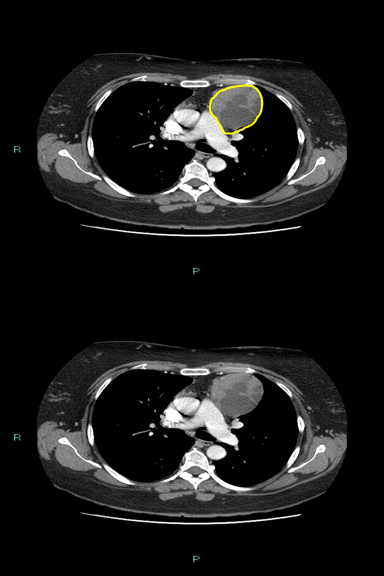
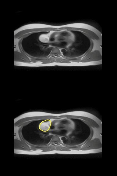
The mediastinum is a non-delineated group of structures in the thorax (chest), surrounded by loose connective tissue. It is the central compartment of the thoracic cavity. It contains the heart, the great vessels of the heart, esophagus, trachea, thymus, and lymph nodes of the central chest. A wide variety of diseases involving mediastinal structures can present as a mediastinal mass or widening of the mediastinum on imaging studies. Mediastinal widening is diagnosed by the mediastinum measuring greater than 8 cm in width on PA chest x-ray. The mediastinal mass may present with symptoms or even without any symptoms. Mediastinal mass may cause a variety of symptoms by the mass pressing against other mediastinal structures, collectively known as mediastinal syndrome. Mediastinal mass can be differentiated depending on their location in mediastinal cavity into: Anterior mediastinal mass, middle mediastinal mass, or posterior mediastinal mass. Mediastinal mass can also be differentiated according to the content of the mass such as: Cystic, fatty or solid (tumor).
Mediastinal Mass Differential Diagnosis
Wide variety of medical conditions can present as a mediastinal mass on radiological imaging.
- Mediastinal mass may cause obstruction, entrapment or infiltration of other mediastinal organs such as: Trachea, bronchi, esophagus, aorta, superior vena cava (SVC) or heart.
- Symptoms caused by any kind of mediastinal mass is collectively known as: Mediastinal syndrome
- Mediastinal syndrome includes:
- Symptoms caused by compression of the trachea: Dyspnea and respiratory insufficiency.
- Symptoms caused by compression of the esophagus: Dysphagia.
- Symptoms caused by compression of SVC causes superior vena cava syndrome: Vein distention, edema of the face or upper extremities and a positive Pemberton's sign.
- Pemberton's sign: Development of suffusion, plethora, or duskiness upon elevation of the arms above the head in patient
- Superior vena cava syndrome is the most severe complication of mediastinal syndrome and a medical emergency.
| ABBREVIATIONS: N/A: Not available, SOB: Shortness of breath, M/C: Most common, RI: Respiratory insufficiency, NM: Neuromuscular system, SVCS: Superior vena cava syndrome, SLE: Systemic lupus erythematosus disease, T3: Triiodothyronine, T4: Thyroxine, TSH: Thyroid stimulating hormone, TFT: Thyroid function test, MRI: Magnetic resonance imaging, CNS: Central nervous system, CSF: Cerebrospinal fluid, FNA: Fine needle aspiration, CBC: Complete blood count, COPD: Chronic obstructive pulmonary disease, AIDS: Acquired immune deficiency syndrome, HIV: Human immunodeficiency virus, Hep C: Hepatitis C virus, HTLV-1: human T-lymphotropic virus, EBV: Epstein Barr virus, HHV-8: Human herpes virus-8 | ||||||||
| Disease | Causes/risk factors | Clinical presentation | Paraclinical findings | |||||
|---|---|---|---|---|---|---|---|---|
| General symptoms | Mediastinal syndrome | |||||||
| Dyspnea/
RI |
Dysphagia | SVCS | Gold standard | Image | Additional findings | |||
| Anterior mediastinal mass | ||||||||
| Tumors | ||||||||
| Thymoma | + | + | + | Biopsy: |  |
Associated condition
| ||
| Disease | Etiology | Symptoms | Dyspnea/
RI |
Dysphagia | SVCS | Gold standard | Image | Additional findings |
| Fatty mass |
|
- | - | - | MRI: |  |
Fatty mass can be: | |
| Non-Hodgkin lymphoma |
(HIV, Hep C, HTLV-1, EBV, HHV-8, H. pylori, psittacosis, Campylobacter jejuni)
(pesticides, methotrexate, TNF inhibitors, trichloroethylene)
|
+/- | +/- | +/- | Excisional lymph node biopsy with immunohistochemical study |  |
| |
| Disease | Etiology | Symptoms | Dyspnea/
RI |
Dysphagia | SVCS | Gold standard | Image | Additional findings |
| Hodgkin's lymphoma | Epstein-Barr virus
|
Other
|
+ | + | +/- | Lymph node biopsy with immunohistochemistry
|
 |
Positron emission tomography (PET)
|
| Mediastinal germ cell tumor
(Non-teratomatous) |
|
+ | - | - | Biopsy: |  |
CT scan:
Laboratory finding: | |
| Teratoma |
|
Benign | +/- | +/- | +/- | Chest CT scan:
|
 |
N/A |
| Disease | Etiology | Symptoms | Dyspnea/
RI |
Dysphagia | SVCS | Gold standard | Image | Additional findings |
| Cystic mass | ||||||||
| Thymic cyst | Congenital
|
- | - | + | Biopsy with histopathology and cytology |  |
CT scan: | |
| Thyroid gland disease | ||||||||
| Mediastinal goiter |
|
+ | + | - | Radioactive iodine scan:
|
 |
Hyperactive gland (hyperthyroid):
Hypoactive gland (hypothyroid): Normal functioning gland (euthyroid): | |
| Disease | Etiology | Symptoms | Dyspnea/
RI |
Dysphagia | SVCS | Gold standard | Image | Additional findings |
| Middle mediastinal mass | ||||||||
| Cardiovascular Disease | ||||||||
| Pericardial effusion |
|
|
+ | +/- | - | Echocardiography guided pericardiocentesis:
|
 |
Physical findings:
EKG:
|
| Aortic dissection | + | +/- | + | MRI: |  |
TEE:
CTA:
| ||
| Disease | Etiology | Symptoms | Dyspnea/
RI |
Dysphagia | SVCS | Gold standard | Image | Additional findings |
| Superior vena cava obstruction | Compression of SVC from: |
|
+ | + | ++ | Contrast-enhanced CT scan:
|
 |
Invasive contrast venography:
|
| Partial anomalous pulmonary venous connection |
|
+ | - | - | MRI with contrast:
|
 |
Associated with
PFT:
| |
| Gastrointestinal tract disease | ||||||||
| Esophageal achalasia |
|
+ | + | - | High resolution manometry (HRM):
|
 |
X ray:
| |
| Disease | Etiology | Symptoms | Dyspnea/
RI |
Dysphagia | SVCS | Gold standard | Image | Additional findings |
| Esophageal cancer |
|
|
- | + | - | Endoscopy with biopsy:
|
 |
Barium swallow:
|
| Esophageal rupture |
|
Other: Patients with cervical perforations can present with
|
+ | + | - | Esophagogram:
|
 |
CT scan:
|
| Hiatus hernia |
|
- | + | - | High resolution manometry with esophageal pressure topography (EPT):
|
 |
Ultrasound:
Ultrasound in pediatric population:
| |
| Disease | Etiology | Symptoms | Dyspnea/
RI |
Dysphagia | SVCS | Gold standard | Image | Additional findings |
| Pulmonary disease | ||||||||
| Hilar lymphadenopathy | Lymphadenopathy:
|
Constituitional symptoms like: | + | - | - | Lymph node biopsy and histopathology |  |
CT scan |
| Pneumomediastinum |
|
|
+ | - | - | CT scan:
Pediatric pneumomediastinum: |
 |
Physical exam:
|
| Disease | Etiology | Symptoms | Dyspnea/
RI |
Dysphagia | SVCS | Gold standard | Image | Additional findings |
| Sarcoidosis |
Immune system
|
Cutaneous sarcoidosis
Renal & electrolyte
|
+ | - | - | Endoscopy with biopsy and histopathology
|
 |
Laboratory findings:
|
| Disease | Etiology | Symptoms | Dyspnea/
RI |
Dysphagia | SVCS | Gold standard | Image | Additional findings |
| Infectious disease | ||||||||
| Mediastinitis |
Risk factors: |
+ | - | - | Culture and sensitivity of mediastinal tissue collected by biopsy/aspiration |  |
Physical exam
| |
| Anthrax | B. anthracis
People at higher risk
|
+ | - | - | Culture and sensitivity: |  |
CT scan
| |
| Tuberculosis | M. tuberculosis
Traveling or living in endemic regions (Sub-saharan African, Russia, India, Pakistan, China)
The risk of contracting TB increases in:
|
|
+ | - | - | Culture and sensitivity |  |
Chest X-ray
|
| Disease | Etiology | Symptoms | Dyspnea/
RI |
Dysphagia | SVCS | Gold standard | Image | Additional findings |
| Cystic mass | ||||||||
| Bronchogenic cyst |
|
|
+ | - | - | CT scan |  |
CT scan:
|
| Esophageal duplication cysts |
|
|
- | + | - | Endoscopic ultrasound (EUS)
|
 |
Endoscopic ultrasound-guided FNA
|
| Disease | Etiology | Symptoms | Dyspnea/
RI |
Dysphagia | SVCS | Gold standard | Image | Additional findings |
| Lymphangioma |
|
|
+ | + | - | Histopathology and cytology |  |
|
| Chronic inflammatory disease | ||||||||
| Churg-Strauss syndrome | + | +/- | - | Lung biopsy
4 out of 6 positive :
|
 |
High-resolution computerized tomography (HRCT):
| ||
| Disease | Etiology | Symptoms | Dyspnea/
RI |
Dysphagia | SVCS | Gold standard | Image | Additional findings |
| Posterior mediastinal mass | ||||||||
| Cystic mass | ||||||||
| Mediastinal neurenteric cyst | + | +/- | - | CT scan:
|
 |
Postnatal chest X-ray:
| ||
| Pancreatic pseudocyst |
|
- | - | - | Histopathology and cytology of cyst and fluid content |  |
CT scan
| |
| Disease | Etiology | Symptoms | Dyspnea/
RI |
Dysphagia | SVCS | Gold standard | Image | Additional findings |
| Central nervous system disease | ||||||||
| Meningocele |
Congenial defect: Maternal nutrition factors:
2. Environmental factors:
|
Symptoms depend on the severity of the defect
Difficulties with executive functions including:
|
- | - | - | Prenatal 2D/3D ultrasound:
|
 |
Laboratory tests:
MRI:
|
| Neurilemmoma |
|
- | - | - | Biopsy with histopathology |  |
MRI | |
| ABBREVIATIONS: N/A: Not available, SOB: Shortness of breath, M/C: Most common, RI: Respiratory insufficiency, NM: Neuromuscular system, SVCS: Superior vena cava syndrome, SLE: Systemic lupus erythematosus disease, T3: Triiodothyronine, T4: Thyroxine, TSH: Thyroid stimulating hormone, TFT: Thyroid function test, MRI: Magnetic resonance imaging, CNS: Central nervous system, CSF: Cerebrospinal fluid, FNA: Fine needle aspiration, CBC: Complete blood count, COPD: Chronic obstructive pulmonary disease, AIDS: Acquired immune deficiency syndrome, HIV: Human immunodeficiency virus, Hep C: Hepatitis C virus, HTLV-1: human T-lymphotropic virus, EBV: Epstein Barr virus, HHV-8: Human herpes virus-8 | ||||||||
References
- ↑ 1.00 1.01 1.02 1.03 1.04 1.05 1.06 1.07 1.08 1.09 1.10 1.11 1.12 1.13 1.14 Juanpere S, Cañete N, Ortuño P, Martínez S, Sanchez G, Bernado L (February 2013). "A diagnostic approach to the mediastinal masses". Insights Imaging. 4 (1): 29–52. doi:10.1007/s13244-012-0201-0. PMID 23225215.
- ↑ Riccio, John C.; Abbott, Jean (1990). "A simple sore throat?: Retropharyngeal emphysema secondary to free-basing cocaine". The Journal of Emergency Medicine. 8 (6): 709–712. doi:10.1016/0736-4679(90)90283-2. ISSN 0736-4679.
- ↑ Abbas, Paulette I.; Akinkuotu, Adesola C.; Peterson, Michelle L.; Mazziotti, Mark V. (2015). "Spontaneous pneumomediastinum in the pediatric patient". The American Journal of Surgery. 210 (6): 1031–1036. doi:10.1016/j.amjsurg.2015.08.002. ISSN 0002-9610.
- ↑ Andrade Semedo, Flávia Helena Monteiro; Silva, Rosário Santos; Pereira, Sónia; Alfaiate, Teresa; Costa, Teresa; Fernandez, Pilar; Pereira, Amélia (2012). "Pneumomediastino espontâneo: relato de um caso". Revista da Associação Médica Brasileira. 58 (3): 355–357. doi:10.1590/S0104-42302012000300017. ISSN 0104-4230.
- ↑ Hammond DI (March 1984). "The "ring-around-the-artery" sign in pneumomediastinum". J Can Assoc Radiol. 35 (1): 88–9. PMID 6725378.
- ↑ Levin, Bertram (1973). "The continuous diaphragm sign". Clinical Radiology. 24 (3): 337–338. doi:10.1016/S0009-9260(73)80050-9. ISSN 0009-9260.
- ↑ Lillard, Richard L.; Allen, Parker (1965). "The Extrapleural Air Sign in Pneumomediastinum". Radiology. 85 (6): 1093–1098. doi:10.1148/85.6.1093. ISSN 0033-8419.