Right atrium
|
WikiDoc Resources for Right atrium |
|
Articles |
|---|
|
Most recent articles on Right atrium Most cited articles on Right atrium |
|
Media |
|
Powerpoint slides on Right atrium |
|
Evidence Based Medicine |
|
Clinical Trials |
|
Ongoing Trials on Right atrium at Clinical Trials.gov Clinical Trials on Right atrium at Google
|
|
Guidelines / Policies / Govt |
|
US National Guidelines Clearinghouse on Right atrium
|
|
Books |
|
News |
|
Commentary |
|
Definitions |
|
Patient Resources / Community |
|
Patient resources on Right atrium Discussion groups on Right atrium Patient Handouts on Right atrium Directions to Hospitals Treating Right atrium Risk calculators and risk factors for Right atrium
|
|
Healthcare Provider Resources |
|
Causes & Risk Factors for Right atrium |
|
Continuing Medical Education (CME) |
|
International |
|
|
|
Business |
|
Experimental / Informatics |
Editor-In-Chief: C. Michael Gibson, M.S., M.D. [1]
Overview
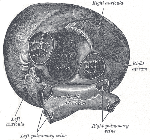
The right atrium (in older texts termed the right auricle) is one of four chambers (two atria and two ventricles) in the human heart. It receives de-oxygenated blood from the superior and inferior vena cavae and the coronary sinus, and pumps it into the right ventricle through the tricuspid valve.
The sinoatrial node (SAN) is located within this chamber next to the vena cava. This is a group of pacemaker cells which spontaneously depolarise to create an action potential. The cardiac action potential then spreads across both atria causing them to contract forcing the blood they hold into their corresponding ventricles.
In early life, when a fetus is in the womb, the right atrium has a hole within its septum through to the left atrium, this makes them continuous with each other which is essential for fetal circulation. This junction is called the foramen ovale. Once born (usually within a year's time) the foreman ovale seals over and it is renamed as the fossa ovalis. The fossa ovalis is seen as an embryonic remnant. In some cases, the formane ovale fails to close and is present in 20% of the general population, however it does not cause problems in the vast majority. This is known as patent foramen ovale.
Within the fetal right atrium, blood from the inferior vena cava and the superior vena flow in separate streams to different locations in the heart, and this has been reported to occur through the Coanda effect.[1]
The right atrium also holds the coronary sinus which is the opening of the vein that drains the myocardium itself. Attached to the right atrium is the right auricular appendix.
Additional images
-
Heart seen from above.

See also
References
- ↑ Ashrafian H. The Coanda effect and preferential right atrial streaming. Chest. 2006 Jul;130(1):300.