Shoulder
Editor-In-Chief: C. Michael Gibson, M.S., M.D. [1]
In human anatomy, the shoulder comprises the part of the body where the arm attaches to the torso. It is made up of three bones: the clavicle (collarbone), the scapula (shoulder blade), and the humerus (upper arm bone) as well as associated muscles, ligaments and tendons. The articulations between the bones of the shoulder make up the shoulder joints. The shoulder must be flexible for the wide range of motion required in the arms and hands and also strong enough to allow for actions such as lifting, pushing and pulling. The compromise between these two functions results in a large number of shoulder problems not faced by other joints such as the hip.
Joints of the shoulder
There are three joints of the shoulder: The glenohumeral, acromioclavicular, and the sternoclavicular joints.
Glenohumeral joint
The glenohumeral joint is the main joint of the shoulder and the generic term "shoulder joint" usually refers to it. It is a ball and socket joint that allows the arm to rotate in a circular fashion or to hinge out and up away from the body. It is formed by the articulation between the head of the humerus and the lateral scapula. The "ball" of the joint is the rounded, medial anterior surface of the humerus and the "socket" is formed by the glenoid fossa, the dish-shaped portion of the lateral scapula. The shallowness of the fossa and relatively loose connections between the shoulder and the rest of the body allows the arm to have tremendous mobility, at the expense of being much easier to dislocate than most other joints in the body.
The capsule is a soft tissue envelope that encircles the glenohumeral joint and attaches to the scapula, humerus, and head of the biceps. It is lined by a thin, smooth synovial membrane. This capsule is strengthened by the coracohumeral ligament which attaches the coracoid process of the scapula to the greater tubercle of the humerus. There are also three other ligaments attaching the lesser tubercle of the humerus to lateral scapula and are collectively called the glenohumeral ligaments.
There is also a ligament called semicirculare humeri which is a transversal band between the posterior sides of the tuberculum minus and majus of the humerus. This band is one of the most important strengtening ligaments of the joint capsule.
Acromioclavicular joint
The acromioclavicular (AC) joint is located between the acromion process of the scapula (part of the scapula that forms the highest point of the shoulder) and the distal end of the clavicle.
The capsule of this joint is reinforced by the coracoclavicular ligament between the scapula and clavicle at the point of articulation. The coracoclavicular ligament in further detail is created by the conoid ligament, medial from the coracoid process of the scapula and inserts on the conoid tubercle of the clavicle. Lateral to the conoid ligament is the trapezoid ligament, which runs from the coracoid process of the scapula to the trapezoid line of the clavicle. One more ligament, the coracoacromial ligament, running from the corocoid process to the acromion of the scapula contributes to the integrity of the acromioclavicular joint.
Sternoclavicular joint
The sternoclavicular occurs at the medial end of the clavicle with the manubrium or top most portion of the sternum. The clavicle is triangular and rounded and the manubrium is convex the two bones articulate.
Movements of the shoulder
The muscles and joints of the shoulder allow it to move through a remarkable range of motion, making it the most mobile joint in the human body. The shoulder can abduct, adduct (such as during the shoulder fly), rotate, be raised in front of and behind the torso and move through a full 360° in the sagittal plane. This tremendous range of motion also makes the shoulder extremely unstable, far more prone to dislocation and injury than other joints.
Major muscles
The muscles that are responsible for movement in the shoulder attach to the scapula, humerus, and clavicle. The muscles that surround the shoulder form the shoulder cap and underarm.
| Name | Attachments | Function |
| serratus anterior | Originates on the surface of the upper eight ribs at the side of the chest and inserts along the entire anterior length of the medial border of the scapula. | It fixes the scapula into the thoracic wall and aids in rotation and abduction of the shoulders. |
| subclavius | Located inferior to the clavicle, originating on the first rib and inserting on the subclavian groove of the clavicle. | It depresses the lateral clavicle and also acts to stabilize the clavicle. |
| pectoralis minor | Arises from the third, fourth, and fifth ribs, near their cartilage and inserts into the medial border and upper surface of the coracoid process of the scapula. | This muscle aids in respiration, medially rotates the scapula, protracts the scapula, and also draws the scapula inferiorly. |
| sternocleidomastoid | Attaches to the sternum (sterno-), the clavicle (cleido-), and the mastoid process of the temporal bone of the skull. | Most of its actions flex and rotate the head. In regards to the shoulder, however, it also aids in respiration by elevating the sternoclavicular joint when the head is fixed. |
| levator scapulae | Arises from the transverse processes of the first four cervical vertebrae and inserts into the vertebral border of the scapula. | It is capable of rotating the scapula downward and elevating the scapula. |
| rhomboid major and rhomboid minor (work together) | They arise from the spinous processes of the thoracic vertebrae T1 to T5 as well as from the spinous processes of the seventh cervical and first thoracic vertebrae. They insert on the medial border of the scapula, from about the level of the scapular spine to the scapula's inferior angle. | They are responsible for downward rotation of the scapula with the levator scapulae, as well as adduction of the scapula. |
| trapezius | Arises from the occipital bone, the ligamentum nuchae, the spinous process of the seventh cervical, and the spinous processes of all the thoracic vertebrae, and from the corresponding portion of the supraspinal ligament. It inserts on the lateral clavicle, the acromion process, and into the spine of the scapula. | Different portions of the fibers perform different actions on the scapula: depression, upward rotation, elevation, and adductions. |
| deltoid, anterior fibers | Arises from the anterior border and upper surface of the lateral third of the clavicle. | The anterior fibres are involved in shoulder abduction when the shoulder is externally rotated. The anterior deltoid is weak in strict transverse flexion but assists the pectoralis major during shoulder transverse flexion / shoulder flexion (elbow slightly inferior to shoulders). |
| deltoid, middle fibers | Arises from the lateral margin and upper surface of the acromion. | The middle fibres are involved in shoulder abduction when the shoulder is internally rotated, are involved in shoulder flexion when the shoulder is internally rotated, and are involved in shoulder transverse abduction (shoulder externally rotated) -- but are not utilized significantly during strict transverse extension (shoulder internally rotated). |
| deltoid, posterior fibers | Arises from the lower lip of the posterior border of the spine of the scapula, as far back as the triangular surface at its medial end. | The posterior fibres are strongly involved in transverse extension particularly since the latissimus dorsi muscle is very weak in strict transverse extension. The posterior deltoid is also the primary shoulder hyperextensor. |
Rotator cuff
The rotator cuff is a structure composed of tendons that, with associated muscles (supraspinatus, infraspinatus, teres minor and subscapularis), holds the ball at the top of the humerus in the glenoid socket and provideoulder joint. The tendons of the rotator cuff muscles also connect to the capsule of the glenohumeral joint.
Two filmy sac-like structures called bursae permit smooth gliding between bone, muscle, and tendon. They cushion and protect the rotator cuff from the bony arch of the acromion.
Measurement of shoulder loads

For understanding normal and pathologic shoulder function knowledge of forces in the glenohumeral joint is essential. It forms the basis for performing fracture treatment or joint replacement surgery, for optimizing implant design and fixation and for improving and verifying analytical biomechanical models of the shoulder. With instrumented shoulder implants the joint contact forces and moments can be measured in vivo during different activities.
Additional images
-
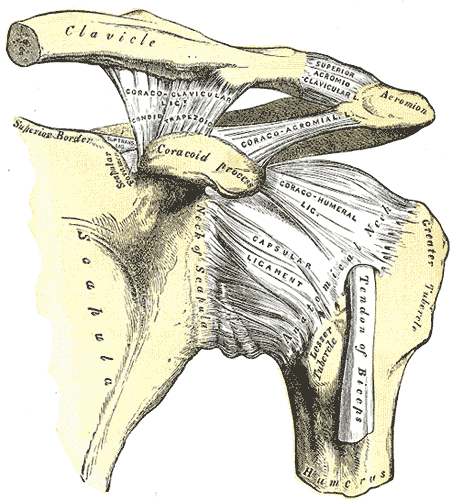
The left shoulder and acromioclavicular joints, and the proper ligaments of the scapula.

Medical problems
References
- Calais-Germain, Blandine. "Anatomy of Movement", Eastland Press, 1993. ISBN 0-939616-17-3
- Martini, Frederic; Timmons, Michael; McKinnley, Michael. "Human Anatomy", 3rd Edition, Prentice-Hall, 2000. ISBN 0-13-010011-0
External links
- Shoulder Resurfacing pt 1
- Shoulder Resurfacing pt 2
- Shoulder Resurfacing pt 3
- Video of the shoulder carriage in motion
- NIH (article includes text from this source)
Template:Joints of upper limbs Template:Human anatomical features
ar:كتف ast:Costazu ca:Espatlla pdc:Schulter de:Schulter eo:Ŝultro it:Spalla he:כתף la:Umerus ln:Libɛkɛ nl:Schouder nn:Skulder simple:Shoulder fi:Olkapää sv:Axel (kroppsdel) vls:Schoere yi:אקסל