Heart

|
WikiDoc Resources for Heart |
|
Articles |
|---|
|
Media |
|
Evidence Based Medicine |
|
Clinical Trials |
|
Ongoing Trials on Heart at Clinical Trials.gov Clinical Trials on Heart at Google
|
|
Guidelines / Policies / Govt |
|
US National Guidelines Clearinghouse on Heart
|
|
Books |
|
News |
|
Commentary |
|
Definitions |
|
Patient Resources / Community |
|
Directions to Hospitals Treating Heart Risk calculators and risk factors for Heart
|
|
Healthcare Provider Resources |
|
Continuing Medical Education (CME) |
|
International |
|
|
|
Business |
|
Experimental / Informatics |
Editor-In-Chief: C. Michael Gibson, M.S., M.D. [1]
Overview
The heart is a muscular organ responsible for pumping blood through the blood vessels by repeated, rhythmic contractions, or a similar structure in the annelids, mollusks, and arthropods.[1] The term cardiac (as in cardiology) means "related to the heart" and comes from the Greek καρδία, kardia, for "heart." The heart is composed of cardiac muscle, an involuntary muscle tissue which is found only within this organ.[2] The average human heart beating at 72 BPM, will beat approximately 2.5 billion times during a lifetime of 66 years.
Early development
The human embryonic heart begins beating about 21 days after conception, or five weeks after the last normal menstrual period (LMP), which is the date normally used to date pregnancy. The human heart begins beating at a rate near the mother’s, about 75-80 beats per minute (BPM). The embryonic heart rate (EHR) then accelerates linearly for the first month of beating, peaking at 165-185 BPM during the early 7th week, (early 9th week after the LMP). This acceleration is approximately 3.3 BPM per day, or about 10 BPM every three days, an increase of 100 BPM in the first month.[3]
After peaking at about 9.5 weeks after the LMP, it decelerates to about 152 BPM (+/-25 BPM) during the 15th week after the LMP. After the 15th week the deceleration slows reaching an average rate of about 145 (+/-25 BPM) BPM at term. The regression formula which describes this acceleration before the embryo reaches 25 mm in crown-rump length or 9.2 LMP weeks is Age in days = EHR(0.3)+6
There is no difference in male and female heart rates before birth.[4]
Structure
In the human body, the heart is usually situated in the middle of the thorax with the largest part of the heart slightly offset to the left (although sometimes it is on the right, see dextrocardia), underneath the breastbone (see diagrams). The heart is usually felt to be on the left side because the left heart (left ventricle) is stronger (it pumps to all body parts). The left lung is smaller than the right lung because the heart occupies more of the left hemithorax. The heart is enclosed by a sac known as the pericardium and is surrounded by the lungs. The pericardium is a double membrane structure containing a serous fluid to reduce friction during heart contractions. The mediastinum, a subdivision of the thoracic cavity, is the name of the heart cavity.
The apex is the blunt point situated in an inferior (pointing down and left) direction. A stethoscope can be placed directly over the apex so that the beats can be counted. It is located posterior to the 5th intercostal space in the left mid-clavicular line. In normal adults, the mass of the heart is 250-350 g (9-12 oz), or about three quarters the size of a clenched fist, but extremely diseased hearts can be up to 1000 g (2 lb) in mass due to hypertrophy. It consists of four chambers, the two upper atria (singular: atrium ) and the two lower ventricles. Below, on the right, is a picture of a fresh human heart which was removed from a 64-year-old British male.
-
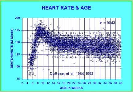
At 21 days after conception, the human heart begins beating at 70 to 80 beats per minute and accelerates linearly for the first month of beating.
-
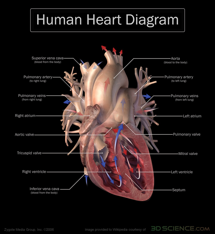
Anterior (frontal) view of the opened heart. Arrows indicate normal blood flow. Image provided courtesy of www.3dscience.com.
-
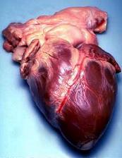
Human heart


Functioning
The function of the right side of the heart (see right heart) is to collect de-oxygenated blood, in the right atrium, from the body and pump it, via the right ventricle, into the lungs (pulmonary circulation) so that carbon dioxide can be dropped off and oxygen picked up (gas exchange). This happens through a passive process called diffusion. The left side (see left heart) collects oxygenated blood from the lungs into the left atrium. From the left atrium the blood moves to the left ventricle which pumps it out to the body. On both sides, the lower ventricles are thicker and stronger than the upper atria. The muscle wall surrounding the left ventricle is thicker than the wall surrounding the right ventricle due to the higher force needed to pump the blood through the systemic circulation.
Starting in the right atrium, the blood flows through the tricuspid valve to the right ventricle. Here it is pumped out the pulmonary semilunar valve and travels through the pulmonary artery to the lungs. From there, blood flows back through the pulmonary vein to the left atrium. It then travels through the bicuspid valve to the left ventricle and on to through the aortic semilunar valve to the aorta. The aorta forks, and the blood is divided between major arteries which supply the upper and lower body. The blood travels the arteries to the smaller arterioles, then finally to the tiny capillaries which feed each cell. The (relatively) deoxygenated blood then travels to the venules, which coalesce into veins, then to the inferior and superior vena cavae and finally back to the right atrium where the process began.
The heart is effectively a syncytium, a meshwork of cardiac muscle cells interconnected by contiguous cytoplasmic bridges. This relates to electrical stimulation of one cell spreading to neighboring cells
First aid
See cardiac arrest for emergencies involving the heart
If a person is encountered in cardiac arrest (no heartbeat), cardiopulmonary resuscitation (CPR) should be started and help called. If an automated external defibrillator is available, this device may automatically administer defibrillation if this is indicated. Usually, if there is enough time, the person can be rushed to the hospital where he or she will be cared for by a cardiologist, a doctor who specializes in the heart and lungs.
References
- ↑ The American Heritage Stedman's Medical Dictionary. "KMLE Medical Dictionary Definition of heart"..
- ↑ The American Heritage Stedman's Medical Dictionary. "KMLE Medical Dictionary Definition of cardiac".
- ↑ http://www.obgyn.net/us/us.asp?page=/us/cotm/0001/ehr2000
- ↑ Terry J. DuBose http://www.obgyn.net/english/pubs/features/dubose/ehr-age.htm Sex, Heart Rate and Age]
See also
- Artificial heart
- Atrium
- Blood pressure
- Cardiology
- Cardiothoracic Surgery
- Cardiovascular pathology
- Circulatory system
- Echocardiography
- Electrical conduction system of the heart
- Haemodynamics
- Heart cancer
- Heart defects
- Heart rate
- Heart transplantation
- Heart valve
- Human anatomy
- Pulse
- Ventricle
- Aorta
- Ventricular hypertrophy
- Holiday heart syndrome
- Circle map — simplified mathematical model of the beating heart.
- MUGA scan
- Cardiac stress test
External links
- eMedicine: Surgical anatomy of the heart
- Very Comprehensive Heart Site
- Self Improvement Wednesday - ABC 702 Drive audio
- The circulatory system
- The position of the heart
- Interactive 3D heart This realistic heart can be rotated, and all its components can be studied from any angle.