Nerve

|
WikiDoc Resources for Nerve |
|
Articles |
|---|
|
Media |
|
Evidence Based Medicine |
|
Clinical Trials |
|
Ongoing Trials on Nerve at Clinical Trials.gov Clinical Trials on Nerve at Google
|
|
Guidelines / Policies / Govt |
|
US National Guidelines Clearinghouse on Nerve
|
|
Books |
|
News |
|
Commentary |
|
Definitions |
|
Patient Resources / Community |
|
Directions to Hospitals Treating Nerve Risk calculators and risk factors for Nerve
|
|
Healthcare Provider Resources |
|
Continuing Medical Education (CME) |
|
International |
|
|
|
Business |
|
Experimental / Informatics |
Editor-In-Chief: C. Michael Gibson, M.S., M.D. [1]
Overview
A nerve is an enclosed, cable-like bundle of axons (the long, slender projection of a neuron). Neurons are sometimes called nerve cells, though this term is technically imprecise since many neurons do not form nerves, and nerves also include the glial cells that ensheath the axons in myelin.
Anatomy
Nerves are part of the peripheral nervous system. Afferent nerves convey sensory signals to the central nervous system, for example from skin or organs, while efferent nerves conduct stimulatory signals from the central nervous system to the muscles and glands. Afferent and efferent nerves are often arranged together, forming mixed nerves. The median nerve controls motor and sensory function in the hand.
Each peripheral nerve is covered externally by a dense sheath of connective tissue, the epineurium. Underlying this is a layer of flat cells forming a complete sleeve, the perineurium. Perineurial septa extend into the nerve and subdivide it into several bundles of fibres. Surrounding each such fibre is the endoneurial sheath. This is a tube which extends, unbroken, from the surface of the spinal cord to the level at which the axon synapses with its muscle fibres or ends in sensory endings. The endoneurial sheath consists of an inner sleeve of material called the glycocalyx and an outer, delicate, meshwork of collagen fibres. Peripheral nerves are richly supplied with blood.
Most nerves connect to the middle systems through the spinal cord. The twelve cranial nerves, however, connect directly to parts of the brain. Spinal nerves are given letter-number combinations according to the vertebra through which they connect to the spinal column. Cranial nerves are assigned numbers, usually expressed as Roman numerals from I to XII. In addition, most nerves and major branches of nerves have descriptive names. Inside the central nervous system, bundles of axons are termed tracts rather than nerves.
The signals that nerves carry, sometimes called nerve impulses, are also known as action potentials: rapidly (up to 120 m/s) traveling electrical waves, which begin typically in the cell body of a neuron and propagate rapidly down the axon to its tip or "terminus." The signals cross over from the terminus to the adjacent neurotransmitter receptor through a gap called the synapse. Motor neurons innervate or activate muscles groups. The nerve system runs through the spinal cord.
Clinical importance
Damage to nerves can be caused by physical injury, swelling (e.g. carpal tunnel syndrome), autoimmune diseases (e.g. Guillain-Barré syndrome), infection (neuritis), diabetes, or failure of the blood vessels surrounding the nerve. Pinched nerves occur when pressure is placed on a nerve, usually from swelling due to an injury or pregnancy. Nerve damage or pinched nerves are usually accompanied by pain, numbness, weakness, or paralysis. Patients may feel these symptoms in areas far from the actual site of damage, a phenomenon called referred pain. Referred pain occurs because when a nerve is damaged, signaling is defective from all parts of the area which the nerve receives input, not just the site of the damage. Neurologists usually diagnose disorders of the nerves by a physical examination, including the testing of reflexes, walking and other directed movements, muscle weakness, proprioception, and the sense of touch. This initial exam can be followed with tests such as nerve conduction study and electromyography (EMG).
See also
Additional images
-
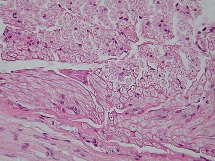
Peripheral nerve, cross section

Template:Nerves Template:Cranial nerves Template:Cervical plexus Template:Brachial plexus Template:Autonomic Template:Lumbosacral plexus
ar:عصب
cs:Nerv
de:Nerv
he:עצב
ko:신경
io:Nervo
id:Saraf
is:Taug
it:Nervo
la:Nervus
lt:Nervas
mk:Нерв
nl:Zenuw
simple:Nerve
fi:Hermo
sv:Nerv
yi:נערוו