Venography
|
WikiDoc Resources for Venography |
|
Articles |
|---|
|
Most recent articles on Venography |
|
Media |
|
Evidence Based Medicine |
|
Clinical Trials |
|
Ongoing Trials on Venography at Clinical Trials.gov Clinical Trials on Venography at Google
|
|
Guidelines / Policies / Govt |
|
US National Guidelines Clearinghouse on Venography
|
|
Books |
|
News |
|
Commentary |
|
Definitions |
|
Patient Resources / Community |
|
Patient resources on Venography Discussion groups on Venography Patient Handouts on Venography Directions to Hospitals Treating Venography Risk calculators and risk factors for Venography
|
|
Healthcare Provider Resources |
|
Causes & Risk Factors for Venography |
|
Continuing Medical Education (CME) |
|
International |
|
|
|
Business |
|
Experimental / Informatics |
Editor-In-Chief: C. Michael Gibson, M.S., M.D. [1]
Overview
Venography (also called phlebography) is a procedure in which an x-ray of the veins, a venogram, is taken after a special dye is injected into the vein or even bone marrow. It is the gold standard for diagnosing acute deep venous thrombosis although its use has been largely supplanted by the less invasive Duplex ultrasound scanning. Venography can also be used to distinguish blood clots from obstructions in the veins, to evaluate congenital vein problems, to evaluate veins prior to treatment of chronic venous insufficiency, to see how well the deep leg vein valves are functioning or to identify a vein for coronary artery bypass grafting.
Indications
A venogram is indicated to evaluate the following entities:
- Deep vein thrombosis
- Lower limb edema of unknown origin
- Congenital venous abnormalities
- Incompetent perforating veins
- Evaluation of veins prior to coronary artery bypass grafting
Contraindication
Performance of a venogram or administration of iodinated contrast in a patient taking metformin can result in metabolic acidosis.
Procedure
- Tile the flouroscopic couch feet down approximately 30 degrees to slow contrast media travel and help dilate the lower veins. Have the patient perform the Valsalva maneuver to further delay the dye transit.
- Apply a tourniquet is applied just above the ankle.
- Cannulate a vein on the dorsum of the foot with a 19 or 21 gauge needle.
- Inject up to 40 cc of contrast media and obtain a series of films from the foot to the pelvis.
Complications
- Thrombophlebits
- Contrast media extravasation
- Arrhythmias in patients with pulmonary hypertension
- General contrast media complications allergic reaction or anaphylaxis
- Hematoma at site of injection site
- Pulmonary embolus
Images
-
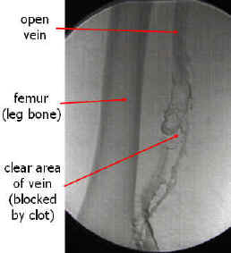
A venographic image of a deep vein thrombosis. Source: www.lakeridgehealth.on.ca
-
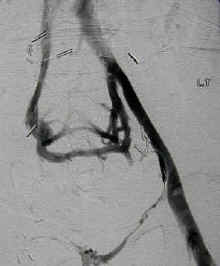
An occluded vein with formation of a collateral vessel. Source: www.lakeridgehealth.on.ca


For Patients
In order for the dye to show up on the x-ray it contains iodine. X-rays cannot penetrate iodine and this casts a white shadow on the x-ray film. Because iodine is being injected, you will need let your doctor know if you have had any allergies to iodine in the past. If you have, your doctor may not perform the procedure or your doctor may premedicate you with Benadryl or with steroids. You should also let your doctor know if you are pregnant because the x-ray could harm the fetus.
There are certain diseases that put the patient at risk for kidney problems after the procedure. You should let your doctor know if you have any of the following:
- Advanced age
- Diabetes mellitus
- Multiple myeloma
- Dehydration
- Poor kidney function
The night before the procedure, your doctor will ask you to drink plenty of fluids. This is because the dye can be hard on the kidneys and drinking plenty of fluids will help flush the dye out of your body and reduce the concentration of the dye going through the kidneys. Other than this, there is usually very little preparation for a venogram.
On the day of the procedure, you should expect to spend 30 to 90 minutes having the test done.
Prior to the procedure, your doctor will usually shave the area where the catheter will be inserted and will usually create a sterile area for the procedure to be done. Most often dye is injected through the back of the foot to evaluate the veins in the leg. If your doctor is evaluating the veins in your arm, the catheter may be inserted in the back of your hand.
Next, your doctor will numb up the area with a local anesthetic. A catheter will then be inserted into the vein under the skin. During the insertion of the catheter you may feel a pinch or something that feels like a bee sting. Your doctor will next inject the dye. During the injection you may feel a sense of warmth in your leg traveling up and going throughout your body. Rarely, some patients become nauseated during this time. Please let your not doctor know if you have any itching, rash, swelling of your common lips or mouth, or difficulty breathing as these may indicate that you have an allergy to the dye. Prompt treatment is necessary for any allergic reaction. It will also be necessary for you to lie very still when the dye is injected so that there is no movement of your leg to blur the image on the x-ray. Once the injection is done, your doctor will remove the catheter, which is painless and will apply pressure for about 10 minutes to stop any bleeding that occurs. You may also be given fluids to clear the dye from your body.
In general venography is very safe. There are a few complications that you should be aware of. A blood clot can form at the site of the injection, a blood clot that was already there can be dislodged and travel upstream in your body to the lungs, you can develop an allergic reaction as described above, and your kidney function can decline.