Aortic valve
.svg.png)
|
WikiDoc Resources for Aortic valve |
|
Articles |
|---|
|
Most recent articles on Aortic valve Most cited articles on Aortic valve |
|
Media |
|
Powerpoint slides on Aortic valve |
|
Evidence Based Medicine |
|
Clinical Trials |
|
Ongoing Trials on Aortic valve at Clinical Trials.gov Clinical Trials on Aortic valve at Google
|
|
Guidelines / Policies / Govt |
|
US National Guidelines Clearinghouse on Aortic valve
|
|
Books |
|
News |
|
Commentary |
|
Definitions |
|
Patient Resources / Community |
|
Patient resources on Aortic valve Discussion groups on Aortic valve Patient Handouts on Aortic valve Directions to Hospitals Treating Aortic valve Risk calculators and risk factors for Aortic valve
|
|
Healthcare Provider Resources |
|
Causes & Risk Factors for Aortic valve |
|
Continuing Medical Education (CME) |
|
International |
|
|
|
Business |
|
Experimental / Informatics |
Editor-In-Chief: C. Michael Gibson, M.S., M.D. [1]
The aortic valve is one of the valves of the heart. It lies between the left ventricle and the aorta.
Morphology
The aortic valve has three cusps. These cusps are half moon shaped hence also called aortic semilunar valve. Each cusp has a small swelling in the center called the nodule. Dilatation of the wall of the aorta behind these cusps is called aortic sinus. When the aortic valve is open, the normal size of the orifice is 3-4 cm² in adults.
Function & Physiology
During ventricular systole, pressure rises in the left ventricle. When the pressure in the left ventricle rises above the pressure in the aorta, the aortic valve opens, allowing blood to exit the left ventricle into the aorta. When ventricular systole ends, pressure in the left ventricle rapidly drops. When the pressure in the left ventricle decreases, the aortic pressure forces the aortic valve to close. The closure of the aortic valve contributes the A2 component of the second heart sound (S2).
Disease of the aortic valve
There are two protypical processes that can affect the aortic valve - aortic stenosis in which the valve fails to open fully, thereby obstructing blood flow out from the heart, and aortic insufficiency, also called aortic regurgitation, in which the aortic valve is incompetent and blood flows passively back to the heart in the wrong direction. These two conditions frequently co-exist.
- Common causes of aortic stenosis include rheumatic fever, degenerative calcification, and congenital diseases such as unicuspid aortic valve, bicuspid aortic valve, tricuspid aortic valve with commissural fushion, quadricuspid aortic valve and hypoplastic aortic annulus.
- Common causes of aortic regurgitation include dilation of the aorta, previous rheumatic fever, infection, i.e. infective endocarditis, myxomatous degeneration of the aortic valve, Marfan's syndrome and rupture of a congenitally fenestrated aortic cusp.
Bicuspid aortic valve
The most common congenital abnormality of the heart is the bicuspid aortic valve. In this condition, instead of three cusps, the aortic valve has two cusps. This condition is often undiagnosed until later in life when the person develops symptomatic aortic stenosis. Aortic stenosis occurs in this condition usually in patients in their 40s or 50s, an average of 10 years earlier than can occur in people with congenitally normal aortic valves.
Aortic Valve Replacement
Aortic valve replacement means that a patient's aortic valve is replaced by a different valve. The aortic valve can be affected by a range of diseases and require aortic valve replacement. The valve can either become leaky (regurgitant or insufficient) or stuck partially shut (stenotic). Aortic valve replacement currently requires open heart surgery. Research is being done now to develop valves that can be implanted using a catheter without open heart surgery. There are two basic types of artificial heart valve, mechanical valves and tissue valves. Tissue heart valves are usually made from animal tissues, either animal heart valve tissue or animal pericardial tissue. The tissue is treated to prevent rejection and to prevent calcification.
There are alternatives to animal tissue valves. In some cases a human aortic valve can be implanted. These are called homografts. Homograft valves are donated by patients and harvested after the patient expires. The durability of homograft valves is probably the same for porcine tissue valves. Another procedure for aortic valve replacement is the Ross procedure (after Donald Ross) or pulmonary autograft. The Ross procedure involves going to surgery to have the aortic valve removed and replacing it with the patient's own pulmonary valve. A pulmonary homograft (a pulmonary valve taken from a cadaver) or a valvular prothesis is then used to replace the patient's own pulmonary valve.
The first minimally invasive aortic valve surgery took place at the Cleveland Clinic in 1996. [1]
Additional images
-
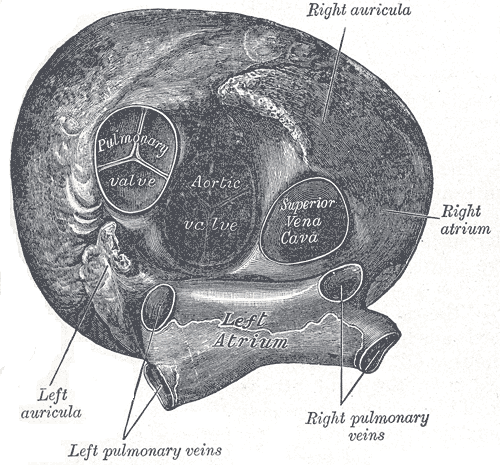 Heart seen from above.
 -
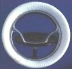 A mechanical artificial heart valve with a pivoting disc.
 -
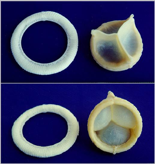 A replaceable model of Cardiac Biological Valve Prosthesis.


See also
References
External links
de:Aortenklappe no:Aortaklaff nn:Aortaklaff uk:Аортальний клапан