Trabeculae carneae
Editor-In-Chief: C. Michael Gibson, M.S., M.D. [1]
Associate Editor-In-Chief: Cafer Zorkun, M.D., Ph.D. [2]
Overview
The trabeculae carneae (columnae carneae or fleshy beams) are rounded or irregular muscular columns which project from the whole of the inner surface of the ventricle, with the exception of the conus arteriosus.
They are of three kinds:
- some are attached along their entire length on one side and merely form prominent ridges,
- others are fixed at their extremities but free in the middle,
- while a third set (musculi papillares) are continuous by their bases with the wall of the ventricle, while their apices give origin to the chordæ tendineæ which pass to be attached to the segments of the tricuspid valve.
The purpose of the trabeculae carnae is most likely to prevent suction that would occur with a flat surfaced membrane and thus impair the heart's ability to pump efficiently.
The trabeculae carnae also serve a similar function to papillary muscles in that their contraction pulls on the chordae tendinae, preventing inversion of the mitral (bicuspid) and tricuspid valves. This prevents backflow of blood from the ventricles into the atriums.
Echocardiographical and Angiographical Demonstration of Trabeculae Carnae
Images shown below are taken from a case report titled "Atrioventricular and ventriculoarterial discordance associated with Ebstein-like displacement of mitral valve"
-
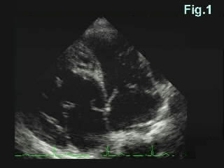 The atrioventricular valve of the left-sided ventricle (right facing to the screen) is attached closer to the apex. The trabeculae carneae of the left-sided ventricle are finer than those of the right ventricle. (Courtesy of the National Cardiovascular Center - Japan}

-
 Regurgitation through the left-sided atrioventricular valve is mild. (Courtesy of the National Cardiovascular Center - Japan}

-
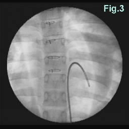 The catheter passes through the inferior vena cava located on the left side of the spine with its tip located in the left pulmonary ventricle. The right structure is found to be the morphological RV (systemic ventricle) and the left one is the morphological LV from the shape of the ventricles and the fineness of the trabeculae carneae. (Courtesy of the National Cardiovascular Center - Japan}
