Esophageal varices
| Resident Survival Guide |
| Esophageal varices | |
 | |
|---|---|
| Gastroscopy image of esophageal varices with prominent red wale spots | |
| ICD-10 | I85 |
| ICD-9 | 456.0-456.2 |
| DiseasesDB | 9177 |
| MedlinePlus | 000268 |
| MeSH | D004932 |
Template:Search infobox Editor-In-Chief: C. Michael Gibson, M.S., M.D. [1]
Overview
In medicine (gastroenterology), esophageal varices are extremely dilated sub-mucosal veins in the esophagus. They are most often a consequence of portal hypertension, such as may be seen with cirrhosis; patients with esophageal varices have a strong tendency to develop bleeding.
Esophageal varices are diagnosed with endoscopy.[1]
Pathogenesis
The majority of blood from the esophagus is drained away via the esophageal veins, which drain deoxygenated blood from the esophagus to the azygos vein which in turn, directly drains into the superior vena cava. These veins have no part in the development of esophageal varices. The remaining blood from the esophagus is drained away via the superficial veins lining the esophagus interior, which drain into the coronary vein (left gastric vein) which in turn, drains directly into the portal vein. These superficial veins lining the esophagus interior (normally only approximately 1mm in diameter) become distended up to 1-2 cm in diameter in association with portal hypertension.
Normal portal pressure is approximately 9 mmHg compared to an inferior vena cava pressure of 2-6 mmHg. This creates a normal pressure gradient of 3-7 mmHg. If the portal pressure rises above 12mmHg, this gradient rises to 7-10 mmHg.[2] A gradient greater than 10 mmHg is considered portal hypertension. At gradients greater than 10 mmHg, blood flow though the hepatic portal system is redirected from the liver into areas with lower venous pressures. This means that collateral circulation develops in the lower esophagus, abdominal wall, stomach and rectum. The small blood vessels in these areas become distended, becoming more thin-walled, and appear as varicosities. In addition, these vessels are poorly supported by other structures, as they are not designed for high pressures.
In situations where portal pressures increase, such as with cirrhosis, there is dilation of veins in the anastomosis, leading to esophageal varices.
Varices can also form in other areas of the body, including the stomach (gastric varices), duodenum (duodenal varices), and rectum (rectal varices). Treatment of these types of varices may differ.
Treatment
Recommendations for the treatment of Esophageal Varices (DO NOT EDIT)
The following is from clinical practice guidelines published in 2007 by the American Association for Study of Liver Diseases and American College of Gastroenterology.[3]
Patients with Cirrhosis and No Varices:
| “ |
|
” |
Patients with Cirrhosis and Small Varices That Have Not Bled:
| “ |
|
” |
Patients with Cirrhosis and Medium/Large Varices That Have Not Bled:
| “ |
|
” |
Patients with Cirrhosis and an Acute Episode of Variceal Hemorrhage:
| “ |
|
” |
Patients with Cirrhosis Who Have Recovered from Acute Variceal Hemorrhage:
| “ |
|
” |
Medical Therapy
In emergency situations, the care is directed at stopping blood loss, maintaining plasma volume, correcting disorders in coagulation induced by cirrhosis, and appropriate use of antibiotics (as infection is either concomitant, or a precipitant).
Therapeutic endoscopy is considered the mainstay of urgent treatment. Two main therapeutic approaches exist:
- Variceal ligation, or banding
- Sclerotherapy
In cases of refractory bleeding, balloon tamponade may be necessary, usually as a bridge to further endoscopy, a transjugular intrahepatic portosystemic shunt (TIPS), or a distal splenorenal shunt procedure or a liver transplantation.
Nutritional supplementation is not necessary if the patient is not eating for four days or less.[4]
-
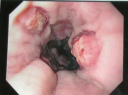 Esophageal varices seven days post banding, showing ulceration at the site of banding.

Prevention
Ideally, patients with known varices should receive treatment to reduce their risk of bleeding.[5][6] The non-selective β-blockers (e.g., propranolol, timolol or nadolol) and nitrates have been evaluated for secondary prophylaxis. The effectiveness of this treatment has been summarized in meta-analyses.[7] However, there may be a 'window effect':
- Patients with cirrhosis and small or no varices may not benefit.[8]
- Patients who are more severelly ill and have had spontaneous bacterial peritonitis may not benefit.[9]
Combining esophageal band ligation with β-blockers is better than either therapy alone.[5]
Unfortunately, non-selective β-blockers do not prevent the formation of esophageal varices.[10]
Variceal bleeding, prophylaxis
- 1. Short-term antibiotic prophylaxis[11]
- Preferred regimen (1): Norfloxacin 400mg PO bid for max. of 7 days
- Preferred regimen (2): Ciprofloxacin 400mg IV q12h max. of 7 days (in patients in whom oral administration is not possible)
- 2. In advanced cirrhosis and in a setting with high prevalence of quinolone-resistant organisms[11]
- Preferred regimen: Ceftriaxone 1g IV q24h for max. of 7 days
References
- ↑ Biecker E, Schepke M, Sauerbruch T (2005). "The role of endoscopy in portal hypertension". Dig Dis. 23 (1): 11–7. PMID 15920321.
- ↑ Arguedas M (2003). "The critically ill liver patient: the variceal bleeder". Semin Gastrointest Dis. 14 (1): 34–8. PMID 12610853.
- ↑ Garcia-Tsao G, Sanyal AJ, Grace ND, Carey WD, Practice Guidelines Committee of American Association for Study of Liver Diseases. Practice Parameters Committee of American College of Gastroenterology (2007). "Prevention and management of gastroesophageal varices and variceal hemorrhage in cirrhosis". Am J Gastroenterol. 102 (9): 2086–102. doi:10.1111/j.1572-0241.2007.01481.x. PMID 17727436.
- ↑ de Lédinghen V, Beau P, Mannant PR; et al. (1997). "Early feeding or enteral nutrition in patients with cirrhosis after bleeding from esophageal varices? A randomized controlled study". Dig. Dis. Sci. 42 (3): 536–41. PMID 9073135.
- ↑ 5.0 5.1 Gonzalez R, Zamora J, Gomez-Camarero J, Molinero LM, Bañares R, Albillos A (2008). "Meta-analysis: Combination endoscopic and drug therapy to prevent variceal rebleeding in cirrhosis". Ann Intern Med. 149 (2): 109–22. PMID 18626050. Review in: ACP J Club. 2008 Nov 18;149(5):10
- ↑ Lebrec D, Poynard T, Hillon P, Benhamou J-P (1981). "Propranolol for prevention of recurrent gastrointestinal bleeding in patients with cirrhosis: a controlled study". N Engl J Med. 305: 1371&ndash, 1374. PMID 7029276.
- ↑ Pagliaro L, D'Amico G, Sörensen TI, Lebrec D, Burroughs AK, Morabito A; et al. (1992). "Prevention of first bleeding in cirrhosis. A meta-analysis of randomized trials of nonsurgical treatment". Ann Intern Med. 117 (1): 59–70. PMID 1350716.
- ↑ Qi XS, Bao YX, Bai M, Xu WD, Dai JN, Guo XZ (2015). "Nonselective beta-blockers in cirrhotic patients with no or small varices: A meta-analysis". World J Gastroenterol. 21 (10): 3100–8. doi:10.3748/wjg.v21.i10.3100. PMC 4356933. PMID 25780311.
- ↑ Mandorfer M, Bota S, Schwabl P, Bucsics T, Pfisterer N, Kruzik M; et al. (2014). "Nonselective β blockers increase risk for hepatorenal syndrome and death in patients with cirrhosis and spontaneous bacterial peritonitis". Gastroenterology. 146 (7): 1680–90.e1. doi:10.1053/j.gastro.2014.03.005. PMID 24631577.
- ↑ Groszmann RJ, Garcia-Tsao G, Bosch J, Grace ND, Burroughs AK, Planas R; et al. (2005). "Beta-blockers to prevent gastroesophageal varices in patients with cirrhosis". N Engl J Med. 353 (21): 2254–61. doi:10.1056/NEJMoa044456. PMID 16306522. Review in: ACP J Club. 2006 May-Jun;144(3):68
- ↑ 11.0 11.1 Garcia-Tsao G, Sanyal AJ, Grace ND, Carey W, Practice Guidelines Committee of the American Association for the Study of Liver Diseases. Practice Parameters Committee of the American College of Gastroenterology (2007). "Prevention and management of gastroesophageal varices and variceal hemorrhage in cirrhosis". Hepatology. 46 (3): 922–38. doi:10.1002/hep.21907. PMID 17879356.
See also
- Portal hypertensive gastropathy
- gastric varices
- intestinal varices
- esophagitis
- Mallory-Weiss syndrome
- peptic ulcer