Superior vena cava
Editor-In-Chief: C. Michael Gibson, M.S., M.D. [1]
Overview
The superior vena cava is a large, yet short vein that carries de-oxygenated blood from the upper half of the body to the heart's right atrium.
It is formed by the left and right brachiocephalic veins, (also referred to as the innominate veins) which receive blood from the upper limbs and the head and neck, behind the lower border of the first right costal cartilage. The azygous vein (which receives blood from the rib cage) joins it just before it enters the right atrium, at the upper right front portion of the heart.
In the adult, no valve separates the superior vena cava from the right atrium. As a result, the (right) atrial and (right) ventricular contractions are conducted up into the internal jugular vein and, through the sternocleidomastoid muscle, can be seen as the jugular venous pressure. In tricuspid valve regurgitation, these pulsations are very strong.
Additional images
-
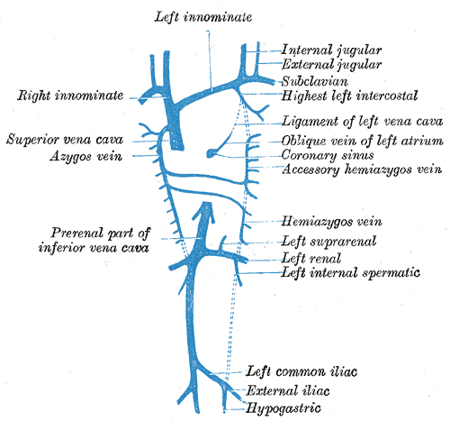 Diagram showing completion of development of the parietal veins.
 -
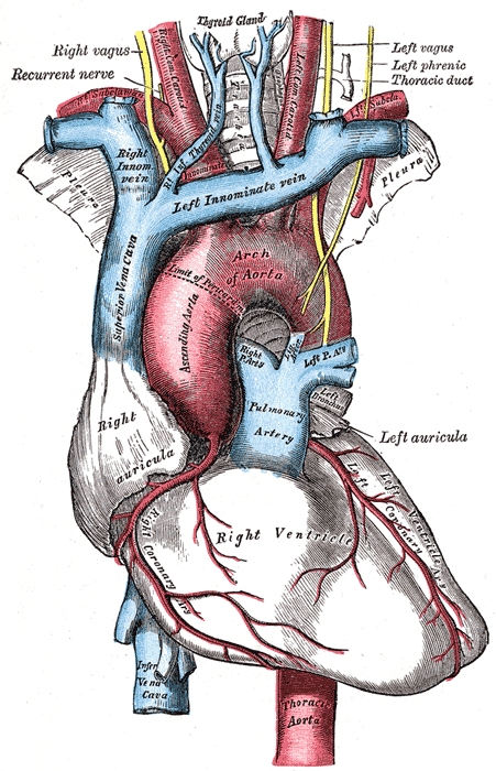
 Front view of heart and lungs.
 -
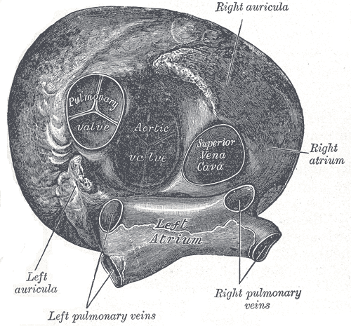 Heart seen from above.
 -
 Transverse section of thorax, showing relations of pulmonary artery.
 -
 The arch of the aorta, and its branches.
 -
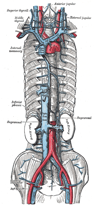 The brachiocephalic veins, superior vena cava, inferior vena cava, azygos vein and their tributaries
 -
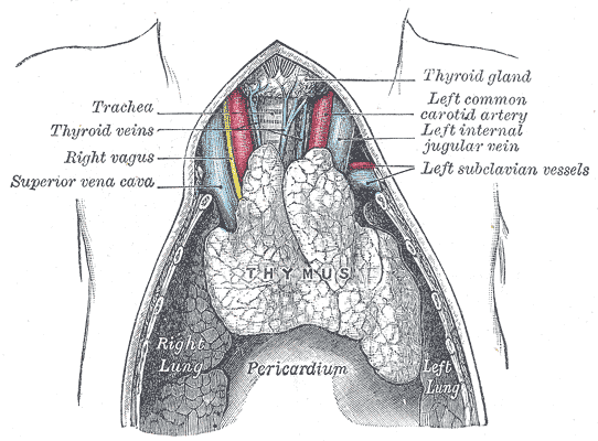 The thymus of a full-time fetus, exposed in situ.


See also
Template:Veins
eu:Goiko kaba
it:Vena cava superiore
nl:Vena cava superior
sk:Horná dutá žila
sr:Горња шупља вена
fi:Yläonttolaskimo