Bowen's disease: Difference between revisions
Sara Mohsin (talk | contribs) |
Sara Mohsin (talk | contribs) No edit summary |
||
| (5 intermediate revisions by the same user not shown) | |||
| Line 1: | Line 1: | ||
__NOTOC__ | __NOTOC__ | ||
{{ | {{Bowen's disease}} | ||
{{CMG}} {{AE}} {{S.M.}}, {{JH}} | |||
}} | |||
{{ | |||
{{SK}} Bowen's carcinoma; [[squamous cell carcinoma]] in situ of skin; intraepidermal carcinoma skin, Intraepithelial neoplasia | |||
{{SK}} Bowen's carcinoma; squamous cell carcinoma in situ of skin; intraepidermal carcinoma skin, Intraepithelial neoplasia | |||
==Overview== | ==Overview== | ||
| Line 23: | Line 11: | ||
==Historical Perspective== | ==Historical Perspective== | ||
* In 1912, Dr John T. Bowen was the first one to describe this [[disease]], hence, it is [[Name reaction|named]] after him | *In 1912, Dr John T. Bowen was the first one to describe this [[disease]], hence, it is [[Name reaction|named]] after him | ||
==Pathophysiology== | ==Pathophysiology== | ||
| Line 32: | Line 20: | ||
*In many [[Case-based reasoning|cases]], [[Cells (biology)|cells]] [[Lookahead|look]] worse under the [[microscope]] than the [[Cells (biology)|cells]] of many outright and [[Invasive (medical)|invading]] [[squamous cell carcinomas]] | *In many [[Case-based reasoning|cases]], [[Cells (biology)|cells]] [[Lookahead|look]] worse under the [[microscope]] than the [[Cells (biology)|cells]] of many outright and [[Invasive (medical)|invading]] [[squamous cell carcinomas]] | ||
*[[Degree (angle)|Degree]] of [[atypia]] ([[Strange matter|strangeness]], unusualness) seen under the [[microscope]] [[Best practice|best]] tells how [[Cells (biology)|cells]] may [[Behavior|behave]] (should they [[Invasive (medical)|invade]] another portion of the [[Human body|body]])<ref name="pmid3195216">{{cite journal| author=Nemkaeva RM, Kurmashov NA, Oslopova S| title=[Bowen's disease]. | journal=Vestn Dermatol Venerol | year= 1988 | volume= | issue= 8 | pages= 71-2 | pmid=3195216 | doi= | pmc= | url=https://www.ncbi.nlm.nih.gov/entrez/eutils/elink.fcgi?dbfrom=pubmed&tool=sumsearch.org/cite&retmode=ref&cmd=prlinks&id=3195216 }} </ref><ref name="pmid7138058">{{cite journal| author=Scarborough DA, Bisaccia EP, Yoder FW| title=Solitary pigmented Bowen's disease. | journal=Arch Dermatol | year= 1982 | volume= 118 | issue= 11 | pages= 954-5 | pmid=7138058 | doi= | pmc= | url=https://www.ncbi.nlm.nih.gov/entrez/eutils/elink.fcgi?dbfrom=pubmed&tool=sumsearch.org/cite&retmode=ref&cmd=prlinks&id=7138058 }} </ref> | *[[Degree (angle)|Degree]] of [[atypia]] ([[Strange matter|strangeness]], unusualness) seen under the [[microscope]] [[Best practice|best]] tells how [[Cells (biology)|cells]] may [[Behavior|behave]] (should they [[Invasive (medical)|invade]] another portion of the [[Human body|body]])<ref name="pmid3195216">{{cite journal| author=Nemkaeva RM, Kurmashov NA, Oslopova S| title=[Bowen's disease]. | journal=Vestn Dermatol Venerol | year= 1988 | volume= | issue= 8 | pages= 71-2 | pmid=3195216 | doi= | pmc= | url=https://www.ncbi.nlm.nih.gov/entrez/eutils/elink.fcgi?dbfrom=pubmed&tool=sumsearch.org/cite&retmode=ref&cmd=prlinks&id=3195216 }} </ref><ref name="pmid7138058">{{cite journal| author=Scarborough DA, Bisaccia EP, Yoder FW| title=Solitary pigmented Bowen's disease. | journal=Arch Dermatol | year= 1982 | volume= 118 | issue= 11 | pages= 954-5 | pmid=7138058 | doi= | pmc= | url=https://www.ncbi.nlm.nih.gov/entrez/eutils/elink.fcgi?dbfrom=pubmed&tool=sumsearch.org/cite&retmode=ref&cmd=prlinks&id=7138058 }} </ref> | ||
===Borst-Jadassohn phenomenon=== | ===Borst-Jadassohn phenomenon=== | ||
*Bowen's [[disease]] can also occur as a part of '''Borst-Jadassohn [[Phenomenology|phenomenon]]''' (previously known as '''intraepidermal epithelioma)''' which is a [[Heterogeneous mixture|heterogeneous]] [[Group (sociology)|group]] of following intraepithelial [[lesions]]:<ref name="pmid31145080">{{cite journal| author=Yanagihara S, Oiso N, Hirota N, Kato M, Miyake S, Kawada A| title=Acantholytic Bowen's disease histopathologically showing the Borst-Jadassohn phenomenon. | journal=Eur J Dermatol | year= 2019 | volume= | issue= | pages= | pmid=31145080 | doi=10.1684/ejd.2019.3545 | pmc= | url=https://www.ncbi.nlm.nih.gov/entrez/eutils/elink.fcgi?dbfrom=pubmed&tool=sumsearch.org/cite&retmode=ref&cmd=prlinks&id=31145080 }} </ref><ref name="pmid26373350">{{cite journal| author=Baykal C, Buyukbabani N, Babuna G, Polat Ekinci A, Kurul S| title=Giant Bowen's disease histologically showing Borst-Jadassohn phenomenon and complicated with squamous cell carcinoma development. | journal=J Eur Acad Dermatol Venereol | year= 2016 | volume= 30 | issue= 10 | pages= e88-e89 | pmid=26373350 | doi=10.1111/jdv.13335 | pmc= | url=https://www.ncbi.nlm.nih.gov/entrez/eutils/elink.fcgi?dbfrom=pubmed&tool=sumsearch.org/cite&retmode=ref&cmd=prlinks&id=26373350 }} </ref><ref name="urlBorst-Jadassohn phenomenon | definition of Borst-Jadassohn phenomenon by Medical dictionary">{{cite web |url=https://medical-dictionary.thefreedictionary.com/Borst-Jadassohn+phenomenon |title=Borst-Jadassohn phenomenon | definition of Borst-Jadassohn phenomenon by Medical dictionary |format= |work= |accessdate=}}</ref><ref name="urlPathology Outlines - Borst-Jadassohn phenomenon">{{cite web |url=https://www.pathologyoutlines.com/topic/skintumornonmelanocyticborstjadassohn.html |title=Pathology Outlines - Borst-Jadassohn phenomenon |format= |work= |accessdate=}}</ref><ref name="urlPayPerView: Jadassohn’s Intraepidermal Epithelioma - Karger Publishers">{{cite web |url=https://www.karger.com/Article/PDF/249658 |title=PayPerView: Jadassohn’s Intraepidermal Epithelioma - Karger Publishers |format= |work= |accessdate=}}</ref><ref name="pmid21587033">{{cite journal| author=Lora V, Chouvet B, Kanitakis J| title=The "intraepidermal epithelioma" revisited: immunohistochemical study of the borst-jadassohn phenomenon. | journal=Am J Dermatopathol | year= 2011 | volume= 33 | issue= 5 | pages= 492-7 | pmid=21587033 | doi=10.1097/DAD.0b013e3181fe6f90 | pmc= | url=https://www.ncbi.nlm.nih.gov/entrez/eutils/elink.fcgi?dbfrom=pubmed&tool=sumsearch.org/cite&retmode=ref&cmd=prlinks&id=21587033 }} </ref> | *Bowen's [[disease]] can also occur as a part of '''Borst-Jadassohn [[Phenomenology|phenomenon]]''' (previously known as '''intraepidermal epithelioma)''' which is a [[Heterogeneous mixture|heterogeneous]] [[Group (sociology)|group]] of following intraepithelial [[lesions]]:<ref name="pmid31145080">{{cite journal| author=Yanagihara S, Oiso N, Hirota N, Kato M, Miyake S, Kawada A| title=Acantholytic Bowen's disease histopathologically showing the Borst-Jadassohn phenomenon. | journal=Eur J Dermatol | year= 2019 | volume= | issue= | pages= | pmid=31145080 | doi=10.1684/ejd.2019.3545 | pmc= | url=https://www.ncbi.nlm.nih.gov/entrez/eutils/elink.fcgi?dbfrom=pubmed&tool=sumsearch.org/cite&retmode=ref&cmd=prlinks&id=31145080 }} </ref><ref name="pmid26373350">{{cite journal| author=Baykal C, Buyukbabani N, Babuna G, Polat Ekinci A, Kurul S| title=Giant Bowen's disease histologically showing Borst-Jadassohn phenomenon and complicated with squamous cell carcinoma development. | journal=J Eur Acad Dermatol Venereol | year= 2016 | volume= 30 | issue= 10 | pages= e88-e89 | pmid=26373350 | doi=10.1111/jdv.13335 | pmc= | url=https://www.ncbi.nlm.nih.gov/entrez/eutils/elink.fcgi?dbfrom=pubmed&tool=sumsearch.org/cite&retmode=ref&cmd=prlinks&id=26373350 }} </ref><ref name="urlBorst-Jadassohn phenomenon | definition of Borst-Jadassohn phenomenon by Medical dictionary">{{cite web |url=https://medical-dictionary.thefreedictionary.com/Borst-Jadassohn+phenomenon |title=Borst-Jadassohn phenomenon | definition of Borst-Jadassohn phenomenon by Medical dictionary |format= |work= |accessdate=}}</ref><ref name="urlPathology Outlines - Borst-Jadassohn phenomenon">{{cite web |url=https://www.pathologyoutlines.com/topic/skintumornonmelanocyticborstjadassohn.html |title=Pathology Outlines - Borst-Jadassohn phenomenon |format= |work= |accessdate=}}</ref><ref name="urlPayPerView: Jadassohn’s Intraepidermal Epithelioma - Karger Publishers">{{cite web |url=https://www.karger.com/Article/PDF/249658 |title=PayPerView: Jadassohn’s Intraepidermal Epithelioma - Karger Publishers |format= |work= |accessdate=}}</ref><ref name="pmid21587033">{{cite journal| author=Lora V, Chouvet B, Kanitakis J| title=The "intraepidermal epithelioma" revisited: immunohistochemical study of the borst-jadassohn phenomenon. | journal=Am J Dermatopathol | year= 2011 | volume= 33 | issue= 5 | pages= 492-7 | pmid=21587033 | doi=10.1097/DAD.0b013e3181fe6f90 | pmc= | url=https://www.ncbi.nlm.nih.gov/entrez/eutils/elink.fcgi?dbfrom=pubmed&tool=sumsearch.org/cite&retmode=ref&cmd=prlinks&id=21587033 }} </ref> | ||
**[[Irritation|Irritated]] [[seborrheic keratosis]] | **[[Irritation|Irritated]] [[seborrheic keratosis]] | ||
| Line 42: | Line 32: | ||
{| | {| | ||
| | | | ||
[[File:Bowen disease (2).jpg|thumb|200px|none|H & E stain of'''Bowen's disease''' as seen under a microscope [https://commons.wikimedia.org/wiki/Category:Histopathology_of_Bowen%27s_disease#/media/File:Bowen_disease_(2).jpg Source: Wikimedia Commons]] | [[File:Bowen disease (2).jpg|thumb|200px|none|H & E stain of'''Bowen's disease''' as seen under a microscope [https://commons.wikimedia.org/wiki/Category:Histopathology_of_Bowen%27s_disease#/media/File:Bowen_disease_(2).jpg Source: Wikimedia Commons]] | ||
| | | | ||
[[File:Bowen disease (3).jpg|thumb|200px|none|Histopathology of squamous cell carcinoma in situ of the skin (Bowen disease). H & E stain [https://commons.wikimedia.org/wiki/Category:Histopathology_of_Bowen%27s_disease#/media/File:Bowen_disease_(3).jpg Source: Wikimedia Commons]] | [[File:Bowen disease (3).jpg|thumb|200px|none|Histopathology of squamous cell carcinoma in situ of the skin (Bowen disease). H & E stain [https://commons.wikimedia.org/wiki/Category:Histopathology_of_Bowen%27s_disease#/media/File:Bowen_disease_(3).jpg Source: Wikimedia Commons]] | ||
| | |||
[[image:Bowen disease (1).jpg|thumb|200px|none|'''Bowen's disease''' as seen under a microscope]] | |||
| | | | ||
|} | |} | ||
| Line 62: | Line 52: | ||
==Causes== | ==Causes== | ||
Bowen's [[disease]] is a non-[[infectious]], non-[[familial]] [[disease]] with common [[causes]] as mentioned below: | Bowen's [[disease]] is a non-[[infectious]], non-[[familial]] [[disease]] with common [[causes]] as mentioned below: | ||
*'''[[Solar nebula|Solar]] damage''' due to [[Long-term potentiation|long-term]] [[sun exposure]] or [[Usage analysis|use]] of sunbeds, especially in [[People's Solidarity|people]] with [[Fair use|fair]] [[skin]] | *'''[[Solar nebula|Solar]] damage''' due to [[Long-term potentiation|long-term]] [[sun exposure]] or [[Usage analysis|use]] of sunbeds, especially in [[People's Solidarity|people]] with [[Fair use|fair]] [[skin]] | ||
*'''[[Aging]]''' | *'''[[Aging]]''' | ||
| Line 87: | Line 78: | ||
==Epidemiology and Demographics== | ==Epidemiology and Demographics== | ||
===Age=== | ===Age=== | ||
*Bowen's [[disease]] can [[affect]] [[Adult|adults]] of any [[age]], most commonly involves [[Old age|older]] [[patients]] in their 60s or 70s | *Bowen's [[disease]] can [[affect]] [[Adult|adults]] of any [[age]], most commonly involves [[Old age|older]] [[patients]] in their 60s or 70s | ||
*It is [[rare]] before the [[age]] of 30 [[Year|years]] | *It is [[rare]] before the [[age]] of 30 [[Year|years]] | ||
===Gender=== | ===Gender=== | ||
*Bowen's [[disease]] occurs more [[Predominance diagram|predominantly]] in [[men]] than in [[Womens Pack|women]] (70-85% of [[Case-based reasoning|cases]]) | *Bowen's [[disease]] occurs more [[Predominance diagram|predominantly]] in [[men]] than in [[Womens Pack|women]] (70-85% of [[Case-based reasoning|cases]]) | ||
===Race=== | ===Race=== | ||
*[[Caucasian honey bee|Caucasians]] are the ones most commonly [[Affect|affected]] by Bowen's [[disease]] | *[[Caucasian honey bee|Caucasians]] are the ones most commonly [[Affect|affected]] by Bowen's [[disease]] | ||
==Natural History, Complications and Prognosis== | ==Natural History, Complications and Prognosis== | ||
*Bowen's [[disease]] [[Growth|grows]] very [[Slow|slowly]] over the [[period]] of months or even [[Year|years]]<ref name="urlBowens disease - NHS">{{cite web |url=https://www.nhs.uk/conditions/bowens-disease/ |title=Bowen's disease - NHS |format= |work= |accessdate=}}</ref> | *Bowen's [[disease]] [[Growth|grows]] very [[Slow|slowly]] over the [[period]] of months or even [[Year|years]]<ref name="urlBowens disease - NHS">{{cite web |url=https://www.nhs.uk/conditions/bowens-disease/ |title=Bowen's disease - NHS |format= |work= |accessdate=}}</ref> | ||
*It is easily [[Treatments|treatable]] if [[Diagnose|diagnosed]] in [[Time constant|time]] | *It is easily [[Treatments|treatable]] if [[Diagnose|diagnosed]] in [[Time constant|time]] | ||
| Line 104: | Line 99: | ||
==Diagnosis== | ==Diagnosis== | ||
===Common symptoms=== | ===Common symptoms=== | ||
*It usually [[Appearance|appears]] as one or more [[skin]] [[Patching|patches]] with following [[Characteristic function (probability theory)|characteristics]]:<ref name="urlBowens disease - NHS">{{cite web |url=https://www.nhs.uk/conditions/bowens-disease/ |title=Bowen's disease - NHS |format= |work= |accessdate=}}</ref> | *It usually [[Appearance|appears]] as one or more [[skin]] [[Patching|patches]] with following [[Characteristic function (probability theory)|characteristics]]:<ref name="urlBowens disease - NHS">{{cite web |url=https://www.nhs.uk/conditions/bowens-disease/ |title=Bowen's disease - NHS |format= |work= |accessdate=}}</ref> | ||
**Persistent | **Persistent | ||
| Line 120: | Line 116: | ||
**[[SplitsTree|Splits]] open ([[fissured]]) | **[[SplitsTree|Splits]] open ([[fissured]]) | ||
**[[Dark matter|Darkly]] [[Color|colored]] ([[Pigmented lesions|pigmented]]) | **[[Dark matter|Darkly]] [[Color|colored]] ([[Pigmented lesions|pigmented]]) | ||
===Signs and symptoms of malignant transformation=== | ===Signs and symptoms of malignant transformation=== | ||
*Following [[Change detection|changes]] in the [[skin]] [[Patched|patch]] are the [[signs]] that bowen's [[disease]] has [[Turn (biochemistry)|turned]] into [[Invasive (medical)|invasive]] [[squamous cell carcinoma of the skin]]: | *Following [[Change detection|changes]] in the [[skin]] [[Patched|patch]] are the [[signs]] that bowen's [[disease]] has [[Turn (biochemistry)|turned]] into [[Invasive (medical)|invasive]] [[squamous cell carcinoma of the skin]]: | ||
**Easy [[bleeding]] | **Easy [[bleeding]] | ||
**[[Tenderness]] | **[[Tenderness]] | ||
**[[Turn (biochemistry)|Turns]] into an open [[sore]] i.e. [[Ulcerated lesion|ulcerates]] | **[[Turn (biochemistry)|Turns]] into an open [[sore]] i.e. [[Ulcerated lesion|ulcerates]] | ||
**[[Hard science|Hardening]] ([[induration]]) | **[[Hard science|Hardening]] ([[induration]]) | ||
**[[Development|Develops]] into a [[lump]]/[[Fleshy beams|fleshy]] [[Nodule (medicine)|nodule]] | **[[Development|Develops]] into a [[lump]]/[[Fleshy beams|fleshy]] [[Nodule (medicine)|nodule]] | ||
===Common sites of involvement=== | ===Common sites of involvement=== | ||
*Common sites involved are:<ref name="pmid3760318">{{cite journal| author=Wagner RF, Grande DJ| title=Solitary pigmented Bowen's disease of the scrotum. | journal=J Dermatol Surg Oncol | year= 1986 | volume= 12 | issue= 10 | pages= 1114-5 | pmid=3760318 | doi= | pmc= | url=https://www.ncbi.nlm.nih.gov/entrez/eutils/elink.fcgi?dbfrom=pubmed&tool=sumsearch.org/cite&retmode=ref&cmd=prlinks&id=3760318 }} </ref><ref name="pmid24746300">{{cite journal| author=Al-Dawsari NA, Raslan W, Dawamneh MF| title=Pigmented Bowen's disease of the penis and scrotum in a patient with AIDS. | journal=Dermatol Online J | year= 2014 | volume= 20 | issue= 4 | pages= 22337 | pmid=24746300 | doi= | pmc= | url=https://www.ncbi.nlm.nih.gov/entrez/eutils/elink.fcgi?dbfrom=pubmed&tool=sumsearch.org/cite&retmode=ref&cmd=prlinks&id=24746300 }} </ref><ref name="pmid31141219">{{cite journal| author=Narahira A, Yanagi T, Kitamura S, Hata H, Shimizu H| title=Dermoscopic features of genital pigmented Bowen's disease: Report of a case and review of the published work. | journal=J Dermatol | year= 2019 | volume= | issue= | pages= | pmid=31141219 | doi=10.1111/1346-8138.14938 | pmc= | url=https://www.ncbi.nlm.nih.gov/entrez/eutils/elink.fcgi?dbfrom=pubmed&tool=sumsearch.org/cite&retmode=ref&cmd=prlinks&id=31141219 }} </ref> | *Common sites involved are:<ref name="pmid3760318">{{cite journal| author=Wagner RF, Grande DJ| title=Solitary pigmented Bowen's disease of the scrotum. | journal=J Dermatol Surg Oncol | year= 1986 | volume= 12 | issue= 10 | pages= 1114-5 | pmid=3760318 | doi= | pmc= | url=https://www.ncbi.nlm.nih.gov/entrez/eutils/elink.fcgi?dbfrom=pubmed&tool=sumsearch.org/cite&retmode=ref&cmd=prlinks&id=3760318 }} </ref><ref name="pmid24746300">{{cite journal| author=Al-Dawsari NA, Raslan W, Dawamneh MF| title=Pigmented Bowen's disease of the penis and scrotum in a patient with AIDS. | journal=Dermatol Online J | year= 2014 | volume= 20 | issue= 4 | pages= 22337 | pmid=24746300 | doi= | pmc= | url=https://www.ncbi.nlm.nih.gov/entrez/eutils/elink.fcgi?dbfrom=pubmed&tool=sumsearch.org/cite&retmode=ref&cmd=prlinks&id=24746300 }} </ref><ref name="pmid31141219">{{cite journal| author=Narahira A, Yanagi T, Kitamura S, Hata H, Shimizu H| title=Dermoscopic features of genital pigmented Bowen's disease: Report of a case and review of the published work. | journal=J Dermatol | year= 2019 | volume= | issue= | pages= | pmid=31141219 | doi=10.1111/1346-8138.14938 | pmc= | url=https://www.ncbi.nlm.nih.gov/entrez/eutils/elink.fcgi?dbfrom=pubmed&tool=sumsearch.org/cite&retmode=ref&cmd=prlinks&id=31141219 }} </ref> | ||
====Skin==== | ====Skin==== | ||
*[[Lesions]] can occur anywhere on the [[skin]] [[Surface area|surface]] or on [[mucosal]] [[Surface area|surfaces]], although the involvement of [[Palms of the hands|palms]] or [[Sole of the foot|soles]] is uncommon | *[[Lesions]] can occur anywhere on the [[skin]] [[Surface area|surface]] or on [[mucosal]] [[Surface area|surfaces]], although the involvement of [[Palms of the hands|palms]] or [[Sole of the foot|soles]] is uncommon | ||
* A persistent progressive non-elevated [[Red-Al|red]] [[Scale (social sciences)|scaly]] or [[Crustacean|crusted]] [[plaque]] which is due to an [[intradermal]] [[carcinoma]] and is [[Potential|potentially]] [[malignant]] | *A persistent progressive non-elevated [[Red-Al|red]] [[Scale (social sciences)|scaly]] or [[Crustacean|crusted]] [[plaque]] which is due to an [[intradermal]] [[carcinoma]] and is [[Potential|potentially]] [[malignant]] | ||
* Atypical [[squamous]] (resembling [[Fish scale disease|fish scales]]) [[Cells (biology)|cells]] [[proliferate]] through the whole [[Thickener|thickness]] of the [[epidermis]] | *Atypical [[squamous]] (resembling [[Fish scale disease|fish scales]]) [[Cells (biology)|cells]] [[proliferate]] through the whole [[Thickener|thickness]] of the [[epidermis]] | ||
{| | {| | ||
| Line 148: | Line 149: | ||
====Extremities==== | ====Extremities==== | ||
* About three-quarters of the [[patients]] have [[lesions]] on the [[lower leg]] (60-85%), usually in previously or [[Presenting symptom|presently]] [[Sun exposure|sun-exposed]] [[Area|areas]] of [[skin]] | *About three-quarters of the [[patients]] have [[lesions]] on the [[lower leg]] (60-85%), usually in previously or [[Presenting symptom|presently]] [[Sun exposure|sun-exposed]] [[Area|areas]] of [[skin]] | ||
====Head and neck area==== | ====Head and neck area==== | ||
*Being [[prone]] to the [[sun exposure]], [[head]] and [[neck]] [[area]] is also one of the common sites to be [[Affect (philosophy)|affected]] by Bowen's [[disease]] | *Being [[prone]] to the [[sun exposure]], [[head]] and [[neck]] [[area]] is also one of the common sites to be [[Affect (philosophy)|affected]] by Bowen's [[disease]] | ||
| Line 163: | Line 165: | ||
====Subungal, periungal region==== | ====Subungal, periungal region==== | ||
*Bowen's [[disease]] also commonly involves [[Subungal exostosis|subungal]] or [[Periungual warts|periungal]] [[Area|areas]] (i.e. either under or around [[fingernails]] or [[toenails]]) | *Bowen's [[disease]] also commonly involves [[Subungal exostosis|subungal]] or [[Periungual warts|periungal]] [[Area|areas]] (i.e. either under or around [[fingernails]] or [[toenails]]) | ||
| Line 174: | Line 177: | ||
====Genitourinary system==== | ====Genitourinary system==== | ||
* Bowen's [[disease]] can also involve [[Genital area|genital]] and perianal [[Area|areas]] | *Bowen's [[disease]] can also involve [[Genital area|genital]] and perianal [[Area|areas]] | ||
* It [[Appearance|appears]] in following 3 forms in [[genital area]]:<ref name="urlBowen’s Disease: Skin Cancer Linked to HPV Infection">{{cite web |url=https://www.webmd.com/cancer/what-is-bowens-disease#1 |title=Bowen’s Disease: Skin Cancer Linked to HPV Infection |format= |work= |accessdate=}}</ref> | *It [[Appearance|appears]] in following 3 forms in [[genital area]]:<ref name="urlBowen’s Disease: Skin Cancer Linked to HPV Infection">{{cite web |url=https://www.webmd.com/cancer/what-is-bowens-disease#1 |title=Bowen’s Disease: Skin Cancer Linked to HPV Infection |format= |work= |accessdate=}}</ref> | ||
{| class="wikitable" | {| class="wikitable" | ||
|+Multiple forms of Bowen's disease involving genital region | |+Multiple forms of Bowen's disease involving genital region | ||
! style="background: #4479BA; width: 200px;" | {{fontcolor|#FFF|Bowen's disease form}} | ! style="background: #4479BA; width: 200px;" |{{fontcolor|#FFF|Bowen's disease form}} | ||
! style="background: #4479BA; width: 400px;" | {{fontcolor|#FFF|Characteristic features}} | ! style="background: #4479BA; width: 400px;" |{{fontcolor|#FFF|Characteristic features}} | ||
|- | |- | ||
| style="padding: 5px 5px; background: #DCDCDC; font-weight: bold" |[[Bowenoid papulosis]] | | style="padding: 5px 5px; background: #DCDCDC; font-weight: bold" |[[Bowenoid papulosis]] | ||
| | | | ||
* [[Affect|Affects]] both [[men]] and [[Womens Pack|women]] | *[[Affect|Affects]] both [[men]] and [[Womens Pack|women]] | ||
* [[Lesions]] can last from 2 weeks to several [[Year|years]] | *[[Lesions]] can last from 2 weeks to several [[Year|years]] | ||
|- | |- | ||
| style="padding: 5px 5px; background: #DCDCDC; font-weight: bold" |[[Erythroplasia of Queyrat]] | | style="padding: 5px 5px; background: #DCDCDC; font-weight: bold" |[[Erythroplasia of Queyrat]] | ||
| | | | ||
* [[Affect|Affects]] tip of the [[penis]] ([[glans penis]]-[[Outer coat|outer]] [[keratinized]] [[Surface area|surface]] of the [[penis]]) | *[[Affect|Affects]] tip of the [[penis]] ([[glans penis]]-[[Outer coat|outer]] [[keratinized]] [[Surface area|surface]] of the [[penis]]) | ||
*Occurs in [[Middle age|middle-aged]] uncircumscribed [[men]] due to unknown [[Reasoning|reasons]] | *Occurs in [[Middle age|middle-aged]] uncircumscribed [[men]] due to unknown [[Reasoning|reasons]] | ||
* May [[Causes|cause]] any of the following: | *May [[Causes|cause]] any of the following: | ||
**[[Red-Al|Reddish]], [[Velvet (fish disease)|velvety]] or [[Smooth pursuit|smooth]] [[plaque]] | **[[Red-Al|Reddish]], [[Velvet (fish disease)|velvety]] or [[Smooth pursuit|smooth]] [[plaque]] | ||
**[[Ulcers]] | **[[Ulcers]] | ||
| Line 217: | Line 221: | ||
===Physical Examination=== | ===Physical Examination=== | ||
*Bowen's [[disease]] [[Typical set|typically]] [[Presenting symptom|presents]] as a gradually enlarging, well [[Demarcation problem|demarcated]] [[erythematous]] [[plaque]] with an [[Irregular lesion|irregular]] border and [[Surface area|surface]] [[Crustacea|crusting]] or [[Scaling skin|scaling]]<ref name="pmid20811602">{{cite journal| author=Inoue T, Kobayashi K, Sawada M, Ishizaki S, Ito H, Fujibayashi M et al.| title=Dermoscopic Features of Pigmented Bowen's Disease in a Japanese Female Mimicking Malignant Melanoma. | journal=Dermatol Res Pract | year= 2010 | volume= 2010 | issue= | pages= | pmid=20811602 | doi=10.1155/2010/543091 | pmc=2929512 | url=https://www.ncbi.nlm.nih.gov/entrez/eutils/elink.fcgi?dbfrom=pubmed&tool=sumsearch.org/cite&retmode=ref&cmd=prlinks&id=20811602 }} </ref><ref name="pmid20652107">{{cite journal| author=Hayashi Y, Tanaka M, Suzaki R, Mori N, Konohana I| title=Dermoscopy of Pigmented Bowen's Disease Mimicking Early Superficial Spreading Melanoma. | journal=Case Rep Dermatol | year= 2009 | volume= 1 | issue= 1 | pages= 11-15 | pmid=20652107 | doi=10.1159/000227284 | pmc=2895203 | url=https://www.ncbi.nlm.nih.gov/entrez/eutils/elink.fcgi?dbfrom=pubmed&tool=sumsearch.org/cite&retmode=ref&cmd=prlinks&id=20652107 }} </ref><ref name="pmid28832993">{{cite journal| author=Yang Y, Lin J, Fang S, Han S, Song Z| title=What's new in dermoscopy of Bowen's disease: two new dermoscopic signs and its differential diagnosis. | journal=Int J Dermatol | year= 2017 | volume= 56 | issue= 10 | pages= 1022-1025 | pmid=28832993 | doi=10.1111/ijd.13734 | pmc= | url=https://www.ncbi.nlm.nih.gov/entrez/eutils/elink.fcgi?dbfrom=pubmed&tool=sumsearch.org/cite&retmode=ref&cmd=prlinks&id=28832993 }} </ref> | *Bowen's [[disease]] [[Typical set|typically]] [[Presenting symptom|presents]] as a gradually enlarging, well [[Demarcation problem|demarcated]] [[erythematous]] [[plaque]] with an [[Irregular lesion|irregular]] border and [[Surface area|surface]] [[Crustacea|crusting]] or [[Scaling skin|scaling]]<ref name="pmid20811602">{{cite journal| author=Inoue T, Kobayashi K, Sawada M, Ishizaki S, Ito H, Fujibayashi M et al.| title=Dermoscopic Features of Pigmented Bowen's Disease in a Japanese Female Mimicking Malignant Melanoma. | journal=Dermatol Res Pract | year= 2010 | volume= 2010 | issue= | pages= | pmid=20811602 | doi=10.1155/2010/543091 | pmc=2929512 | url=https://www.ncbi.nlm.nih.gov/entrez/eutils/elink.fcgi?dbfrom=pubmed&tool=sumsearch.org/cite&retmode=ref&cmd=prlinks&id=20811602 }} </ref><ref name="pmid20652107">{{cite journal| author=Hayashi Y, Tanaka M, Suzaki R, Mori N, Konohana I| title=Dermoscopy of Pigmented Bowen's Disease Mimicking Early Superficial Spreading Melanoma. | journal=Case Rep Dermatol | year= 2009 | volume= 1 | issue= 1 | pages= 11-15 | pmid=20652107 | doi=10.1159/000227284 | pmc=2895203 | url=https://www.ncbi.nlm.nih.gov/entrez/eutils/elink.fcgi?dbfrom=pubmed&tool=sumsearch.org/cite&retmode=ref&cmd=prlinks&id=20652107 }} </ref><ref name="pmid28832993">{{cite journal| author=Yang Y, Lin J, Fang S, Han S, Song Z| title=What's new in dermoscopy of Bowen's disease: two new dermoscopic signs and its differential diagnosis. | journal=Int J Dermatol | year= 2017 | volume= 56 | issue= 10 | pages= 1022-1025 | pmid=28832993 | doi=10.1111/ijd.13734 | pmc= | url=https://www.ncbi.nlm.nih.gov/entrez/eutils/elink.fcgi?dbfrom=pubmed&tool=sumsearch.org/cite&retmode=ref&cmd=prlinks&id=28832993 }} </ref> | ||
*[[Lesion]] might [[Feeling|feel]] [[Tenderness (medicine)|tender]] [[Representative agent|representing]] its [[malignant transformation]] | *[[Lesion]] might [[Feeling|feel]] [[Tenderness (medicine)|tender]] [[Representative agent|representing]] its [[malignant transformation]] | ||
| Line 229: | Line 234: | ||
===Dermoscopy=== | ===Dermoscopy=== | ||
*[[Dermoscopy]] is the [[skin]] [[examination]] via [[skin]] [[Surface area|surface]] [[microscopy]] | *[[Dermoscopy]] is the [[skin]] [[examination]] via [[skin]] [[Surface area|surface]] [[microscopy]] | ||
*Also known as: | *Also known as: | ||
| Line 242: | Line 248: | ||
===Skin biopsy=== | ===Skin biopsy=== | ||
*[[Skin biopsy]] must be [[done]] to: | *[[Skin biopsy]] must be [[done]] to: | ||
**Confirm the [[diagnosis]] Bowen's [[disease]] | **Confirm the [[diagnosis]] Bowen's [[disease]] | ||
| Line 248: | Line 255: | ||
==Treatment== | ==Treatment== | ||
*[[Specific activity|Specific]] [[Treatments|treatment]] for an [[Individual growth|individual]] [[Case-based reasoning|case]] [[Dependent variable|depends]] upon various factors, such as:<ref name="urlBowen Disease - NORD (National Organization for Rare Disorders)">{{cite web |url=https://rarediseases.org/rare-diseases/bowen-disease/ |title=Bowen Disease - NORD (National Organization for Rare Disorders) |format= |work= |accessdate=}}</ref> | *[[Specific activity|Specific]] [[Treatments|treatment]] for an [[Individual growth|individual]] [[Case-based reasoning|case]] [[Dependent variable|depends]] upon various factors, such as:<ref name="urlBowen Disease - NORD (National Organization for Rare Disorders)">{{cite web |url=https://rarediseases.org/rare-diseases/bowen-disease/ |title=Bowen Disease - NORD (National Organization for Rare Disorders) |format= |work= |accessdate=}}</ref> | ||
**[[Affect|Affected]] [[Human body|body]] site | **[[Affect|Affected]] [[Human body|body]] site | ||
| Line 259: | Line 267: | ||
**[[Possibility theory|Possible]] long [[Term logic|term]] [[Effect size|effects]] of the [[Treatments|treatment]] | **[[Possibility theory|Possible]] long [[Term logic|term]] [[Effect size|effects]] of the [[Treatments|treatment]] | ||
**[[Patient|Patient's]] [[Preferences|preference]] | **[[Patient|Patient's]] [[Preferences|preference]] | ||
* Different [[Treatments|treatment]] options for Bowen's [[disease]] include the following: | *Different [[Treatments|treatment]] options for Bowen's [[disease]] include the following: | ||
{| class="wikitable" | {| class="wikitable" | ||
| Line 268: | Line 276: | ||
| style="padding: 5px 5px; background: #DCDCDC; font-weight: bold" |'''[[Watch and wait]]''' | | style="padding: 5px 5px; background: #DCDCDC; font-weight: bold" |'''[[Watch and wait]]''' | ||
| | | | ||
* [[Patient]] with a [[slow]]-[[Growth|growing]] [[disease]] is followed up without any [[Treatments|treatment]] given until the [[disease]] progression occurs | *[[Patient]] with a [[slow]]-[[Growth|growing]] [[disease]] is followed up without any [[Treatments|treatment]] given until the [[disease]] progression occurs | ||
*[[Done]] in elderly [[patients]] having a [[Slow|slowly]] [[Growth|growing]] [[lesion]] in poor [[healing]] [[area]] such as [[lower leg]] | *[[Done]] in elderly [[patients]] having a [[Slow|slowly]] [[Growth|growing]] [[lesion]] in poor [[healing]] [[area]] such as [[lower leg]] | ||
|- | |- | ||
| Line 274: | Line 282: | ||
| | | | ||
*[[Cells (biology)|Cells]] of the [[lesion]] are destroyed by [[freezing]] them with either [[liquid nitrogen]] or [[argon]] [[gas]] | *[[Cells (biology)|Cells]] of the [[lesion]] are destroyed by [[freezing]] them with either [[liquid nitrogen]] or [[argon]] [[gas]] | ||
* Later on, the [[Dead space|dead]] [[Tissue (biology)|tissue]] sloughs off on thawing within [[Fewmets|few]] weeks | *Later on, the [[Dead space|dead]] [[Tissue (biology)|tissue]] sloughs off on thawing within [[Fewmets|few]] weeks | ||
* Favored by some [[clinicians]] over [[excision]] | *Favored by some [[clinicians]] over [[excision]] | ||
*Mostly [[Effective method|effective]] for single or small [[lesions]] | *Mostly [[Effective method|effective]] for single or small [[lesions]] | ||
|- | |- | ||
| style="padding: 5px 5px; background: #DCDCDC; font-weight: bold" |'''[[Topical application|Topical]] [[Cream (pharmaceutical)|cream]]''' | | style="padding: 5px 5px; background: #DCDCDC; font-weight: bold" |'''[[Topical application|Topical]] [[Cream (pharmaceutical)|cream]]''' | ||
| | | | ||
* Includes following 2 options: | *Includes following 2 options: | ||
**5% [[Imiquimod]] [[Cream (pharmaceutical)|cream]] | **5% [[Imiquimod]] [[Cream (pharmaceutical)|cream]] | ||
**[[Chemotherapy]] [[Cream (pharmaceutical)|cream]] ([[5-fluorouracil]]) | **[[Chemotherapy]] [[Cream (pharmaceutical)|cream]] ([[5-fluorouracil]]) | ||
* Favored by some [[clinicians]] over [[excision]] | *Favored by some [[clinicians]] over [[excision]] | ||
*[[Cream (pharmaceutical)|Cream]] needs to be [[Applied probability|applied]] once or twice for atleast two weeks if not longer | *[[Cream (pharmaceutical)|Cream]] needs to be [[Applied probability|applied]] once or twice for atleast two weeks if not longer | ||
|- | |- | ||
| Line 290: | Line 298: | ||
| | | | ||
*[[Affect|Affected]] [[skin]] [[area]] is [[Scrapie|scraped]] away under [[local anaesthetic]] (which [[Numbed|numbs]] the [[skin]]) [[Usage analysis|using]] a device having a [[Circular folds|circular]] blade ([[Curette|curet]]) | *[[Affect|Affected]] [[skin]] [[area]] is [[Scrapie|scraped]] away under [[local anaesthetic]] (which [[Numbed|numbs]] the [[skin]]) [[Usage analysis|using]] a device having a [[Circular folds|circular]] blade ([[Curette|curet]]) | ||
* Then an [[electric current]] is [[Passing (disability)|passed]] through the remaining of the [[lesion]] via an [[Electric current|electric]] [[needle]] which destroys any of the remaining [[Cancer cells|cells]] | *Then an [[electric current]] is [[Passing (disability)|passed]] through the remaining of the [[lesion]] via an [[Electric current|electric]] [[needle]] which destroys any of the remaining [[Cancer cells|cells]] | ||
*[[Liquid nitrogen]] can be [[Usage analysis|used]] in order to [[Freezing|freeze]] the [[base]] and [[Edge detection|edges]] of the [[Treatments|treated]] [[area]] | *[[Liquid nitrogen]] can be [[Usage analysis|used]] in order to [[Freezing|freeze]] the [[base]] and [[Edge detection|edges]] of the [[Treatments|treated]] [[area]] | ||
|- | |- | ||
| style="padding: 5px 5px; background: #DCDCDC; font-weight: bold" |'''[[Cauterization]] or [[diathermy]] [[coagulation]]'''<ref name="pmid19209288">{{cite journal| author=Neubert T, Lehmann P| title=Bowen's disease - a review of newer treatment options. | journal=Ther Clin Risk Manag | year= 2008 | volume= 4 | issue= 5 | pages= 1085-95 | pmid=19209288 | doi= | pmc=2621408 | url=https://www.ncbi.nlm.nih.gov/entrez/eutils/elink.fcgi?dbfrom=pubmed&tool=sumsearch.org/cite&retmode=ref&cmd=prlinks&id=19209288 }} </ref> | | style="padding: 5px 5px; background: #DCDCDC; font-weight: bold" |'''[[Cauterization]] or [[diathermy]] [[coagulation]]'''<ref name="pmid19209288">{{cite journal| author=Neubert T, Lehmann P| title=Bowen's disease - a review of newer treatment options. | journal=Ther Clin Risk Manag | year= 2008 | volume= 4 | issue= 5 | pages= 1085-95 | pmid=19209288 | doi= | pmc=2621408 | url=https://www.ncbi.nlm.nih.gov/entrez/eutils/elink.fcgi?dbfrom=pubmed&tool=sumsearch.org/cite&retmode=ref&cmd=prlinks&id=19209288 }} </ref> | ||
| | | | ||
* After [[Scrapie|scraping]] the [[Affect|affected]] [[area]], [[cautery]] is [[Applied probability|applied]] to the remaining of [[lesion]] and to [[Stop signs|stop]] any [[bleeding]] | *After [[Scrapie|scraping]] the [[Affect|affected]] [[area]], [[cautery]] is [[Applied probability|applied]] to the remaining of [[lesion]] and to [[Stop signs|stop]] any [[bleeding]] | ||
|- | |- | ||
| style="padding: 5px 5px; background: #DCDCDC; font-weight: bold" |'''[[Photodynamic therapy]] (PDT)'''<ref name="pmid11359493">{{cite journal| author=Wong TW, Sheu HM, Lee JY, Fletcher RJ| title=Photodynamic therapy for Bowen's disease (squamous cell carcinoma in situ) of the digit. | journal=Dermatol Surg | year= 2001 | volume= 27 | issue= 5 | pages= 452-6 | pmid=11359493 | doi= | pmc= | url=https://www.ncbi.nlm.nih.gov/entrez/eutils/elink.fcgi?dbfrom=pubmed&tool=sumsearch.org/cite&retmode=ref&cmd=prlinks&id=11359493 }} </ref><ref name="pmid16181460">{{cite journal| author=Britton JE, Goulden V, Stables G, Stringer M, Sheehan-Dare R| title=Investigation of the use of the pulsed dye laser in the treatment of Bowen's disease using 5-aminolaevulinic acid phototherapy. | journal=Br J Dermatol | year= 2005 | volume= 153 | issue= 4 | pages= 780-4 | pmid=16181460 | doi=10.1111/j.1365-2133.2005.06830.x | pmc= | url=https://www.ncbi.nlm.nih.gov/entrez/eutils/elink.fcgi?dbfrom=pubmed&tool=sumsearch.org/cite&retmode=ref&cmd=prlinks&id=16181460 }} </ref><ref name="pmid24882981">{{cite journal| author=Kang HK, Yun JH, Son YM, Roh JY, Lee JR| title=Photodynamic Therapy for Bowen's Disease of the Vulva Area. | journal=Ann Dermatol | year= 2014 | volume= 26 | issue= 2 | pages= 241-5 | pmid=24882981 | doi=10.5021/ad.2014.26.2.241 | pmc=4037679 | url=https://www.ncbi.nlm.nih.gov/entrez/eutils/elink.fcgi?dbfrom=pubmed&tool=sumsearch.org/cite&retmode=ref&cmd=prlinks&id=24882981 }} </ref><ref name="pmid11843215">{{cite journal| author=Dijkstra AT, Majoie IM, van Dongen JW, van Weelden H, van Vloten WA| title=Photodynamic therapy with violet light and topical 6-aminolaevulinic acid in the treatment of actinic keratosis, Bowen's disease and basal cell carcinoma. | journal=J Eur Acad Dermatol Venereol | year= 2001 | volume= 15 | issue= 6 | pages= 550-4 | pmid=11843215 | doi= | pmc= | url=https://www.ncbi.nlm.nih.gov/entrez/eutils/elink.fcgi?dbfrom=pubmed&tool=sumsearch.org/cite&retmode=ref&cmd=prlinks&id=11843215 }} </ref><ref name="pmid9693665">{{cite journal| author=Harth Y, Hirshowitz B, Kaplan B| title=Modified topical photodynamic therapy of superficial skin tumors, utilizing aminolevulinic acid, penetration enhancers, red light, and hyperthermia. | journal=Dermatol Surg | year= 1998 | volume= 24 | issue= 7 | pages= 723-6 | pmid=9693665 | doi= | pmc= | url=https://www.ncbi.nlm.nih.gov/entrez/eutils/elink.fcgi?dbfrom=pubmed&tool=sumsearch.org/cite&retmode=ref&cmd=prlinks&id=9693665 }} </ref><ref name="pmid18031503">{{cite journal| author=de Haas ER, de Vijlder HC, Sterenborg HJ, Neumann HA, Robinson DJ| title=Fractionated aminolevulinic acid-photodynamic therapy provides additional evidence for the use of PDT for non-melanoma skin cancer. | journal=J Eur Acad Dermatol Venereol | year= 2008 | volume= 22 | issue= 4 | pages= 426-30 | pmid=18031503 | doi=10.1111/j.1468-3083.2007.02445.x | pmc= | url=https://www.ncbi.nlm.nih.gov/entrez/eutils/elink.fcgi?dbfrom=pubmed&tool=sumsearch.org/cite&retmode=ref&cmd=prlinks&id=18031503 }} </ref> | | style="padding: 5px 5px; background: #DCDCDC; font-weight: bold" |'''[[Photodynamic therapy]] (PDT)'''<ref name="pmid11359493">{{cite journal| author=Wong TW, Sheu HM, Lee JY, Fletcher RJ| title=Photodynamic therapy for Bowen's disease (squamous cell carcinoma in situ) of the digit. | journal=Dermatol Surg | year= 2001 | volume= 27 | issue= 5 | pages= 452-6 | pmid=11359493 | doi= | pmc= | url=https://www.ncbi.nlm.nih.gov/entrez/eutils/elink.fcgi?dbfrom=pubmed&tool=sumsearch.org/cite&retmode=ref&cmd=prlinks&id=11359493 }} </ref><ref name="pmid16181460">{{cite journal| author=Britton JE, Goulden V, Stables G, Stringer M, Sheehan-Dare R| title=Investigation of the use of the pulsed dye laser in the treatment of Bowen's disease using 5-aminolaevulinic acid phototherapy. | journal=Br J Dermatol | year= 2005 | volume= 153 | issue= 4 | pages= 780-4 | pmid=16181460 | doi=10.1111/j.1365-2133.2005.06830.x | pmc= | url=https://www.ncbi.nlm.nih.gov/entrez/eutils/elink.fcgi?dbfrom=pubmed&tool=sumsearch.org/cite&retmode=ref&cmd=prlinks&id=16181460 }} </ref><ref name="pmid24882981">{{cite journal| author=Kang HK, Yun JH, Son YM, Roh JY, Lee JR| title=Photodynamic Therapy for Bowen's Disease of the Vulva Area. | journal=Ann Dermatol | year= 2014 | volume= 26 | issue= 2 | pages= 241-5 | pmid=24882981 | doi=10.5021/ad.2014.26.2.241 | pmc=4037679 | url=https://www.ncbi.nlm.nih.gov/entrez/eutils/elink.fcgi?dbfrom=pubmed&tool=sumsearch.org/cite&retmode=ref&cmd=prlinks&id=24882981 }} </ref><ref name="pmid11843215">{{cite journal| author=Dijkstra AT, Majoie IM, van Dongen JW, van Weelden H, van Vloten WA| title=Photodynamic therapy with violet light and topical 6-aminolaevulinic acid in the treatment of actinic keratosis, Bowen's disease and basal cell carcinoma. | journal=J Eur Acad Dermatol Venereol | year= 2001 | volume= 15 | issue= 6 | pages= 550-4 | pmid=11843215 | doi= | pmc= | url=https://www.ncbi.nlm.nih.gov/entrez/eutils/elink.fcgi?dbfrom=pubmed&tool=sumsearch.org/cite&retmode=ref&cmd=prlinks&id=11843215 }} </ref><ref name="pmid9693665">{{cite journal| author=Harth Y, Hirshowitz B, Kaplan B| title=Modified topical photodynamic therapy of superficial skin tumors, utilizing aminolevulinic acid, penetration enhancers, red light, and hyperthermia. | journal=Dermatol Surg | year= 1998 | volume= 24 | issue= 7 | pages= 723-6 | pmid=9693665 | doi= | pmc= | url=https://www.ncbi.nlm.nih.gov/entrez/eutils/elink.fcgi?dbfrom=pubmed&tool=sumsearch.org/cite&retmode=ref&cmd=prlinks&id=9693665 }} </ref><ref name="pmid18031503">{{cite journal| author=de Haas ER, de Vijlder HC, Sterenborg HJ, Neumann HA, Robinson DJ| title=Fractionated aminolevulinic acid-photodynamic therapy provides additional evidence for the use of PDT for non-melanoma skin cancer. | journal=J Eur Acad Dermatol Venereol | year= 2008 | volume= 22 | issue= 4 | pages= 426-30 | pmid=18031503 | doi=10.1111/j.1468-3083.2007.02445.x | pmc= | url=https://www.ncbi.nlm.nih.gov/entrez/eutils/elink.fcgi?dbfrom=pubmed&tool=sumsearch.org/cite&retmode=ref&cmd=prlinks&id=18031503 }} </ref> | ||
| Line 306: | Line 314: | ||
| | | | ||
* High-[[Power (communication)|powered]] [[Energy (biology)|energy]] [[Beamline|beams]] such as [[X-rays]] are [[Usage analysis|used]] to [[Killer cell|kill]] [[Affect|affected]] [[Cells (biology)|cells]] | *High-[[Power (communication)|powered]] [[Energy (biology)|energy]] [[Beamline|beams]] such as [[X-rays]] are [[Usage analysis|used]] to [[Killer cell|kill]] [[Affect|affected]] [[Cells (biology)|cells]] | ||
* Two types of [[radiation therapy]] can be [[Usage analysis|used]]: | *Two types of [[radiation therapy]] can be [[Usage analysis|used]]: | ||
**[[External beam radiation therapy|External radiation therapy]] (a [[Machine perception|machine]] outside the [[Human body|body]] is [[Usage analysis|used]] to send [[radiation]] towards the [[diseased]] [[Cells (biology)|cells]]) | **[[External beam radiation therapy|External radiation therapy]] (a [[Machine perception|machine]] outside the [[Human body|body]] is [[Usage analysis|used]] to send [[radiation]] towards the [[diseased]] [[Cells (biology)|cells]]) | ||
**[[Internal]] [[radiation therapy]] ([[radioactive]] [[substance]] is [[Usage analysis|used]] which is [[Sealed source radiotherapy|sealed]] in [[Needle|needles]], [[Seed|seeds]], [[Wire|wires]], or [[catheters]] which are [[Place cell|placed]] [[Directly observed treatment|directly]] into or near the [[lesion]]) | **[[Internal]] [[radiation therapy]] ([[radioactive]] [[substance]] is [[Usage analysis|used]] which is [[Sealed source radiotherapy|sealed]] in [[Needle|needles]], [[Seed|seeds]], [[Wire|wires]], or [[catheters]] which are [[Place cell|placed]] [[Directly observed treatment|directly]] into or near the [[lesion]]) | ||
| Line 313: | Line 321: | ||
| style="padding: 5px 5px; background: #DCDCDC; font-weight: bold" |'''[[Excision]]''' | | style="padding: 5px 5px; background: #DCDCDC; font-weight: bold" |'''[[Excision]]''' | ||
| | | | ||
* It includes [[Cut|cutting]] off the [[Affect|affected]] [[Tissue (biology)|tissue]] with a surrounding [[Marginal distribution|margin]] of [[healthy]] [[skin]] under [[local anesthetic]] and might require [[Stitch|stitches]] afterwards | *It includes [[Cut|cutting]] off the [[Affect|affected]] [[Tissue (biology)|tissue]] with a surrounding [[Marginal distribution|margin]] of [[healthy]] [[skin]] under [[local anesthetic]] and might require [[Stitch|stitches]] afterwards | ||
*Highly successful | *Highly successful | ||
*[[Complications]] include [[Surgery|surgical]] [[scar]] | *[[Complications]] include [[Surgery|surgical]] [[scar]] | ||
| Line 319: | Line 327: | ||
| style="padding: 5px 5px; background: #DCDCDC; font-weight: bold" |'''[[Mohs micrographic surgery]]''' | | style="padding: 5px 5px; background: #DCDCDC; font-weight: bold" |'''[[Mohs micrographic surgery]]''' | ||
| | | | ||
* [[Usage analysis|Uses]] [[Special function|special]] [[training]] & technique involving the [[skin]] removal, layer by layer with subsequent [[examination]] of each layer under [[microscope]], until no [[abnormal]] [[Cells (biology)|cells]] are remaining | *[[Usage analysis|Uses]] [[Special function|special]] [[training]] & technique involving the [[skin]] removal, layer by layer with subsequent [[examination]] of each layer under [[microscope]], until no [[abnormal]] [[Cells (biology)|cells]] are remaining | ||
* [[Least squares|Least]] [[Amount of substance|amount]] of the surrounding [[healthy]] [[Tissue (biology)|tissue]] is removed | *[[Least squares|Least]] [[Amount of substance|amount]] of the surrounding [[healthy]] [[Tissue (biology)|tissue]] is removed | ||
* [[Edge detection|Edges]] are immediately [[Check|checked]] to [[See Clearly Method|see]] if [[tumor]] is found | *[[Edge detection|Edges]] are immediately [[Check|checked]] to [[See Clearly Method|see]] if [[tumor]] is found | ||
* Provides [[Best practice|best]] [[Cosmetics|cosmetically]] favorable [[Result|results]] especially on the [[skin]] [[Area|areas]] with: | *Provides [[Best practice|best]] [[Cosmetics|cosmetically]] favorable [[Result|results]] especially on the [[skin]] [[Area|areas]] with: | ||
*[[Recurrence plot|Recurrent]] [[lesions]] of [[head]] and [[neck]] region with [[Limiting factor|limited]] [[Excess risk|excess]] [[skin]] such as [[face]] | *[[Recurrence plot|Recurrent]] [[lesions]] of [[head]] and [[neck]] region with [[Limiting factor|limited]] [[Excess risk|excess]] [[skin]] such as [[face]] | ||
*Larger [[lesions]] | *Larger [[lesions]] | ||
*[[Lesions]] involving the [[Area|areas]] requiring as much [[Tissue (biology)|tissue]] [[Preservative|preservation]] as [[Possibility theory|possible]] such as around the [[Nail (anatomy)|nail]] | *[[Lesions]] involving the [[Area|areas]] requiring as much [[Tissue (biology)|tissue]] [[Preservative|preservation]] as [[Possibility theory|possible]] such as around the [[Nail (anatomy)|nail]] | ||
* Highest [[cure]] [[Rates|rate]] among all the available [[Treatments|treatment]] options | *Highest [[cure]] [[Rates|rate]] among all the available [[Treatments|treatment]] options | ||
|} | |} | ||
| Line 334: | Line 342: | ||
{| class="wikitable" | {| class="wikitable" | ||
|+Primary preventive measures for Bowen's disease | |+Primary preventive measures for Bowen's disease | ||
! style="background: #4479BA; width: 200px;" | {{fontcolor|#FFF|Preventive method}} | ! style="background: #4479BA; width: 200px;" |{{fontcolor|#FFF|Preventive method}} | ||
! style="background: #4479BA; width: 400px;" | {{fontcolor|#FFF|Details}} | ! style="background: #4479BA; width: 400px;" |{{fontcolor|#FFF|Details}} | ||
|- | |- | ||
| style="padding: 5px 5px; background: #DCDCDC; font-weight: bold" |[[Avoidance reaction|Avoiding]] [[Sunburn|sunburns]] and [[Suntanning|suntans]] | | style="padding: 5px 5px; background: #DCDCDC; font-weight: bold" |[[Avoidance reaction|Avoiding]] [[Sunburn|sunburns]] and [[Suntanning|suntans]] | ||
| | | | ||
*[[Avoidance response|Avoid]] [[sun exposure]]: | *[[Avoidance response|Avoid]] [[sun exposure]]: | ||
** During the middle of the [[Day spa|day]], usually from 10 AM to 4 PM, especially in North America | **During the middle of the [[Day spa|day]], usually from 10 AM to 4 PM, especially in North America | ||
** Even during [[Cloud|cloudy]] [[Day spa|days]] or winters | **Even during [[Cloud|cloudy]] [[Day spa|days]] or winters | ||
|- | |- | ||
| style="padding: 5px 5px; background: #DCDCDC; font-weight: bold" |[[Wear red day|Wearing]] [[Protective group|protective]] clothing | | style="padding: 5px 5px; background: #DCDCDC; font-weight: bold" |[[Wear red day|Wearing]] [[Protective group|protective]] clothing | ||
|[[Wear red day|Wear]] the following while being in the outdoor [[Environment (biophysical)|environment]]: | |[[Wear red day|Wear]] the following while being in the outdoor [[Environment (biophysical)|environment]]: | ||
* Tightly woven, [[Dark matter|dark]] clothing | *Tightly woven, [[Dark matter|dark]] clothing | ||
* Shirts with long sleeves | *Shirts with long sleeves | ||
*[[Broad Institute|Broad]]-brimmed hats | *[[Broad Institute|Broad]]-brimmed hats | ||
* Pants that [[Full service|fully]] [[Cover test|cover]] the [[legs]] ([[Avoidance response|avoid]] shorts) | *Pants that [[Full service|fully]] [[Cover test|cover]] the [[legs]] ([[Avoidance response|avoid]] shorts) | ||
* Photoprotective clothing | *Photoprotective clothing | ||
*[[Sunglasses]] (the ones which [[Block design|block]] both types of [[UV radiation]] i.e. [[UVA radiation|UVA]] & [[UVB radiation|UVB]]) | *[[Sunglasses]] (the ones which [[Block design|block]] both types of [[UV radiation]] i.e. [[UVA radiation|UVA]] & [[UVB radiation|UVB]]) | ||
|- | |- | ||
| Line 357: | Line 365: | ||
*[[Wear red day|Wear]] a [[sunscreen]] generously [[year]]-round, even during winters & cloudy days | *[[Wear red day|Wear]] a [[sunscreen]] generously [[year]]-round, even during winters & cloudy days | ||
* It should be at least [[SPF]] 30 | *It should be at least [[SPF]] 30 | ||
* It should be [[broad-spectrum]] [[Blocking (statistics)|blocking]] both [[UVA radiation|UVA]] and [[UVB radiation|UVB rays]] | *It should be [[broad-spectrum]] [[Blocking (statistics)|blocking]] both [[UVA radiation|UVA]] and [[UVB radiation|UVB rays]] | ||
*[[Cover test|Cover]] all the [[Exposure effect|exposed]] [[Area|areas]] including the [[back]] of [[hands]], [[neck]], [[lips]], tip of [[ears]] | *[[Cover test|Cover]] all the [[Exposure effect|exposed]] [[Area|areas]] including the [[back]] of [[hands]], [[neck]], [[lips]], tip of [[ears]] | ||
* Reapply it every 2 hours & more often after [[swimming]] or if [[Perspiration|perspiring]] a lot | *Reapply it every 2 hours & more often after [[swimming]] or if [[Perspiration|perspiring]] a lot | ||
|- | |- | ||
| style="padding: 5px 5px; background: #DCDCDC; font-weight: bold" |'''[[Avoidance response|Avoiding]] [[Tanning booths|tanning beds]]''' | | style="padding: 5px 5px; background: #DCDCDC; font-weight: bold" |'''[[Avoidance response|Avoiding]] [[Tanning booths|tanning beds]]''' | ||
| Line 370: | Line 378: | ||
*[[Over-the-counter drugs]] and common [[prescriptions]] including [[antibiotics]] may have tendency to increase the [[sensitivity]] of [[skin]] | *[[Over-the-counter drugs]] and common [[prescriptions]] including [[antibiotics]] may have tendency to increase the [[sensitivity]] of [[skin]] | ||
* Always [[Ask a question|ask]] the [[Doctor of Medicine|doctor]] or [[pharmacist]] about the [[Possibility theory|possible]] [[side effects]] of any [[medications]] before taking them | *Always [[Ask a question|ask]] the [[Doctor of Medicine|doctor]] or [[pharmacist]] about the [[Possibility theory|possible]] [[side effects]] of any [[medications]] before taking them | ||
* Take extra [[Precautionary principle|precautions]] to [[Protecting group|protect]] the [[skin]] by staying away from the [[Sun exposure|sun]],in case if the [[medications]] being [[Usage analysis|used]] have tendency to increase the [[skin]] [[Sensitivity (human)|sensitivity]] to [[sunlight]] | *Take extra [[Precautionary principle|precautions]] to [[Protecting group|protect]] the [[skin]] by staying away from the [[Sun exposure|sun]],in case if the [[medications]] being [[Usage analysis|used]] have tendency to increase the [[skin]] [[Sensitivity (human)|sensitivity]] to [[sunlight]] | ||
|- | |- | ||
| style="padding: 5px 5px; background: #DCDCDC; font-weight: bold" |'''[[Check|Checking]] [[skin]] regularly and [[Reporting disease cases|reporting]] any [[new]] or unusual [[skin changes]] to the [[Doctor of Medicine|doctor]]''' | | style="padding: 5px 5px; background: #DCDCDC; font-weight: bold" |'''[[Check|Checking]] [[skin]] regularly and [[Reporting disease cases|reporting]] any [[new]] or unusual [[skin changes]] to the [[Doctor of Medicine|doctor]]''' | ||
| Line 387: | Line 395: | ||
==Differentiating Bowen's disease from other Diseases== | ==Differentiating Bowen's disease from other Diseases== | ||
*Bowen's [[disease]] must be [[Differentiate|differentiated]] from the following: | *Bowen's [[disease]] must be [[Differentiate|differentiated]] from the following: | ||
**[[Psoriasis]] | **[[Psoriasis]] | ||
| Line 399: | Line 408: | ||
{| class="wikitable" | {| class="wikitable" | ||
|+Differentiating Bowen's disease from other diseases | |+Differentiating Bowen's disease from other diseases | ||
! style="background: #4479BA; width: 200px;" | {{fontcolor|#FFF|Disease entity}} | ! style="background: #4479BA; width: 200px;" |{{fontcolor|#FFF|Disease entity}} | ||
! style="background: #4479BA; width: 400px;" | {{fontcolor|#FFF|Etiology}} | ! style="background: #4479BA; width: 400px;" |{{fontcolor|#FFF|Etiology}} | ||
! style="background: #4479BA; width: 200px;" | {{fontcolor|#FFF|Pathophysiology}} | ! style="background: #4479BA; width: 200px;" |{{fontcolor|#FFF|Pathophysiology}} | ||
! style="background: #4479BA; width: 400px;" | {{fontcolor|#FFF|Common sites of involvement}} | ! style="background: #4479BA; width: 400px;" |{{fontcolor|#FFF|Common sites of involvement}} | ||
! style="background: #4479BA; width: 200px;" | {{fontcolor|#FFF|Clinical manifestations}} | ! style="background: #4479BA; width: 200px;" |{{fontcolor|#FFF|Clinical manifestations}} | ||
! style="background: #4479BA; width: 400px;" | {{fontcolor|#FFF|Images}} | ! style="background: #4479BA; width: 400px;" |{{fontcolor|#FFF|Images}} | ||
|- | |- | ||
| style="padding: 5px 5px; background: #DCDCDC; font-weight: bold" |'''Bowen's [[disease]]''' | | style="padding: 5px 5px; background: #DCDCDC; font-weight: bold" |'''Bowen's [[disease]]''' | ||
| Line 421: | Line 430: | ||
|[[Histopathology]] shows: | |[[Histopathology]] shows: | ||
* Unusual or atypical [[Cells (biology)|cells]] | *Unusual or atypical [[Cells (biology)|cells]] | ||
* Borst-Jadassohn [[Phenomenology|phenomenon]] | *Borst-Jadassohn [[Phenomenology|phenomenon]] | ||
<br /> | <br /> | ||
|Most commonly involves: | |Most commonly involves: | ||
* [[Lower leg|Lower legs]] | *[[Lower leg|Lower legs]] | ||
* [[Head]] and [[neck]] [[area]] | *[[Head]] and [[neck]] [[area]] | ||
* [[Skin fold|Skin folds]] | *[[Skin fold|Skin folds]] | ||
* Perianal and [[genital area]] | *Perianal and [[genital area]] | ||
* [[Subungal exostosis|Subungal]] and periungal [[area]] | *[[Subungal exostosis|Subungal]] and periungal [[area]] | ||
| | | | ||
*[[Appearance|Appears]] as one or more persistent, non-[[healing]], non-elevated, [[Scaling skin|scaly]] or [[Crustacea|crusty]], [[itchy]], [[erythematous]] [[Patching|patches]] or [[plaques]] | *[[Appearance|Appears]] as one or more persistent, non-[[healing]], non-elevated, [[Scaling skin|scaly]] or [[Crustacea|crusty]], [[itchy]], [[erythematous]] [[Patching|patches]] or [[plaques]] | ||
*[[Signs]] of [[malignant transformation]] of the [[lesion]] include [[bleeding]], [[ulceration]], [[induration]], [[tenderness]], [[Fleshy beams|fleshy]] [[Bumps on skin|bump]] | *[[Signs]] of [[malignant transformation]] of the [[lesion]] include [[bleeding]], [[ulceration]], [[induration]], [[tenderness]], [[Fleshy beams|fleshy]] [[Bumps on skin|bump]] | ||
| | | | ||
[[image:Bowens.jpg|thumb|200px|none|'''Bowen's disease''']] | |||
{| | {| | ||
|} | |} | ||
|- | |- | ||
| style="padding: 5px 5px; background: #DCDCDC; font-weight: bold" |'''[[Psoriasis]]''' | | style="padding: 5px 5px; background: #DCDCDC; font-weight: bold" |'''[[Psoriasis]]''' | ||
| | | | ||
* PSORS-1 (part of the [[major histocompatibility complex]]) on [[chromosome]] 6p2, is the major [[genetic]] determinant of [[psoriasis]] | *PSORS-1 (part of the [[major histocompatibility complex]]) on [[chromosome]] 6p2, is the major [[genetic]] determinant of [[psoriasis]] | ||
*[[Environmental factor|Environmental factors]] implicated in the [[Development (biology)|development]] or aggravation of [[psoriasis]] are: | *[[Environmental factor|Environmental factors]] implicated in the [[Development (biology)|development]] or aggravation of [[psoriasis]] are: | ||
**[[Stress]] (physical and [[mental]]) | **[[Stress]] (physical and [[mental]]) | ||
| Line 456: | Line 466: | ||
*[[Hyperkeratosis]] | *[[Hyperkeratosis]] | ||
* Parakeratosis | *Parakeratosis | ||
* Munro's microabscess | *Munro's microabscess | ||
* Kogoj [[pustules]] | *Kogoj [[pustules]] | ||
*[[Dermal]] [[Infiltration (medical)|infiltration]] with [[mononuclear cells]] mainly [[myeloid]] and [[T cells]] | *[[Dermal]] [[Infiltration (medical)|infiltration]] with [[mononuclear cells]] mainly [[myeloid]] and [[T cells]] | ||
*[[Capillary]] loop [[dilation]] ([[causes]] [[Red-Al|red]] [[appearance]] of [[psoriatic]] [[lesions]]) | *[[Capillary]] loop [[dilation]] ([[causes]] [[Red-Al|red]] [[appearance]] of [[psoriatic]] [[lesions]]) | ||
|Involves: | |Involves: | ||
* [[Dorsal|Extensor]] [[Surface area|surfaces]] like [[elbows]] and [[knees]] | *[[Dorsal|Extensor]] [[Surface area|surfaces]] like [[elbows]] and [[knees]] | ||
* [[Scalp]] | *[[Scalp]] | ||
* [[Lumbosacral trunk|Lumbosacral area]] | *[[Lumbosacral trunk|Lumbosacral area]] | ||
| | | | ||
*[[Red-Al|Red]] or [[salmon]]-[[Color|colored]] well-defined [[Plaque|plaques]]<nowiki/> with [[Silver|silvery]]-[[White (mutation)|white]] dry [[Scaling skin|scaling]] | *[[Red-Al|Red]] or [[salmon]]-[[Color|colored]] well-defined [[Plaque|plaques]]<nowiki/> with [[Silver|silvery]]-[[White (mutation)|white]] dry [[Scaling skin|scaling]] | ||
| Line 479: | Line 489: | ||
*[[Geographic tongue]] | *[[Geographic tongue]] | ||
| | | | ||
[[File:Psoriasis on back.jpg|thumb|200px|none|A young man whose back and arms are affected by psoriasis, source: wikipedia.org]] | |||
{| | {| | ||
|} | |} | ||
|- | |- | ||
| Line 513: | Line 524: | ||
*[[Infants]] have [[rash]] on [[face]] and [[Scalp rash|scalp]] | *[[Infants]] have [[rash]] on [[face]] and [[Scalp rash|scalp]] | ||
| | | | ||
* Dry, [[Scaling skin|scaly]], [[Erythematous rash|erythematous]] [[skin rash]] | *Dry, [[Scaling skin|scaly]], [[Erythematous rash|erythematous]] [[skin rash]] | ||
*[[Itching]] (worst at night) | *[[Itching]] (worst at night) | ||
*[[Blister|Blistering]] | *[[Blister|Blistering]] | ||
* Peeling of [[skin]] | *Peeling of [[skin]] | ||
*[[Lichenification]] of [[skin]] (leather like) | *[[Lichenification]] of [[skin]] (leather like) | ||
*[[Depression (clinical)|Depression]] | *[[Depression (clinical)|Depression]] | ||
*[[Social anxiety]] | *[[Social anxiety]] | ||
| | | | ||
[[File:Dermatitis.jpg|thumb|200px|none|Typical, mild dermatitis]] | |||
{| | {| | ||
|} | |} | ||
|- | |- | ||
| Line 530: | Line 542: | ||
*[[Radiation exposure]][[Sunlight|(sunlight]] ([[UV light]]), [[Tanning booths|tanning beds]], and [[x-rays]]) | *[[Radiation exposure]][[Sunlight|(sunlight]] ([[UV light]]), [[Tanning booths|tanning beds]], and [[x-rays]]) | ||
*[[TP53 (gene)|TP53 gene]] [[mutations]] | *[[TP53 (gene)|TP53 gene]] [[mutations]] | ||
* Inappropriate [[Activation energy|activation]] of the [[hedgehog signaling pathway]] ([[Loss function|loss-of-function]] [[mutations]] in [[Tumor-suppressor gene|tumor-suppressor]][[protein]] [[patched]] [[Homolog|homologue]] 1 ([[PTCH1]]) | *Inappropriate [[Activation energy|activation]] of the [[hedgehog signaling pathway]] ([[Loss function|loss-of-function]] [[mutations]] in [[Tumor-suppressor gene|tumor-suppressor]][[protein]] [[patched]] [[Homolog|homologue]] 1 ([[PTCH1]]) | ||
*[[Gain-of-function mutation|Gain-of-function mutations]] in [[sonic hedgehog]] (SHH), [[smoothened]] (SMO) | *[[Gain-of-function mutation|Gain-of-function mutations]] in [[sonic hedgehog]] (SHH), [[smoothened]] (SMO) | ||
*[[Xeroderma pigmentosum]] | *[[Xeroderma pigmentosum]] | ||
* Epidermodysplastic verruciformis | *Epidermodysplastic verruciformis | ||
*[[Nevoid basal cell carcinoma syndrome]] | *[[Nevoid basal cell carcinoma syndrome]] | ||
*[[Bazex syndrome|Bazex Syndrome]] | *[[Bazex syndrome|Bazex Syndrome]] | ||
* Rombo [[syndrome]] | *Rombo [[syndrome]] | ||
* | * | ||
| | | | ||
* Multiple [[lobular]] foci of basaloid palisading [[keratinocyte]] [[tumors]] | *Multiple [[lobular]] foci of basaloid palisading [[keratinocyte]] [[tumors]] | ||
* These are usually [[Attachment (psychology)|attached]] [[Superficial|superficially]] to the [[epidermis]] with a myxoid [[stroma]] and band-like [[Lichen|lichenoid]] [[Infiltration (medical)|infiltrate]] | *These are usually [[Attachment (psychology)|attached]] [[Superficial|superficially]] to the [[epidermis]] with a myxoid [[stroma]] and band-like [[Lichen|lichenoid]] [[Infiltration (medical)|infiltrate]] | ||
| | | | ||
* Most commonly involves the [[trunk]] | *Most commonly involves the [[trunk]] | ||
*Can also involve [[Head (anatomy)|head]], [[neck]], and [[face]] | *Can also involve [[Head (anatomy)|head]], [[neck]], and [[face]] | ||
| | | | ||
* Well-circumscribed,[[erythematous]], thin [[plaque]] or [[Patched|patch]] with [[Scaling skin|scaling]] | *Well-circumscribed,[[erythematous]], thin [[plaque]] or [[Patched|patch]] with [[Scaling skin|scaling]] | ||
* [[Central]] clearing and thin rolled borders | *[[Central]] clearing and thin rolled borders | ||
| | | | ||
[[File:Basal cell carcinoma, superficial.jpg|thumb|center|200px|Kelly Nelson (Photographer) [Public domain], via Wikimedia Commons,https://upload.wikimedia.org/wikipedia/commons/3/32/Basal_cell_carcinoma%2C_superficial.jpg]] | |||
{| | {| | ||
|} | |} | ||
|- | |- | ||
| style="padding: 5px 5px; background: #DCDCDC; font-weight: bold" |[[Actinic keratosis|'''Actinic keratosis''']] | | style="padding: 5px 5px; background: #DCDCDC; font-weight: bold" |[[Actinic keratosis|'''Actinic keratosis''']] | ||
| | | | ||
* Solar damage [[Causes|caused]] by [[constant]] [[Repeatability|repeated]] [[sun exposure]] in [[Fair use|fair]]-[[Skin|skinned]] [[Individual growth|individuals]] | *Solar damage [[Causes|caused]] by [[constant]] [[Repeatability|repeated]] [[sun exposure]] in [[Fair use|fair]]-[[Skin|skinned]] [[Individual growth|individuals]] | ||
| | | | ||
* [[Constant]] [[Repeatability|repeated]] [[sun exposure]] [[causes]] dry, thick, [[Scaling skin|scaly]], or [[Crustacea|crusty]] [[Bumps on skin|bumps]] | *[[Constant]] [[Repeatability|repeated]] [[sun exposure]] [[causes]] dry, thick, [[Scaling skin|scaly]], or [[Crustacea|crusty]] [[Bumps on skin|bumps]] | ||
* [[Growth|Growths]] start out as [[Flat affect|flat]] [[Scaling skin|scaly]] [[Area|areas]], and later [[Growth|grow]] into a tough, [[wart]]-like [[area]] | *[[Growth|Growths]] start out as [[Flat affect|flat]] [[Scaling skin|scaly]] [[Area|areas]], and later [[Growth|grow]] into a tough, [[wart]]-like [[area]] | ||
|Can involve any [[Sun exposure|sun-exposed]] [[area]] such as: | |Can involve any [[Sun exposure|sun-exposed]] [[area]] such as: | ||
* [[Face]] | *[[Face]] | ||
* [[Ear|Ears]] | *[[Ear|Ears]] | ||
* [[Neck]] | *[[Neck]] | ||
* [[Scalp]] | *[[Scalp]] | ||
* [[Chest]] | *[[Chest]] | ||
*[[Back]] of [[hands]] | *[[Back]] of [[hands]] | ||
* [[Forearm|Forearms]] | *[[Forearm|Forearms]] | ||
* [[Lip|Lips]] | *[[Lip|Lips]] | ||
| | | | ||
* [[Scaling skin|Scaly]], [[Crustacea|crusty]], dry, thick [[Growth|growths]] or [[Bumps on skin|bumps]] which are [[Dark matter|dark]] or [[light]], [[Tanning oil|tan]], [[Pinks|pink]], [[Red-Al|red]], a [[Combination reaction|combination]] of all of these | *[[Scaling skin|Scaly]], [[Crustacea|crusty]], dry, thick [[Growth|growths]] or [[Bumps on skin|bumps]] which are [[Dark matter|dark]] or [[light]], [[Tanning oil|tan]], [[Pinks|pink]], [[Red-Al|red]], a [[Combination reaction|combination]] of all of these | ||
| | | | ||
[[File:Actinic keratosis 1.jpg|thumb|200px|none| Actinic keratosis [http://peir.path.uab.edu/wiki/Main_Page Source: Professor Peter Anderson DVM PhD]]] | |||
{| | {| | ||
|} | |} | ||
|- | |- | ||
| style="padding: 5px 5px; background: #DCDCDC; font-weight: bold" |[[Seborrheic keratosis|'''Seborrheic keratosis''']] | | style="padding: 5px 5px; background: #DCDCDC; font-weight: bold" |[[Seborrheic keratosis|'''Seborrheic keratosis''']] | ||
| | | | ||
* [[Association (statistics)|Associated]] with [[Activating group|activating]] [[mutation|mutations]] of a [[gene]] [[Coding sequence|coding]] for a [[growth factor receptor]] ([[FGFR3]]) | *[[Association (statistics)|Associated]] with [[Activating group|activating]] [[mutation|mutations]] of a [[gene]] [[Coding sequence|coding]] for a [[growth factor receptor]] ([[FGFR3]]) | ||
| | | | ||
* Increased [[Cell (biology)|cell]] [[replication]] and [[proliferation]] | *Increased [[Cell (biology)|cell]] [[replication]] and [[proliferation]] | ||
|Involves: | |Involves: | ||
* [[Face]] | *[[Face]] | ||
* [[Chest]] | *[[Chest]] | ||
* [[Shoulders]] | *[[Shoulders]] | ||
* [[Back]] | *[[Back]] | ||
| | | | ||
* Round or [[oval]] [[Waxy flexibility|waxy]] [[Growth|growths]] resembling[[Flat affect|flattened]] or raised [[Wart|warts]], giving the [[appearance]] as if they were [[Dripping|dripped]] onto the [[skin]] by a [[Candlelighters|candle]] | *Round or [[oval]] [[Waxy flexibility|waxy]] [[Growth|growths]] resembling[[Flat affect|flattened]] or raised [[Wart|warts]], giving the [[appearance]] as if they were [[Dripping|dripped]] onto the [[skin]] by a [[Candlelighters|candle]] | ||
* Exhibit a variety of [[Color|colors]], from [[Pinks|pink]] or [[Yellow 2G|yellow]] through [[light]] tan, [[brown]] and [[black]] | *Exhibit a variety of [[Color|colors]], from [[Pinks|pink]] or [[Yellow 2G|yellow]] through [[light]] tan, [[brown]] and [[black]] | ||
* "[[Paste (rheology)|Pasted]]-on" [[appearance]] | *"[[Paste (rheology)|Pasted]]-on" [[appearance]] | ||
*[[Local|Localized]] [[infection]] | *[[Local|Localized]] [[infection]] | ||
*[[Excess risk|Excessive]] [[itching]] due to [[irritation]] [[Causes|caused]] by jewelry or clothing | *[[Excess risk|Excessive]] [[itching]] due to [[irritation]] [[Causes|caused]] by jewelry or clothing | ||
*[[Size consistency|Size]] [[Variable|varies]] from very small to more than 1 [[inch]] (2.5 [[Centimeter|centimeters]]) | *[[Size consistency|Size]] [[Variable|varies]] from very small to more than 1 [[inch]] (2.5 [[Centimeter|centimeters]]) | ||
| | | | ||
[[File:Seborrheic keratosis.jpg|thumb|200px|none|Gross natural color photo of face with multiple typical lesions [http://www.peir.net Source: Professor Peter Anderson DVM PhD, University of Alabama at Birmingham, Department of Pathology]]] | |||
{| | {| | ||
|} | |} | ||
|- | |- | ||
| Line 602: | Line 617: | ||
|[[Causes]] include: | |[[Causes]] include: | ||
* [[Allergic reaction]] to[[medications]] for [[high blood pressure]], [[heart disease]] and [[arthritis]] such as [[bendrofluazide]], [[chloroquine]], [[gold salts]], [[hydrochlorothiazide]], [[mepacrine]], [[methyldopa]], [[methyldopate]], [[penicillamine]] | *[[Allergic reaction]] to[[medications]] for [[high blood pressure]], [[heart disease]] and [[arthritis]] such as [[bendrofluazide]], [[chloroquine]], [[gold salts]], [[hydrochlorothiazide]], [[mepacrine]], [[methyldopa]], [[methyldopate]], [[penicillamine]] | ||
* [[Chronic (medical)|Chronic]] [[hepatitis C]] [[Virus (biology)|virus]] [[infection]] | *[[Chronic (medical)|Chronic]] [[hepatitis C]] [[Virus (biology)|virus]] [[infection]] | ||
* [[Chronic (medical)|Chronic]] [[graft-versus-host disease]] of the [[skin]] | *[[Chronic (medical)|Chronic]] [[graft-versus-host disease]] of the [[skin]] | ||
* [[Stress (medicine)|Stress]] | *[[Stress (medicine)|Stress]] | ||
* [[Heart disease]] | *[[Heart disease]] | ||
* [[Lichen planopilaris]] | *[[Lichen planopilaris]] | ||
* [[Lichen ruber moniliformis]] | *[[Lichen ruber moniliformis]] | ||
| | | | ||
* Hyperparakeratosis with [[Thickener|thickening]] of the [[Granular cell|granular cel]]<nowiki/>l layer | *Hyperparakeratosis with [[Thickener|thickening]] of the [[Granular cell|granular cel]]<nowiki/>l layer | ||
* [[Development (biology)|Development]] of a "saw-tooth" [[appearance]] of the [[rete pegs]] | *[[Development (biology)|Development]] of a "saw-tooth" [[appearance]] of the [[rete pegs]] | ||
* [[Degeneration (medical)|Degeneration]] of the [[basal cell layer]] | *[[Degeneration (medical)|Degeneration]] of the [[basal cell layer]] | ||
* [[Infiltration (medical)|Infiltration]] of [[inflammatory cells]] into the subepithelial layer of [[connective tissue]] | *[[Infiltration (medical)|Infiltration]] of [[inflammatory cells]] into the subepithelial layer of [[connective tissue]] | ||
|Involves: | |Involves: | ||
* Sites are near [[wrist]] and [[ankle]] | *Sites are near [[wrist]] and [[ankle]] | ||
* [[Scalp]] ([[Lichen planopilaris|'''lichen Planopilaris''']]''')''' | *[[Scalp]] ([[Lichen planopilaris|'''lichen Planopilaris''']]''')''' | ||
* [[Oral mucosa|Oral]] [[Mucous membranes|mucous membrane]] | *[[Oral mucosa|Oral]] [[Mucous membranes|mucous membrane]] | ||
| | | | ||
* Well-defined [[rash]] well-described by the "5 P's": | *Well-defined [[rash]] well-described by the "5 P's": | ||
* [[Pruritis|Pruritic]] | *[[Pruritis|Pruritic]] | ||
* [[Planar chirality|Planar]] | *[[Planar chirality|Planar]] | ||
* [[Purple haze|Purple]] | *[[Purple haze|Purple]] | ||
* Polygonal | *Polygonal | ||
* [[Papules]] | *[[Papules]] | ||
| | | | ||
[[File:Lichen planus 1.jpg|thumb|200px|none|Lichen planus]] | |||
{| | {| | ||
|} | |} | ||
|- | |- | ||
| style="padding: 5px 5px; background: #DCDCDC; font-weight: bold" |[[Tinea corporis|'''Tinea corporis''']] | | style="padding: 5px 5px; background: #DCDCDC; font-weight: bold" |[[Tinea corporis|'''Tinea corporis''']] | ||
| | | | ||
* [[Causes|Caused]] by a tiny [[fungus]] known as [[dermatophyte]] | *[[Causes|Caused]] by a tiny [[fungus]] known as [[dermatophyte]] | ||
* [[Risk factors]] for [[Acquired|acquiring]] this [[fungal infection]] include: | *[[Risk factors]] for [[Acquired|acquiring]] this [[fungal infection]] include: | ||
** Petting or grooming [[animals]] such as [[Dog odor|dogs]], cats, horses, pigs, [[ferrets]] and cows | **Petting or grooming [[animals]] such as [[Dog odor|dogs]], cats, horses, pigs, [[ferrets]] and cows | ||
** [[Touch|Touching]] inanimate [[Objectivity (science)|objects]] such as personal care [[Product (biology)|products]], bed linen, combs, athletic gear, or [[hair]] [[Brushing|brushes]] [[Contamination|contaminated]] by an [[Affect|affected]] [[person]] | **[[Touch|Touching]] inanimate [[Objectivity (science)|objects]] such as personal care [[Product (biology)|products]], bed linen, combs, athletic gear, or [[hair]] [[Brushing|brushes]] [[Contamination|contaminated]] by an [[Affect|affected]] [[person]] | ||
** Living in crowded, [[Humidity|humid]] [[conditions]] | **Living in crowded, [[Humidity|humid]] [[conditions]] | ||
** Excessively [[sweating]] | **Excessively [[sweating]] | ||
** Participating in close contact [[Sports medicine|sports]] like soccer, rugby, or wrestling | **Participating in close contact [[Sports medicine|sports]] like soccer, rugby, or wrestling | ||
** [[Wear red day|Wearing]] tight, constrictive clothing with poor [[Aerated lagoon|aeration]] | **[[Wear red day|Wearing]] tight, constrictive clothing with poor [[Aerated lagoon|aeration]] | ||
** [[Immunodeficiency|Weakened immune system]] ([[HIV]] [[infection]] or taking [[Immunosuppressive drug|immunosuppressive drugs]]) | **[[Immunodeficiency|Weakened immune system]] ([[HIV]] [[infection]] or taking [[Immunosuppressive drug|immunosuppressive drugs]]) | ||
| | | | ||
* [[Fungal]] [[skin infection]] [[Causes|caused]] by [[ringworm]]/[[dermatophytes]] | *[[Fungal]] [[skin infection]] [[Causes|caused]] by [[ringworm]]/[[dermatophytes]] | ||
*[[Fungi]] inhabit the nonliving, cornified layers of the [[skin]], [[Hair|hair,]] and [[nail]] (attractive and [[Conduct|conducive]] for [[fungal]] [[proliferation]] due to [[Moist skin|moist]], warm [[Environment (biophysical)|environment]]) | *[[Fungi]] inhabit the nonliving, cornified layers of the [[skin]], [[Hair|hair,]] and [[nail]] (attractive and [[Conduct|conducive]] for [[fungal]] [[proliferation]] due to [[Moist skin|moist]], warm [[Environment (biophysical)|environment]]) | ||
* After 1-3 weeks of [[incubation period]], [[dermatophytes]] [[Invasive (medical)|invade]] peripherally in a [[Centrifuge|centrifugal]] [[pattern]] | *After 1-3 weeks of [[incubation period]], [[dermatophytes]] [[Invasive (medical)|invade]] peripherally in a [[Centrifuge|centrifugal]] [[pattern]] | ||
* Increased [[epidermal]] [[cell proliferation]] occurs in active border with [[Result|resultant]] [[Scaling skin|scaling]] in [[Response element|response]] to [[infection]] | *Increased [[epidermal]] [[cell proliferation]] occurs in active border with [[Result|resultant]] [[Scaling skin|scaling]] in [[Response element|response]] to [[infection]] | ||
| | | | ||
* [[Arm|Arms]] especially [[armpits]] in [[People's Solidarity|people]] who [[sweat]] excessively | *[[Arm|Arms]] especially [[armpits]] in [[People's Solidarity|people]] who [[sweat]] excessively | ||
* [[Legs]] | *[[Legs]] | ||
* [[Groin]] creases | *[[Groin]] creases | ||
* [[Skin fold|Skin folds]] of [[Human abdomen|abdomen]] | *[[Skin fold|Skin folds]] of [[Human abdomen|abdomen]] | ||
* [[Hair]] | *[[Hair]] | ||
* [[Nails]] | *[[Nails]] | ||
| | | | ||
* Enlarging, raised [[Red-Al|red]] rings with a [[central]] [[area]] of clearing ([[ringworm]]) | *Enlarging, raised [[Red-Al|red]] rings with a [[central]] [[area]] of clearing ([[ringworm]]) | ||
* [[Scaling skin|Scaly,]] elevated [[Edge detection|edges]] of [[rash]] | *[[Scaling skin|Scaly,]] elevated [[Edge detection|edges]] of [[rash]] | ||
* Dry, flaky surrounding [[skin]] | *Dry, flaky surrounding [[skin]] | ||
* [[Hair loss]] ([[alopecia]]) in the [[Infection|infective]] [[area]] | *[[Hair loss]] ([[alopecia]]) in the [[Infection|infective]] [[area]] | ||
| | | | ||
[[File:Ringworm on the arm, or tinea corporis due to Trichophyton mentagrophytes PHIL 2938 lores.jpg|thumb|200px|none|This patient presented with ringworm on the arm, or tinea corporis due to Trichophyton mentagrophytes.]] | |||
{| | {| | ||
|} | |} | ||
|- | |- | ||
| Line 667: | Line 684: | ||
|Most common [[Causes|cause]] is the [[Underlying representation|underlying]] [[malignancy]] such as: | |Most common [[Causes|cause]] is the [[Underlying representation|underlying]] [[malignancy]] such as: | ||
* [[Underlying representation|Underlying]] [[cutaneous]] [[adnexal]] [[carcinoma]] (mainly [[Apocrine gland|apocrine]] type) | *[[Underlying representation|Underlying]] [[cutaneous]] [[adnexal]] [[carcinoma]] (mainly [[Apocrine gland|apocrine]] type) | ||
* Sometimes arises from [[Periurethral phlegmon|periurethral]], [[Eccrine sweat glands|eccrine]], perianal, or [[Bartholin's glands]] | *Sometimes arises from [[Periurethral phlegmon|periurethral]], [[Eccrine sweat glands|eccrine]], perianal, or [[Bartholin's glands]] | ||
* [[Internal]] [[carcinoma]] of [[Urinary bladder|bladder]], [[rectum]], [[cervix]], [[Prostate Gland|prostate]], or [[urethra]] | *[[Internal]] [[carcinoma]] of [[Urinary bladder|bladder]], [[rectum]], [[cervix]], [[Prostate Gland|prostate]], or [[urethra]] | ||
| | | | ||
* [[Paget's disease|Paget's]] [[Cells (biology)|cells]] (large [[Cells (biology)|cells]] with abundant amphophilic or [[basophilic]], finely [[Granular cell|granular]] [[cytoplasm]], large centrally-[[Location parameter|located]] [[Cell nucleus|nucleus]] with prominent [[nucleolus]]) and [[Signet cell|signet ring cells]] | *[[Paget's disease|Paget's]] [[Cells (biology)|cells]] (large [[Cells (biology)|cells]] with abundant amphophilic or [[basophilic]], finely [[Granular cell|granular]] [[cytoplasm]], large centrally-[[Location parameter|located]] [[Cell nucleus|nucleus]] with prominent [[nucleolus]]) and [[Signet cell|signet ring cells]] | ||
* Arises from [[Keratinocyte|keratinocytic]] [[Stem cell|stem cells]] or from [[apocrine gland]] [[ducts]] | *Arises from [[Keratinocyte|keratinocytic]] [[Stem cell|stem cells]] or from [[apocrine gland]] [[ducts]] | ||
|[[Frequentism|Frequently]] occurs in [[Area|areas]] of [[Abundance (chemistry)|abundance]] of [[apocrine sweat glands]] such as: | |[[Frequentism|Frequently]] occurs in [[Area|areas]] of [[Abundance (chemistry)|abundance]] of [[apocrine sweat glands]] such as: | ||
* [[Vulva]] | *[[Vulva]] | ||
* [[Perineum]] | *[[Perineum]] | ||
* [[Scrotum]] | *[[Scrotum]] | ||
* Perianal [[skin]] | *Perianal [[skin]] | ||
* [[Penis]] | *[[Penis]] | ||
| | | | ||
* Irregular, or ring-[[Shape parameter|shaped]], [[erythematous]], or [[White (mutation)|white]], [[Eczematous Scaling|eczematous]] [[skin]] [[plaque]] | *Irregular, or ring-[[Shape parameter|shaped]], [[erythematous]], or [[White (mutation)|white]], [[Eczematous Scaling|eczematous]] [[skin]] [[plaque]] | ||
*'''“[[Strawberry|Strawberries]] and [[Cream (pharmaceutical)|cream]]” [[appearance]]''' due to scattered [[Area|areas]] of [[Erosion (dental)|erosion]] and [[White (mutation)|white]] [[Scale (social sciences)|scale]] | *'''“[[Strawberry|Strawberries]] and [[Cream (pharmaceutical)|cream]]” [[appearance]]''' due to scattered [[Area|areas]] of [[Erosion (dental)|erosion]] and [[White (mutation)|white]] [[Scale (social sciences)|scale]] | ||
*Weeping, crusting[[Erosion (dental)|erosions]] | *Weeping, crusting[[Erosion (dental)|erosions]] | ||
*[[Ulceration]] | *[[Ulceration]] | ||
*'''‘Underpants-[[pattern]] [[erythema]]’''' in the [[groin area]] which [[Spread of the cancer|spreads]] peripherally to the [[Area|areas]] [[Cover test|covered]] by underwear (due to [[lymphatic system]] [[invasion]]) | *'''‘Underpants-[[pattern]] [[erythema]]’''' in the [[groin area]] which [[Spread of the cancer|spreads]] peripherally to the [[Area|areas]] [[Cover test|covered]] by underwear (due to [[lymphatic system]] [[invasion]]) | ||
* | * | ||
| | | | ||
[[File:Extramammary Paget's Disease.png|thumb|200px|none|Extramammary paget's disease [[https://commons.wikimedia.org/wiki/File:Extramammary_Paget%27s_Disease.png Source: Wikimedia Commons]]] | [[File:Extramammary Paget's Disease.png|thumb|200px|none|Extramammary paget's disease [[https://commons.wikimedia.org/wiki/File:Extramammary_Paget%27s_Disease.png Source: Wikimedia Commons]]] | ||
| Line 693: | Line 710: | ||
==References== | ==References== | ||
{{Reflist|2}} | {{Reflist|2}} | ||
[[Category:Dermatology]] | [[Category:Dermatology]] | ||
Latest revision as of 16:47, 22 January 2021
|
Bowen's disease Microchapters |
Editor-In-Chief: C. Michael Gibson, M.S., M.D. [1] Associate Editor(s)-in-Chief: Sara Mohsin, M.D.[2], Jesus Rosario Hernandez, M.D. [3]
Synonyms and keywords: Bowen's carcinoma; squamous cell carcinoma in situ of skin; intraepidermal carcinoma skin, Intraepithelial neoplasia
Overview
In medicine (dermatology), Bowen's disease (BD) is a sunlight-induced skin disease, classified either as an early noninvasive stage or intraepidermal form of squamous cell carcinoma which if left undiagnosed, untreated, or neglected has <10% chance of malignant transformation into invasive squamous cell carcinoma of the skin. It usually appears as a erythematous, scaly or crusty patch or plaque anywhere on the body but most commonly involves lower legs, with other possible sites of involvement to be head, neck, genitals, and skin folds. It is easily curable by various treatment options such as cryotherapy, curettage, cautery, photodynamic therapy, topical chemotherapy, radiotherapy, or excision of the lesion.
Historical Perspective
Pathophysiology
Microscopic Pathology
- Cells in Bowen's disease are extremely unusual or atypical under the microscope[1]
- In many cases, cells look worse under the microscope than the cells of many outright and invading squamous cell carcinomas
- Degree of atypia (strangeness, unusualness) seen under the microscope best tells how cells may behave (should they invade another portion of the body)[2][3]
Borst-Jadassohn phenomenon
- Bowen's disease can also occur as a part of Borst-Jadassohn phenomenon (previously known as intraepidermal epithelioma) which is a heterogeneous group of following intraepithelial lesions:[4][5][6][7][8][9]
- Irritated seborrheic keratosis
- Clonal seborrheic keratosis
- Eccrine poroma
- Malignant eccrine poroma
- Intraepithelial neoplasia (Bowen's disease)
- Other intraepidermal sweat gland tumors
.jpg) |
.jpg) |
.jpg) |
.jpg) |
 |
 |
Causes
Bowen's disease is a non-infectious, non-familial disease with common causes as mentioned below:
- Solar damage due to long-term sun exposure or use of sunbeds, especially in people with fair skin
- Aging
- Carcinogens such as arsenic:
- Weak immune system due to:
- Viral infection such as:
- Different types HPV/human papillomavirus:[10][11][12][13][14][15][16]
- HPV 16, 18, 34, and 48 are associated with Bowen's disease of perianal & genital region especially bowenoid papulosis and VIN are caused by HPV 16
- HPV 2, 16, 34, and 35 are associated with Bowen's disease in other areas of body (excluding genitals)
- 47% of acral and 24% of nonacral extragenital Bowen's disease lesion contains HPV genome
- Merkel cell polyomavirus
- Different types HPV/human papillomavirus:[10][11][12][13][14][15][16]
- Previous irradiation such as:
- Ultraviolet irradiation
- Radiotherapy treatment
- Photochemotherapy
- Chronic skin injury
- Dermatoses
- Sjögren's syndrome
Epidemiology and Demographics
Age
- Bowen's disease can affect adults of any age, most commonly involves older patients in their 60s or 70s
- It is rare before the age of 30 years
Gender
- Bowen's disease occurs more predominantly in men than in women (70-85% of cases)
Race
- Caucasians are the ones most commonly affected by Bowen's disease
Natural History, Complications and Prognosis
- Bowen's disease grows very slowly over the period of months or even years[17]
- It is easily treatable if diagnosed in time
- If left undiagnosed, untreated or neglected, Bowen's disease can ultimately progressively develop into invasive squamous cell carcinoma of the skin in 1 in 20-30 people (i.e. 3-5% risk)
- SCC is a treatable condition, but if it's deeper or invasive, it means it's very serious
Diagnosis
Common symptoms
- It usually appears as one or more skin patches with following characteristics:[17]
Signs and symptoms of malignant transformation
- Following changes in the skin patch are the signs that bowen's disease has turned into invasive squamous cell carcinoma of the skin:
Common sites of involvement
Skin
- Lesions can occur anywhere on the skin surface or on mucosal surfaces, although the involvement of palms or soles is uncommon
- A persistent progressive non-elevated red scaly or crusted plaque which is due to an intradermal carcinoma and is potentially malignant
- Atypical squamous (resembling fish scales) cells proliferate through the whole thickness of the epidermis
 |
 |
 |
 |
Extremities
- About three-quarters of the patients have lesions on the lower leg (60-85%), usually in previously or presently sun-exposed areas of skin
Head and neck area
- Being prone to the sun exposure, head and neck area is also one of the common sites to be affected by Bowen's disease
 |
 |
 |
Subungal, periungal region
- Bowen's disease also commonly involves subungal or periungal areas (i.e. either under or around fingernails or toenails)
Skinfolds
 |
Genitourinary system
- Bowen's disease can also involve genital and perianal areas
- It appears in following 3 forms in genital area:[21]
| Bowen's disease form | Characteristic features |
|---|---|
| Bowenoid papulosis | |
| Erythroplasia of Queyrat |
|
| Vulvar intraepithelial neoplasia (VIN) |
 |
 |
Physical Examination
- Bowen's disease typically presents as a gradually enlarging, well demarcated erythematous plaque with an irregular border and surface crusting or scaling[22][23][24]
- Lesion might feel tender representing its malignant transformation
 |
 |
Dermoscopy
- Dermoscopy is the skin examination via skin surface microscopy
- Also known as:
- 'Epiluminoscopy'
- 'Epiluminescent microscopy'
- It is usually used for examination of pigmented skin lesions
- Benefits of dermoscopy include:
- Improvement of diagnostic accuracy of pigmented skin lesions
- Evaluation of nonpigmented skin tumors (recognizes vascular structures not visible to naked eye)
- Helpful in diagnosis of Bowen's disease and shows:[25]
- Repetitive morphological findings such as glomerular vessels and scaly surface
- Small brown globules and/or homogeneous pigmentation in case of pigmented Bowen's disease[26]
Skin biopsy
- Skin biopsy must be done to:
- Confirm the diagnosis Bowen's disease
- Differentiate from other skin lesions
- Rule out invasive squamous cell carcinoma of the skin
Treatment
- Specific treatment for an individual case depends upon various factors, such as:[27]
- Different treatment options for Bowen's disease include the following:
| Treatment option | Details |
|---|---|
| Watch and wait | |
| Cryosurgery/Freezing | |
| Topical cream |
|
| Curettage & electrodesiccation or cryotherapy |
|
| Cauterization or diathermy coagulation[28] | |
| Photodynamic therapy (PDT)[29][30][31][32][33][34] | |
| Radiation therapy[35][36][37][38][39][40] | |
| Excision | |
| Mohs micrographic surgery |
|
Prevention
Following are the few preventive measures to lower the risk for developing Bowen's disease:
| Preventive method | Details |
|---|---|
| Avoiding sunburns and suntans | |
| Wearing protective clothing | Wear the following while being in the outdoor environment: |
| Wearing SPF sunscreen | |
| Avoiding tanning beds |
|
| Being aware of sun-sensitizing medications |
|
| Checking skin regularly and reporting any new or unusual skin changes to the doctor |
|
| Reducing the exposure to ultraviolet (UV) radiation, especially during the early years of life |
|
Differentiating Bowen's disease from other Diseases
- Bowen's disease must be differentiated from the following:


.jpg)
.jpg)
.jpg)
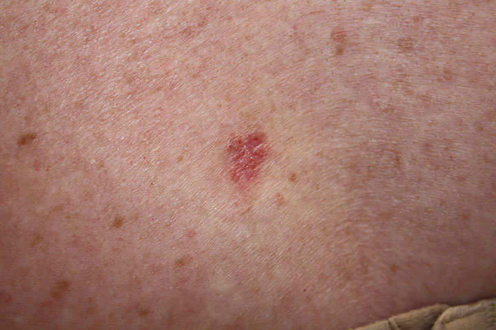
References
- ↑ Neagu TP, Tiglis M, Botezatu D, Enache V, Cobilinschi CO, Vâlcea-Precup MS, GrinTescu IM (2017). "Clinical, histological and therapeutic features of Bowen's disease". Rom J Morphol Embryol. 58 (1): 33–40. PMID 28523295.
- ↑ Nemkaeva RM, Kurmashov NA, Oslopova S (1988). "[Bowen's disease]". Vestn Dermatol Venerol (8): 71–2. PMID 3195216.
- ↑ Scarborough DA, Bisaccia EP, Yoder FW (1982). "Solitary pigmented Bowen's disease". Arch Dermatol. 118 (11): 954–5. PMID 7138058.
- ↑ Yanagihara S, Oiso N, Hirota N, Kato M, Miyake S, Kawada A (2019). "Acantholytic Bowen's disease histopathologically showing the Borst-Jadassohn phenomenon". Eur J Dermatol. doi:10.1684/ejd.2019.3545. PMID 31145080.
- ↑ Baykal C, Buyukbabani N, Babuna G, Polat Ekinci A, Kurul S (2016). "Giant Bowen's disease histologically showing Borst-Jadassohn phenomenon and complicated with squamous cell carcinoma development". J Eur Acad Dermatol Venereol. 30 (10): e88–e89. doi:10.1111/jdv.13335. PMID 26373350.
- ↑ "Borst-Jadassohn phenomenon | definition of Borst-Jadassohn phenomenon by Medical dictionary".
- ↑ "Pathology Outlines - Borst-Jadassohn phenomenon".
- ↑ "PayPerView: Jadassohn's Intraepidermal Epithelioma - Karger Publishers".
- ↑ Lora V, Chouvet B, Kanitakis J (2011). "The "intraepidermal epithelioma" revisited: immunohistochemical study of the borst-jadassohn phenomenon". Am J Dermatopathol. 33 (5): 492–7. doi:10.1097/DAD.0b013e3181fe6f90. PMID 21587033.
- ↑ Clavel CE, Huu VP, Durlach AP, Birembaut PL, Bernard PM, Derancourt CG (1999). "Mucosal oncogenic human papillomaviruses and extragenital Bowen disease". Cancer. 86 (2): 282–7. PMID 10421264.
- ↑ Zheng S, Adachi A, Shimizu M, Shibata SI, Yasue S, Sakakibara A; et al. (2005). "Human papillomaviruses of the mucosal type are present in some cases of extragenital Bowen's disease". Br J Dermatol. 152 (6): 1243–7. doi:10.1111/j.1365-2133.2005.06643.x. PMID 15948988.
- ↑ Lampert A, Pauwels C, Duboucher C, Morel G, Poveda JD, Périé G (2000). "[Detection of human papillomavirus in cutaneous extragenital Bowen's disease in immunocompetent patients]". Ann Dermatol Venereol. 127 (1): 40–5. PMID 10717561.
- ↑ Collina G, Rossi E, Bettelli S, Cook MG, Cesinaro AM, Trentini GP (1995). "Detection of human papillomavirus in extragenital Bowen's disease using in situ hybridization and polymerase chain reaction". Am J Dermatopathol. 17 (3): 236–41. PMID 8599431.
- ↑ Derancourt C, Mougin C, Chopard Lallier M, Coumes-Marquet S, Drobacheff C, Laurent R (2001). "[Oncogenic human papillomaviruses in extra-genital Bowen disease revealed by in situ hybridization]". Ann Dermatol Venereol. 128 (6–7): 715–8. PMID 11460032.
- ↑ Pham-Huu V, Derancourt C, Clavel C, Durlach A, Birembaut P, Bernard P (1999). "[Oncogenic mucosal human papillomaviruses in Bowen's disease of the hands]". Ann Dermatol Venereol. 126 (11): 808–12. PMID 10612858.
- ↑ Murao K, Yoshioka R, Kubo Y (2014). "Human papillomavirus infection in Bowen disease: negative p53 expression, not p16(INK4a) overexpression, is correlated with human papillomavirus-associated Bowen disease". J. Dermatol. 41 (10): 878–84. doi:10.1111/1346-8138.12613. PMID 25201325.
- ↑ 17.0 17.1 "Bowen's disease - NHS".
- ↑ Wagner RF, Grande DJ (1986). "Solitary pigmented Bowen's disease of the scrotum". J Dermatol Surg Oncol. 12 (10): 1114–5. PMID 3760318.
- ↑ Al-Dawsari NA, Raslan W, Dawamneh MF (2014). "Pigmented Bowen's disease of the penis and scrotum in a patient with AIDS". Dermatol Online J. 20 (4): 22337. PMID 24746300.
- ↑ Narahira A, Yanagi T, Kitamura S, Hata H, Shimizu H (2019). "Dermoscopic features of genital pigmented Bowen's disease: Report of a case and review of the published work". J Dermatol. doi:10.1111/1346-8138.14938. PMID 31141219.
- ↑ "Bowen's Disease: Skin Cancer Linked to HPV Infection".
- ↑ Inoue T, Kobayashi K, Sawada M, Ishizaki S, Ito H, Fujibayashi M; et al. (2010). "Dermoscopic Features of Pigmented Bowen's Disease in a Japanese Female Mimicking Malignant Melanoma". Dermatol Res Pract. 2010. doi:10.1155/2010/543091. PMC 2929512. PMID 20811602.
- ↑ Hayashi Y, Tanaka M, Suzaki R, Mori N, Konohana I (2009). "Dermoscopy of Pigmented Bowen's Disease Mimicking Early Superficial Spreading Melanoma". Case Rep Dermatol. 1 (1): 11–15. doi:10.1159/000227284. PMC 2895203. PMID 20652107.
- ↑ Yang Y, Lin J, Fang S, Han S, Song Z (2017). "What's new in dermoscopy of Bowen's disease: two new dermoscopic signs and its differential diagnosis". Int J Dermatol. 56 (10): 1022–1025. doi:10.1111/ijd.13734. PMID 28832993.
- ↑ Zalaudek I, Argenziano G, Leinweber B, Citarella L, Hofmann-Wellenhof R, Malvehy J; et al. (2004). "Dermoscopy of Bowen's disease". Br J Dermatol. 150 (6): 1112–6. doi:10.1111/j.1365-2133.2004.05924.x. PMID 15214896.
- ↑ Cameron A, Rosendahl C, Tschandl P, Riedl E, Kittler H (2010). "Dermatoscopy of pigmented Bowen's disease". J Am Acad Dermatol. 62 (4): 597–604. doi:10.1016/j.jaad.2009.06.008. PMID 20079953.
- ↑ "Bowen Disease - NORD (National Organization for Rare Disorders)".
- ↑ Neubert T, Lehmann P (2008). "Bowen's disease - a review of newer treatment options". Ther Clin Risk Manag. 4 (5): 1085–95. PMC 2621408. PMID 19209288.
- ↑ Wong TW, Sheu HM, Lee JY, Fletcher RJ (2001). "Photodynamic therapy for Bowen's disease (squamous cell carcinoma in situ) of the digit". Dermatol Surg. 27 (5): 452–6. PMID 11359493.
- ↑ Britton JE, Goulden V, Stables G, Stringer M, Sheehan-Dare R (2005). "Investigation of the use of the pulsed dye laser in the treatment of Bowen's disease using 5-aminolaevulinic acid phototherapy". Br J Dermatol. 153 (4): 780–4. doi:10.1111/j.1365-2133.2005.06830.x. PMID 16181460.
- ↑ Kang HK, Yun JH, Son YM, Roh JY, Lee JR (2014). "Photodynamic Therapy for Bowen's Disease of the Vulva Area". Ann Dermatol. 26 (2): 241–5. doi:10.5021/ad.2014.26.2.241. PMC 4037679. PMID 24882981.
- ↑ Dijkstra AT, Majoie IM, van Dongen JW, van Weelden H, van Vloten WA (2001). "Photodynamic therapy with violet light and topical 6-aminolaevulinic acid in the treatment of actinic keratosis, Bowen's disease and basal cell carcinoma". J Eur Acad Dermatol Venereol. 15 (6): 550–4. PMID 11843215.
- ↑ Harth Y, Hirshowitz B, Kaplan B (1998). "Modified topical photodynamic therapy of superficial skin tumors, utilizing aminolevulinic acid, penetration enhancers, red light, and hyperthermia". Dermatol Surg. 24 (7): 723–6. PMID 9693665.
- ↑ de Haas ER, de Vijlder HC, Sterenborg HJ, Neumann HA, Robinson DJ (2008). "Fractionated aminolevulinic acid-photodynamic therapy provides additional evidence for the use of PDT for non-melanoma skin cancer". J Eur Acad Dermatol Venereol. 22 (4): 426–30. doi:10.1111/j.1468-3083.2007.02445.x. PMID 18031503.
- ↑ Anna Z, John K, Maria T, George K, Ivelina B, Ioanna K; et al. (2012). "The potential role of radiation therapy in Bowen's disease: a review of the current literature". Rev Recent Clin Trials. 7 (1): 42–6. PMID 21864250.
- ↑ Lukas VanderSpek LA, Pond GR, Wells W, Tsang RW (2005). "Radiation therapy for Bowen's disease of the skin". Int J Radiat Oncol Biol Phys. 63 (2): 505–10. doi:10.1016/j.ijrobp.2005.02.024. PMID 16168842.
- ↑ Herman JM, Pierce LJ, Sandler HM, Griffith KA, Jabbari S, Hiniker SM; et al. (2008). "Radiotherapy using a water bath in the treatment of Bowen's disease of the digit". Radiother Oncol. 88 (3): 398–402. doi:10.1016/j.radonc.2008.05.025. PMID 18571754.
- ↑ Goodman CR, DeNittis A (2017). "Photon irradiation using a water bath technique for treatment of confluent carcinoma in situ of the hand, digits, and nail bed: a case report". J Med Case Rep. 11 (1): 86. doi:10.1186/s13256-017-1233-3. PMC 5372342. PMID 28356127.
- ↑ Hunt WT, Cameron A, Craig P, de Berker DA (2013). "Multiple-digit periungual Bowen's disease: a novel treatment approach with radiotherapy". Clin Exp Dermatol. 38 (8): 857–61. doi:10.1111/ced.12149. PMID 23937119.
- ↑ Dupree MT, Kiteley RA, Weismantle K, Panos R, Johnstone PA (2001). "Radiation therapy for Bowen's disease: lessons for lesions of the lower extremity". J Am Acad Dermatol. 45 (3): 401–4. doi:10.1067/mjd.2001.116581. PMID 11511838.
- ↑ "Extramammary Paget Disease - StatPearls - NCBI Bookshelf".
- ↑ Yao H, Xie M, Fu S, Guo J, Peng Y, Cai Z; et al. (2018). "Survival analysis of patients with invasive extramammary Paget disease: implications of anatomic sites". BMC Cancer. 18 (1): 403. doi:10.1186/s12885-018-4257-1. PMC 5894213. PMID 29636019.
- ↑ Karam A, Dorigo O (2014). "Increased risk and pattern of secondary malignancies in patients with invasive extramammary Paget disease". Br J Dermatol. 170 (3): 661–71. doi:10.1111/bjd.12635. PMID 24617434.
- ↑ van der Zwan JM, Siesling S, Blokx WA, Pierie JP, Capocaccia R (2012). "Invasive extramammary Paget's disease and the risk for secondary tumours in Europe". Eur J Surg Oncol. 38 (3): 214–21. doi:10.1016/j.ejso.2011.12.008. PMID 22244907.
- ↑ Asel M, LeBoeuf NR (2019). "Extramammary Paget's Disease". Hematol Oncol Clin North Am. 33 (1): 73–85. doi:10.1016/j.hoc.2018.09.003. PMID 30497678.
- ↑ Chumbalkar V, Jennings TA, Ainechi S, Lee EC, Lee H (2016). "Extramammary Paget's Disease of Anal Canal Associated With Rectal Adenoma Without Invasive Carcinoma". Gastroenterology Res. 9 (6): 99–102. doi:10.14740/gr727e. PMC 5191897. PMID 28058078.
- ↑ Jabir S, Khatib M, Ali S, Niranjan N (2013). "Perianal Paget's disease: a diagnostic dilemma". BMJ Case Rep. 2013. doi:10.1136/bcr-2013-200504. PMC 3794179. PMID 24072832.
- ↑ Tarng SJ, Fan HA, Lin SE, Lai CR, Tang RP, You YD; et al. (1990). "Perianal Paget's disease--report of 4 cases". Changgeng Yi Xue Za Zhi. 13 (4): 314–21. PMID 2178040.
- ↑ Lian P, Gu WL, Zhang Z, Cai GX, Wang MH, Xu Y; et al. (2010). "Retrospective analysis of perianal Paget's disease with underlying anorectal carcinoma". World J Gastroenterol. 16 (23): 2943–8. doi:10.3748/wjg.v16.i23.2943. PMC 2887592. PMID 20556842.
- ↑ {{cite journal| author=Rajendran S, Koh CE, Solomon MJ| title=Extramammary Paget's disease of the perianal region: a 20-year experience. | journal=ANZ J Surg | year= 2017 | volume= 87 | issue= 3 | pages= 132-137 | pmid=25113623 | doi=10.1111/ans.12814 | pmc= | <ref name="pmid27746643">Nagai Y, Kazama S, Yamada D, Miyagawa T, Murono K, Yasuda K; et al. (2016). "Perianal and Vulvar Extramammary Paget Disease: A Report of Six Cases and Mapping Biopsy of the Anal Canal". Ann Dermatol. 28 (5): 624–628. doi:10.5021/ad.2016.28.5.624. PMC 5064193. PMID 27746643.
- ↑ Jung JH, Kwak C, Kim HH, Ku JH (2013). "Extramammary Paget Disease of External Genitalia: Surgical Excision and Follow-up Experiences With 19 Patients". Korean J Urol. 54 (12): 834–9. doi:10.4111/kju.2013.54.12.834. PMC 3866286. PMID 24363864.
- ↑ Yang WJ, Kim DS, Im YJ, Cho KS, Rha KH, Cho NH; et al. (2005). "Extramammary Paget's disease of penis and scrotum". Urology. 65 (5): 972–5. doi:10.1016/j.urology.2004.12.010. PMID 15882734.
- ↑ Fu M, Gao S, Wang P (1999). "[19 cases of Paget's disease]". Zhonghua Wai Ke Za Zhi. 37 (7): 429–31. PMID 11829881.
- ↑ "Extramammary Paget's disease of the perianal region: a 20‐year experience - Rajendran - 2017 - ANZ Journal of Surgery - Wiley Online Library".