Internal carotid artery
Template:WikiDoc Cardiology News
Editor-In-Chief: C. Michael Gibson, M.S., M.D. [1]
Overview
In human anatomy, the internal carotid artery is a major artery of the head and neck that helps supply blood to the brain.
Classification
Terminologia Anatomica currently breaks the artery into four parts: "cervical", "petrous", "cavernous", and "cerebral".[1][2] However, a more recent classification system of the internal carotid artery follows Bouthillier,[3] and describes seven anatomical segments of the internal carotid artery. The Bouthillier system is often used clinically by neurosurgeons, neuroradiologists and neurologists. This nomenclature system is a clinical one, based on the angiographic appearance of the artery and its relationship to surrounding anatomy, in contrast to an embryologic classification system. An older clinical classification is based on work by Fischer in 1938 is also commonly used, as well as classification schemes based on the embryologic anatomy of the carotid artery.
The segments of the internal carotid artery are as follows:
- Cervical segment, or C1, identical to the commonly used Cervical portion
- Petrous segment, or C2
- Lacerum segment, or C3
- C2 and C3 comprise the commonly used Petrous portion
- Cavernous segment, or C4, almost identical to the commonly used Cavernous portion
- Clinoid segment, or C5. This segment is not identified in some earlier classifications, and lies between the commonly used Cavernous portion and Cerebral or Supraclinoid portion
- Ophthalamic, or supraclinoid segment, or C6
- Communicating, or terminal segment, or C7
- C6 and C7 together comprise the commonly used Cerebral or Supraclinoid portion
Course
The internal carotid artery is a terminal branch of the common carotid artery; it arises around the level of the third cervical vertebra when the common carotid bifurcates into this artery and its more superficial counterpart, external carotid artery.
C1: Cervical segment

The cervical segment, or C1, of the internal carotid extends from the carotid bifurcation until it enters the carotid canal in the skull anterior to the jugular foramen.
At its origin, the internal carotid artery is somewhat dilated. This part of the artery is known as the carotid sinus or the carotid bulb. The ascending portion of the cervical segment occurs distal to the bulb, when the vessel walls are again parallel.
The internal carotid runs perpendicularly upward in the carotid sheath, and enters the skull through the carotid canal. During this part of its course, it lies in front of the transverse processes of the upper three cervical vertebrae.
It is relatively superficial at its start, where it is contained in the carotid triangle of the neck, and lies behind and lateral to the external carotid, overlapped by the sternocleidomastoid muscle, and covered by the deep fascia, the platysma, and integument: it then passes beneath the parotid gland, being crossed by the hypoglossal nerve, the digastric muscle and the stylohyoid muscle, the occipital artery and the posterior auricular artery. Higher up, it is separated from the external carotid by the styloglossus and stylopharyngeus muscles, the tip of the styloid process and the stylohyoid ligament, the glossopharyngeal nerve and the pharyngeal branch of the vagus nerve. It is in relation, behind, with the longus capitis, the superior cervical ganglion of the sympathetic trunk, and the superior laryngeal nerve; laterally, with the internal jugular vein and vagus nerve, the nerve lying on a plane posterior to the artery; medially, with the pharynx, superior laryngeal nerve, and ascending pharyngeal artery. At the base of the skull the glossopharyngeal, vagus, accessory, and hypoglossal nerves lie between the artery and the internal jugular vein.
Unlike the external carotid artery, the internal carotid normally has no branches in the neck.
C2: Petrous segment
The petrous segment, or C2, of the internal carotid is that which is inside the petrous part of the temporal bone. This segment extends until the foramen lacerum. The petrous portion classically has three sections: an ascending, or vertical portion; the genu, or bend; and the horizontal portion.
When the internal carotid artery enters the canal in the petrous portion of the temporal bone, it first ascends a short distance, then curves anteriorly and medially. The artery lies at first in front of the cochlea and tympanic cavity; from the latter cavity it is separated by a thin, bony lamella, which is cribriform in the young subject, and often partly absorbed in old age. Farther forward it is separated from the trigeminal ganglion by a thin plate of bone, which forms the floor of the fossa for the ganglion and the roof of the horizontal portion of the canal. Frequently this bony plate is more or less deficient, and then the ganglion is separated from the artery by fibrous membrane. The artery is separated from the bony wall of the carotid canal by a prolongation of dura mater, and is surrounded by a number of small veins and by filaments of the carotid plexus, derived from the ascending branch of the superior cervical ganglion of the sympathetic trunk.
The named branches of the petrous segment of the internal carotid artery are:
- the vidian artery or artery of the pterygoid canal
- the caroticotympanic artery
C3: Lacerum segment
The lacerum segment, or C3, is a short segment that begins above the foramen lacerum and ends at the petrolingual ligament, a reflection of periosteum between the lingula and petrous apex (or petrosal process) of the sphenoid. The lacerum portion is still considered to be 'extra-dural', as it is surrounded by periosteum and fibrocartilage along its course. The lacerum segment normally has no named branches, though the vidian artery may occasionally arise from this segment.
C4: Cavernous segment

The cavernous segment, or C4, of the internal carotid artery begins at the petrolingual ligament and extends to the proximal dural ring, which is formed by the medial and inferior periosteum of the anterior clinoid process. The cavernous segment is surrounded by the cavernous sinus.
In this part of its course, the artery is situated between the layers of the dura mater forming the cavernous sinus, but covered by the lining membrane of the sinus. It at first ascends toward the posterior clinoid process, then passes forward by the side of the body of the sphenoid bone, and again curves upward on the medial side of the anterior clinoid process, and perforates the dura mater forming the roof of the sinus. This portion of the artery is surrounded by filaments of the sympathetic trunk, and on its lateral side is the abducent nerve, or cranial nerve VI.
The named branches of the cavernous segment are:
- the meningohypophyseal artery
- the inferolateral trunk
The cavernous segment also gives rise to small capsular arteries that supply the wall of the cavernous sinus.
C5: Clinoid segment
The clinoid segment, or C5, is another short segment of the internal carotid that begins after the artery exits the cavernous sinus at the proximal dural ring and extends distally to the distal dural ring, afterwhich the carotid artery is considered 'intra-dural' and has entered the subarachnoid space.
The clinoid segment normally has no named branches, though the ophthalmic artery may arise from the clinoid segment.
C6: Ophthalmic segment

The ophthalmic segment, or C6, extends from the distal dural ring, which is continuous with the falciform ligament, and extends distally to the origin of the posterior communicating artery. The ophthalmic segment courses roughly horizontally, parallel to the optic nerve which runs superomedially to the carotid at this point.
The named branches of the ophthalmic segment are:
C7: Communicating segment
The communicating segment, or terminal segment, or C7, of the internal carotid artery passes between the optic and oculomotor nerves to the anterior perforated substance at the medial extremity of the lateral cerebral fissure. Angiographically, this segment extends from the origin of the posterior communicating artery to the bifurcation of the internal carotid artery.
The named branches of the communicating segment are:
The internal carotid then divides to form the anterior cerebral artery and middle cerebral artery. The internal carotid artery can receive blood flow via an important collateral pathway supplying the brain, the cerebral arterial circle, which is more commonly known as the Circle of Willis.
Branches
The following are the branches of the internal carotid artery, listed by segment:[4]
- C1: Branches from the cervical portion - none.
- C2: Branches from the petrous portion
- C3: Branches from the lacerum portion - none
- C4: Branches from the cavernous portion
- Branches of the meningohypophyseal trunk:
- Tentorial basal branch
- Tentorial marginal branch
- Meningeal branch - helps supply blood to the meninges of the anterior cranial fossa
- Clivus branches - tiny branches that supply the clivus
- Inferior hypophyseal artery
- Capsular branches - supplies wall of cavernous sinus
- Branches of the inferolateral trunk:
- Branches to trigeminal ganglion - provide blood to trigeminal ganglion
- Artery of the foramen rotundum
- Branches to nerves
- Branches of the meningohypophyseal trunk:
- C5: Branches from the clinoid portion - none
- C6: Branches from the ophthalmic portion
- C7: Branches from the communicating portion
- Posterior communicating artery
- Anterior choroidal artery
- Anterior cerebral artery (a terminal branch)
- Middle cerebral artery (a terminal branch)
Carotid plexus
The sympathetic trunk forms a plexus of nerves around the artery known as the carotid plexus. The internal carotid nerve arises from the superior cervical ganglion, and forms this plexus, which follows the internal carotid into the skull.
Additional images
-
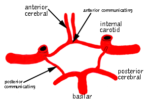 Circle of Willis
 -
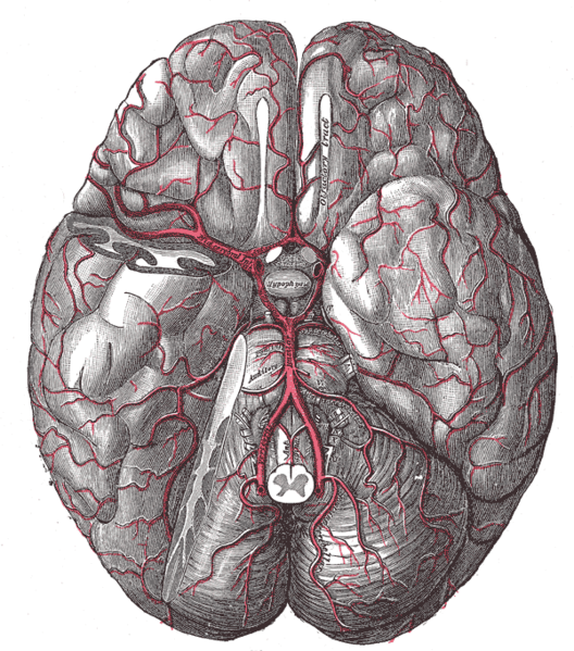 The arteries of the base of the brain.
 -
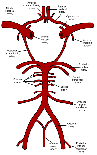 Diagram of the arterial circulation at the base of the brain.
 -
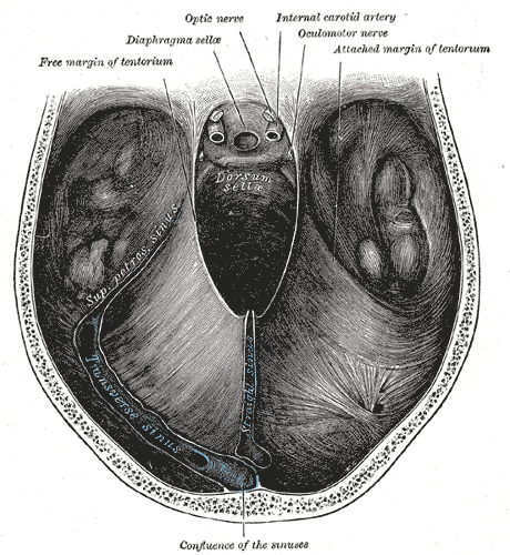 Tentorium cerebelli from above.
 -
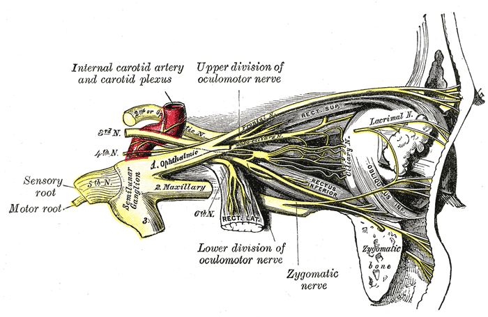 Nerves of the orbit, and the ciliary ganglion. Side view.
 -
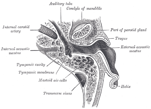 Horizontal section through left ear; upper half of section.
 -
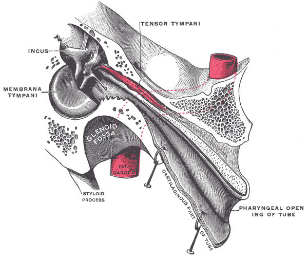 Auditory tube, laid open by a cut in its long axis.
 -
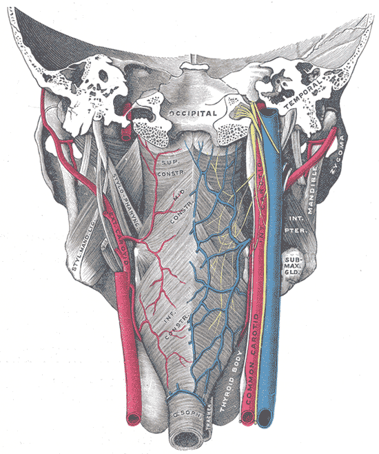 Muscles of the pharynx, viewed from behind, together with the associated vessels and nerves.
 -
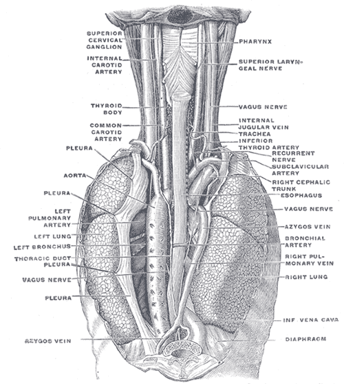 The position and relation of the esophagus in the cervical region and in the posterior mediastinum. Seen from behind.


See also
- External carotid artery
- Carotid endarterectomy
- Carotid sinus
- Carotid body
- Carotid sheath
- Carotid triangle
- Circle of Willis
References
- ↑ Template:LoyolaMedEd
- ↑ Template:Dorlands
- ↑ Bouthillier, Alain (1996). "Segments of the internal carotid artery: a new classification". Neurosurgery. 38 (3): 425–432. PMID 8837792. Unknown parameter
|coauthors=ignored (help); Unknown parameter|month=ignored (help) - ↑ Osborn, Anne (1999). Diagnostic Cerebral Angiography (2nd ed.). Philadelphia, PA, USA: Lippincott Williams & Wilkins. ISBN 0-397-58404-0.
External links
- Template:SUNYAnatomyLabs
- Template:UMichAtlas
- The Anatomy Wiz. Internal Carotid Artery