Pharynx
|
WikiDoc Resources for Pharynx |
|
Articles |
|---|
|
Most recent articles on Pharynx |
|
Media |
|
Evidence Based Medicine |
|
Clinical Trials |
|
Ongoing Trials on Pharynx at Clinical Trials.gov Clinical Trials on Pharynx at Google
|
|
Guidelines / Policies / Govt |
|
US National Guidelines Clearinghouse on Pharynx
|
|
Books |
|
News |
|
Commentary |
|
Definitions |
|
Patient Resources / Community |
|
Directions to Hospitals Treating Pharynx Risk calculators and risk factors for Pharynx
|
|
Healthcare Provider Resources |
|
Causes & Risk Factors for Pharynx |
|
Continuing Medical Education (CME) |
|
International |
|
|
|
Business |
|
Experimental / Informatics |
Editor-In-Chief: C. Michael Gibson, M.S., M.D. [1]
Overview
The pharynx (plural: pharynges) is an organ found in vertebrates and invertebrates, though the structure is not universally the same across the species. In humans the pharynx is part of the digestive system and also of the conducting zone of the respiratory system. (The conducting zone also includes the nose, larynx, trachea, bronchi, and bronchioles, and their function is to filter, warm, and moisten air and conduct it into the lungs.[1]) The pharynx makes up the part of the throat situated immediately posterior to the nasal cavity, posterior to the mouth and superior to the esophagus and larynx. The human pharynx is conventionally divided into three sections: the nasopharynx, the oropharynx and the laryngopharynx. It is also important in vocalization.
There are two sets of pharyngeal muscles that act upon the pharynx. These are arranged as an inner layer of longitudinal muscles and an outer circular layer.
Structure
Nasopharynx
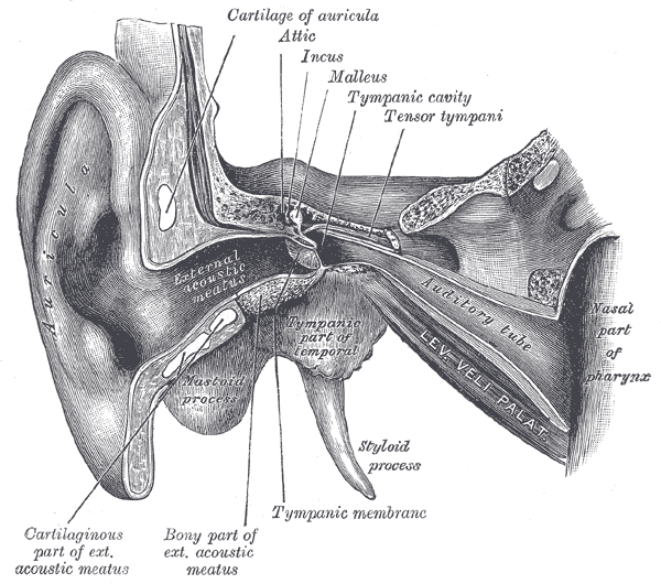
The upper portion of the pharynx, the nasopharynx, extends from the base of the skull to the upper surface of the soft palate.[2] It includes the space between the internal nares and the soft palate and lies above the oral cavity. The adenoids, also known as the pharyngeal tonsils, are lymphoid tissue structures located in the posterior wall of the nasopharynx. Waldeyer's tonsillar ring is an annular arrangement of lymphoid tissue in both the nasopharynx and oropharynx. Polyps or mucus can obstruct the nasopharynx, as can congestion due to an upper respiratory infection. The eustachian tubes, which connect the middle ear to the pharynx, open into the nasopharynx. The opening and closing of the eustachian tubes serves to equalize the barometric pressure in the middle ear with that of the ambient atmosphere.
The anterior aspect of the nasopharynx communicates through the choanae with the nasal cavities. On its lateral walls are the pharyngeal ostia of the auditory tube, somewhat triangular in shape, and bounded behind by a firm prominence, the torus tubarius or cushion, caused by the medial end of the cartilage of the tube that elevates the mucous membrane. Two folds arise from the cartilaginous opening:
- the salpingopharyngeal fold, a vertical fold of mucous membrane extending from the inferior part of the torus and containing the salpingopharyngeus muscle
- the salpingopalatine fold, a smaller fold extending from the superior part of the torus to the palate and containing the levator veli palatini muscle. The tensor veli palatini is lateral to the levator and does not contribute the fold, since the origin is deep to the cartilaginous opening.
Behind the opening of the auditory tube is a deep recess, the pharyngeal recess (also referred to as the fossa of Rosenmüller). On the posterior wall is a prominence, best marked in childhood, produced by a mass of lymphoid tissue, which is known as the pharyngeal tonsil. Superior to the pharyngeal tonsil, in the midline, an irregular flask-shaped depression of the mucous membrane sometimes extends up as far as the basilar process of the occipital bone, this is known as the pharyngeal bursa.
Oropharynx
The oropharynx lies behind the oral cavity, extending from the uvula to the level of the hyoid bone. It opens anteriorly, through the isthmus faucium, into the mouth, while in its lateral wall, between the palatoglossal arch and the palatopharyngeal arch, is the palatine tonsil.[3] The anterior wall consists of the base of the tongue and the epiglottic vallecula; the lateral wall is made up of the tonsil, tonsillar fossa, and tonsillar (faucial) pillars; the superior wall consists of the inferior surface of the soft palate and the uvula. Because both food and air pass through the pharynx, a flap of connective tissue called the epiglottis closes over the glottis when food is swallowed to prevent aspiration. The oropharynx is lined by non-keratinised squamous stratified epithelium.
The HACEK organisms (Haemophilus, Actinobacillus actinomycetemcomitans, Cardiobacterium hominis, Eikenella corrodens, Kingella) are part of the normal oropharyngeal flora, which grow slowly, prefer a carbon dioxide-enriched atmosphere, and share an enhanced capacity to produce endocardial infections, especially in young children.[4] Fusobacterium is a pathogen.[5]
Laryngopharynx
The laryngopharynx, (Latin: pars laryngea pharyngis), also known as hypopharynx, is the caudal part of the pharynx; it is the part of the throat that connects to the esophagus. It lies inferior to the epiglottis and extends to the location where this common pathway diverges into the respiratory (larynx) and digestive (esophagus) pathways. At that point, the laryngopharynx is continuous with the esophagus posteriorly. The esophagus conducts food and fluids to the stomach; air enters the larynx anteriorly. During swallowing, food has the "right of way", and air passage temporarily stops. Corresponding roughly to the area located between the 4th and 6th cervical vertebrae, the superior boundary of the laryngopharynx is at the level of the hyoid bone. The laryngopharynx includes three major sites: the pyriform sinus, postcricoid area, and the posterior pharyngeal wall. Like the oropharynx above it, the laryngopharynx serves as a passageway for food and air and is lined with a stratified squamous epithelium. It is innervated by the pharyngeal plexus.
The vascular supply to the laryngopharynx includes the superior thyroid artery, the lingual artery and the ascending pharyngeal artery. The primary neural supply is from both the vagus and glossopharyngeal nerves. The vagus nerve provides a branch termed "Arnolds Nerve" which also supplies the external auditory canal, thus laryngopharyngeal cancer can result in referred otalgia. This nerve is also responsible for the ear-cough reflex in which stimulation of the ear canal results in a person coughing.
Clinical significance
Inflammation

Pharyngitis is an inflammation of the throat or pharynx.[6] In most cases it is painful, and thus is often referred to as a sore throat. Inflammation of the tonsils (tonsillitis) and/or larynx (laryngitis) may occur simultaneously, which can make eating difficult or painful.
Most cases are caused by virus|viral infections (40%-60%), with the remainder caused by bacterial infections, fungus|fungal infections, or irritants such as pollutants or chemical substances.[7]
Treatment of viral causes are mainly symptomatic while bacterial or fungal causes may be amenable to antibiotics and antifungals respectively.
Pharyngeal cancer
Neck squamous cell carcinoma, an oropharyngeal cancer, occurs in humans.[8]
History
Etymology
The word pharynx (pronounced /ˈfærɪŋks/[9][10]) is derived from the Greek φάρυγξ phárynx, meaning "throat". Its plural form is pharynges /fəˈrɪndʒiːz/ or pharynxes /ˈfærɪŋksəz/, and its adjective form is pharyngeal (/ˌfærɪnˈdʒiːəl/ or /fəˈrɪndʒiəl/).
Other animals
Vertebrates
All vertebrates have a pharynx, used in both feeding and respiration. The pharynx arises during development in all vertebrates through a series of six or more outpocketings on the lateral sides of the head. These outpocketings are pharyngeal arches, and they give rise to a number of different structures in the skeletal, muscular and circulatory systems. The structure of the pharynx varies across the vertebrates. It differs in dogs, horses and ruminants. In dogs a single duct connects the nasopharynx to the nasal cavity. The tonsils are a compact mass which point away from the lumen of the pharynx. In the horse the auditory tube opens into the guttural pouch and the tonsils are diffuse and raised slightly. Horses are unable to breathe through the mouth as the free apex of the rostral epiglottis lies dorsal to the soft palate in a normal horse. In ruminants the tonsils are a compact mass which point towards the lumen of the pharynx.
Pharyngeal arches are characteristic features of vertebrates whose origin can be traced back through chordates to basal deuterostomes who also share endodermal outpocketings of the pharyngeal apparatus. Similar patterns of gene expression can be detected in the developing pharynx of amphioxus and hemichordates. However, the vertebrate pharynx is unique in that it gives rise to endoskeletal support through the contribution of neural crest cells.[11]
Pharyngeal jaws are a "second set" of jaws contained within the pharynx of many species of fish, distinct from the primary (oral) jaws. Pharyngeal jaws have been studied in moray eels where their specific action is noted. When the moray bites prey, it first bites normally with its oral jaws, capturing the prey. Immediately thereafter, the pharyngeal jaws are brought forward and bite down on the prey to grip it; they then retract, pulling the prey down the eel's esophagus, allowing it to be swallowed.[12]
Invertebrates
Invertebrates also have a pharynx. Invertebrates with a pharynx include the tardigrades,[13]annelids and arthropods,[14] and the priapulids (which have an eversible pharynx).[15]
The term "pharynx" is used loosely across the animal kingdom for the portion of the digestive tract loosely analogous to the vertebrate pharynx, but this need not imply deep structural or developmental similarities, or that the structures are homologous.[citation needed]
The "pharynx" of the nematode worm is a muscular food pump in the head, triangular in cross-section, that grinds food and transports it directly to the intestines. A one-way valve connects the pharynx to the excretory canal.
Additional images
-
Conducting passages
-
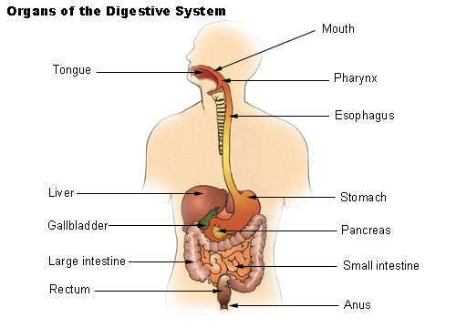 Organs of the digestive system
 -
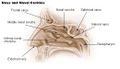 Nose and nasal
 -
-
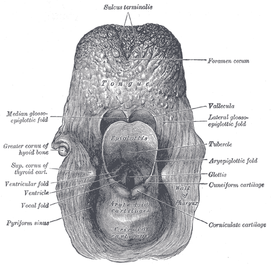 The entrance to the larynx, viewed from behind
 -
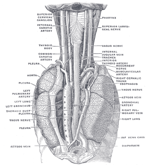 The position and relation of the esophagus in the cervical region and in the posterior mediastinum. Seen from behind.
 -
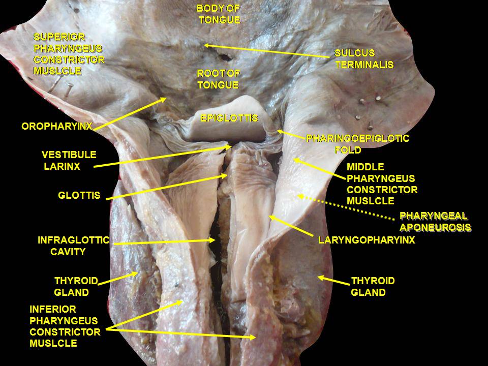 Deep dissection of human larynx, pharynx and tongue seen from behind
 -
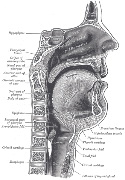 The nasopharynx, oropharynx, and laryngopharynx or larynx can be seen clearly in this sagittal section of the head and neck.


See also
- Nasopharyngeal carcinoma
- Pharyngeal consonant
- Pharyngeal (disambiguation)
- Pharyngitis
- Saccopharynx, a genus of deep sea eel-like fishes with large mouths, distensible stomachs and long scaleless bodies
- Tonsil
- Uvula
References
- ↑ Respiratory Syster (PDF). Benjamin Cummings (Pearson Education, Inc). 2006. p. 1.
- ↑ Clinical Head and Neck and Functional Neuroscience Course Notes, 2008-2009, Uniformed Services University of the Health Sciences School of Medicine, Bethesda, Maryland
- ↑ http://teachmeanatomy.info/neck/viscera/pharynx/
- ↑ Morpeth S, Murdoch D, Cabell CH, et al. (December 2007). "Non-HACEK gram-negative bacillus endocarditis". Ann. Intern. Med. 147 (12): 829–35. doi:10.7326/0003-4819-147-12-200712180-00002. PMID 18087053.
- ↑ Aliyu SH, Marriott RK, Curran MD, et al. (2004). "Real-time PCR investigation into the importance of Fusobacterium necrophorum as a cause of acute pharyngitis in general practice". J Med Microbiol. 53 (Pt 10): 1029&ndash, 35. doi:10.1099/jmm.0.45648-0. PMID 15358827.
- ↑ Dorlands Dictionary, volume six, pharyngitis
- ↑ "eMedicine - Pharyngitis : Article by John R Acerra".
- ↑ Almadori G, Bussu F, Galli J, Limongelli A, Persichilli S, Zappacosta B, Minucci A, Paludetti G, Giardina B (July 2007). "Salivary glutathione and uric acid levels in patients with head and neck squamous cell carcinoma". Head Neck. 29 (7): 648–54. PMID 17274058.
- ↑ OED 2nd edition, 1989.
- ↑ Entry "pharynx" in Merriam-Webster Online Dictionary, retrieved 2012-07-28.
- ↑ Graham, A; Richardson, J (2012). "Developmental and evolutionary origins of the pharyngeal apparatus". EvoDevo. 3 (3): 24. doi:10.1186/2041-9139-3-24. PMC 3564725. PMID 23020903.
- ↑ Mehta, Rita S.; Peter C. Wainwright (2007-09-06). "Raptorial jaws in the throat help moray eels swallow large prey". Nature. 449 (7158): 79–82. doi:10.1038/nature06062. PMID 17805293. Retrieved 2007-09-06.
- ↑ Eibye-Jacobsen (2001). "Are the supportive structures of the tardigrade pharynx homologous throughout the entire group?". Journal of Zoological Systematics and Evolutionary Research. 39: 1. doi:10.1046/j.1439-0469.2001.00140.x.
- ↑ Elzinga, R. J. (1998). "Microspines in the alimentary canal of arthropoda, onychophora, annelida". International Journal of Insect Morphology and Embryology. 27 (4): 341. doi:10.1016/S0020-7322(98)00027-0.
- ↑ Morse, M. Patricia (1981). "Meiopriapulus fijiensis n. gen., n. sp.: An Interstitial Priapulid from Coarse Sand in Fiji". Transactions of the American Microscopical Society. 100 (3): 239. doi:10.2307/3225549. JSTOR 3225549.
General
- Pharynx, Stedman's Online Medical Dictionary at Lippincott Williams and Wilkins
- Human Anatomy and Physiology Elaine N. Marieb and Katja Hoehn, Seventh Edition.
- TNM Classification of Malignant Tumours Sobin LH & Wittekind Ch (eds)Sixth edition UICC 2002 ISBN 0-471-22288-7
External links
| File:Wiktionary-logo-en-v2.svg | Look up pharynx in Wiktionary, the free dictionary. |
| Wikimedia Commons has media related to Pharynx. |
Template:Human system and organs Template:Nose anatomy Template:Mouth anatomy Template:Digestive tract