Internal jugular vein
Editor-In-Chief: C. Michael Gibson, M.S., M.D. [1]
Overview
The internal jugular vein collects the blood from the brain, from the superficial parts of the face, and from the neck.
Path
It is directly continuous with the sigmoid sinus, and begins in the posterior compartment of the jugular foramen, at the base of the skull.
At its origin it is somewhat dilated, and this dilatation is called the superior bulb.
It runs down the side of the neck in a vertical direction, lying at first lateral to the internal carotid artery, and then lateral to the common carotid, and at the root of the neck unites with the subclavian vein to form the brachiocephalic vein (innominate vein); a little above its termination is a second dilatation, the inferior bulb.
Above, it lies upon the Rectus capitis lateralis, behind the internal carotid artery and the nerves passing through the jugular foramen; lower down, the vein and artery lie upon the same plane, the glossopharyngeal and hypoglossal nerves passing forward between them; the vagus descends between and behind the vein and the artery in the same sheath (the carotid sheath), and the accessory runs obliquely backward, superficial or deep to the vein.
At the root of the neck the right internal jugular vein is placed at a little distance from the common carotid artery, and crosses the first part of the subclavian artery, while the left internal jugular vein usually overlaps the common carotid artery.
The left vein is generally smaller than the right, and each contains a pair of valves, which are placed about 2.5 cm. above the termination of the vessel.
Clinical Relevance
The jugular veins are relatively superficial and not protected by tissues such as bone or cartilage. This makes it susceptible to damage. Due to the large volumes of blood that flow though the jugular veins, damage to the jugulars can quickly cause significant blood loss which can lead to hypovolæmic shock and then death if not treated.
JVP
As there are no valves between the right atrium of the heart and the internal jugular, blood can flow back into the internal jugular when the pressure in the atrium is sufficiently high. This can be seen from the outside, and allows one to estimate the pressure in the atrium. The pulsation seen is called the jugular venous pressure, or JVP. This is normally viewed with the patient at 45 degrees turning their head slightly away from the observer. The JVP can be raised in a number of conditions:[1]
- right ventricular failure (heart failure),
- tricuspid stenosis
- tricuspid regurgitation
- cardiac tamponade
The JVP can also be artificially raised by applying pressure to the liver (the hepatojugular reflux). This method is used to locate the JVP and distinguish it from the carotid pulse. Unlike the carotid pulse, the JVP is impalpable.
Catheterization
As the internal jugular is large, central and relatively superficial, it is often used to place venous lines. Such a line may be inserted for several reasons, such as to accurately measure the central venous pressure or to administer fluids when a line in a peripheral vein would be unsuitable (such as during resuscitation when peripheral veins are hard to locate).
Because the internal jugular rarely varies in its location, it is easier to find than other veins. However sometimes when a line is inserted the jugular is missed and other structures such as the carotid artery or the vagus nerve (CN X) are punctured, causing damage to those structures.
Additional images
-
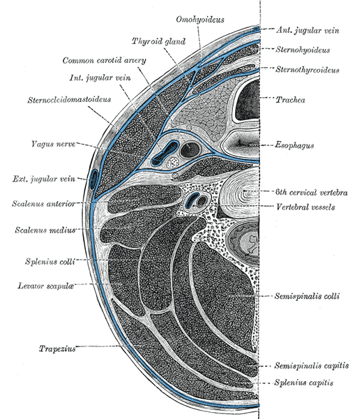 Section of the neck at about the level of the sixth cervical vertebra.
 -
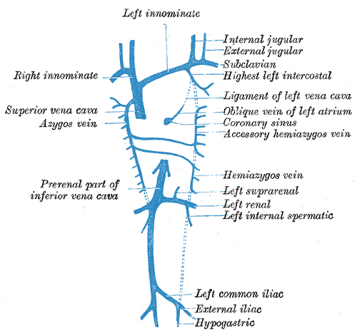 Diagram showing completion of development of the parietal veins.
 -
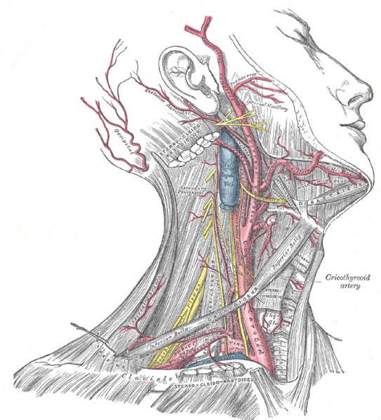 Superficial dissection of the right side of the neck, showing the carotid and subclavian arteries.
 -
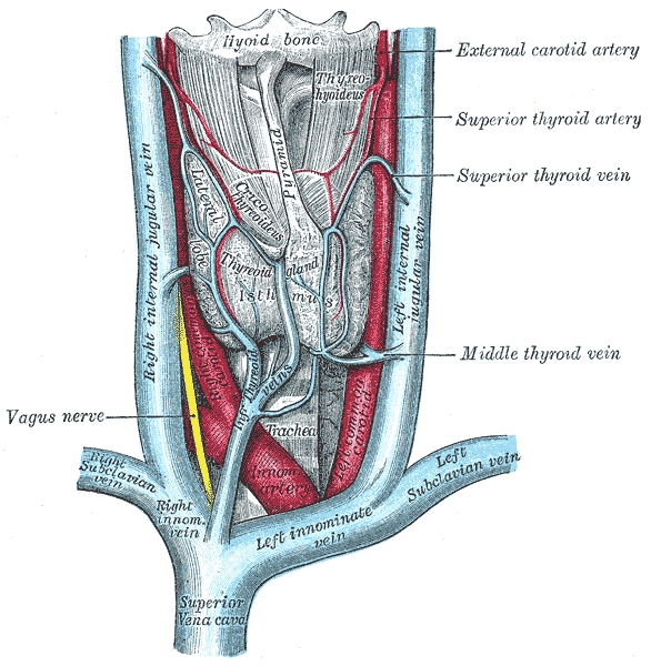
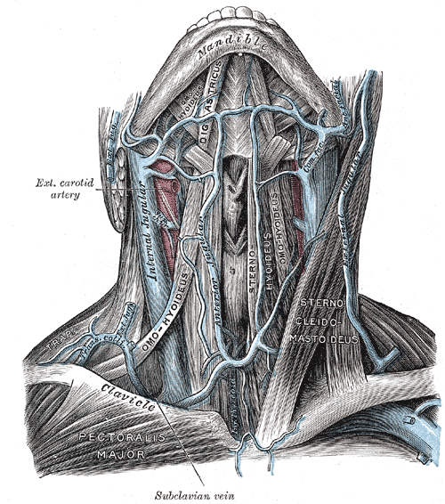 The veins of the neck, viewed from in front.
 -
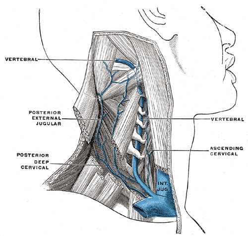 The vertebral vein.
 -
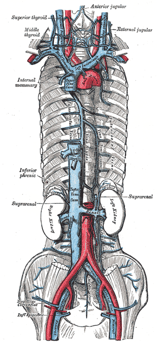 The venæ cavæ and azygos veins, with their tributaries.
 -
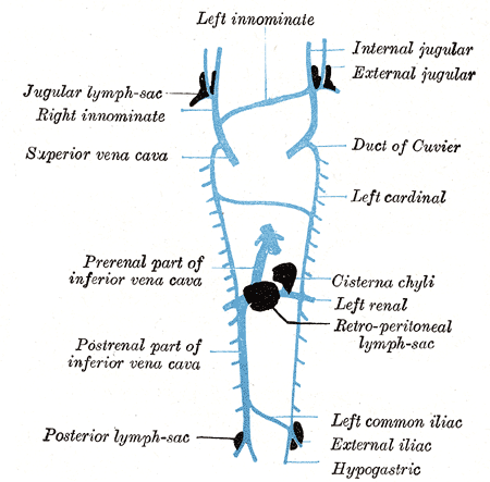 Scheme showing relative positions of primary lymph sacs.
 -
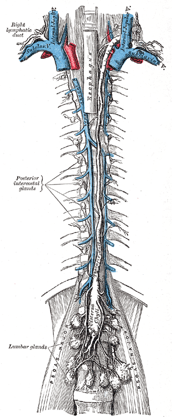 The thoracic and right lymphatic ducts.
 -
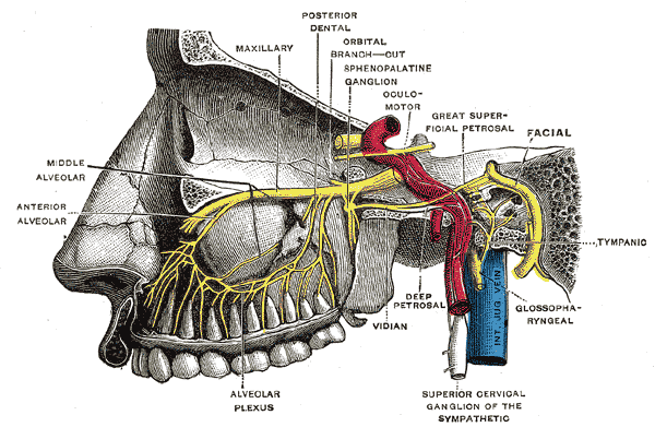 Alveolar branches of superior maxillary nerve and sphenopalatine ganglion.
 -
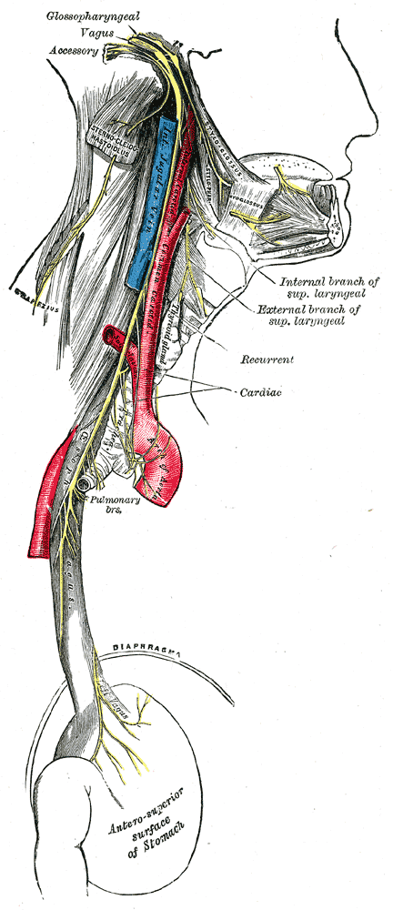 Course and distribution of the glossopharyngeal, vagus, and accessory nerves.
 -
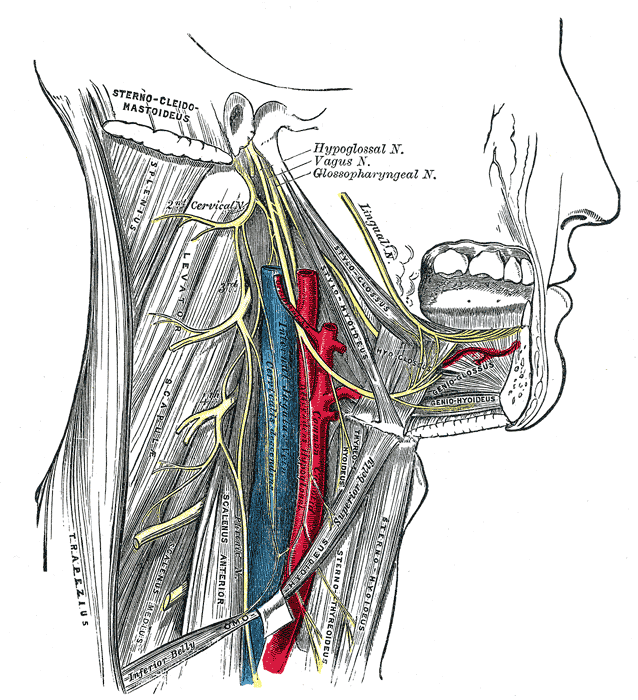 Hypoglossal nerve, cervical plexus, and their branches.
 -
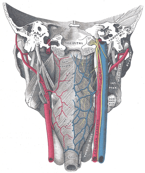 Muscles of the pharynx, viewed from behind, together with the associated vessels and nerves.
 -
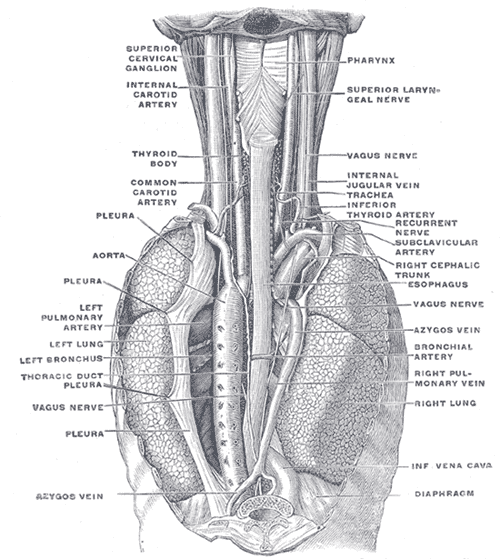 The position and relation of the esophagus in the cervical region and in the posterior mediastinum. Seen from behind.
 -
 The thyroid gland and its relations.
 -
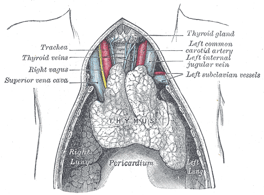 The thymus of a full-term fetus, exposed in situ.


See also
References
Template:Gray's Template:VeinsHeadNeck Template:Veins of the torso no:Vena jugularis interna sk:Vnútorná hrdlová žila