Occipital artery
|
WikiDoc Resources for Occipital artery |
|
Articles |
|---|
|
Most recent articles on Occipital artery Most cited articles on Occipital artery |
|
Media |
|
Powerpoint slides on Occipital artery |
|
Evidence Based Medicine |
|
Clinical Trials |
|
Ongoing Trials on Occipital artery at Clinical Trials.gov Trial results on Occipital artery Clinical Trials on Occipital artery at Google
|
|
Guidelines / Policies / Govt |
|
US National Guidelines Clearinghouse on Occipital artery NICE Guidance on Occipital artery
|
|
Books |
|
News |
|
Commentary |
|
Definitions |
|
Patient Resources / Community |
|
Patient resources on Occipital artery Discussion groups on Occipital artery Patient Handouts on Occipital artery Directions to Hospitals Treating Occipital artery Risk calculators and risk factors for Occipital artery
|
|
Healthcare Provider Resources |
|
Causes & Risk Factors for Occipital artery |
|
Continuing Medical Education (CME) |
|
International |
|
|
|
Business |
|
Experimental / Informatics |
Editor-In-Chief: C. Michael Gibson, M.S., M.D. [1]
The occipital artery arise opposite the facial artery, its path is below the posterior belly of digastic to the occipital region. This artery supplies blood to the back of the scalp and sterno-mastoid muscles. Other muscles it supplies are deep muscles in the back and neck.
Course and Relations
At its origin, it is covered by the posterior belly of the Digastricus and the Stylohyoideus, and the hypoglossal nerve winds around it from behind forward; higher up, it crosses the internal carotid artery, the internal jugular vein, and the vagus and accessory nerves.
It next ascends to the interval between the transverse process of the atlas and the mastoid process of the temporal bone, and passes horizontally backward, grooving the surface of the latter bone, being covered by the Sternocleidomastoideus, Splenius capitis, Longissimus capitis, and Digastricus, and resting upon the Rectus capitis lateralis, the Obliquus superior, and Semispinalis capitis.
It then changes its course and runs vertically upward, pierces the fascia connecting the cranial attachment of the Trapezius with the Sternocleidomastoideus, and ascends in a tortuous course in the superficial fascia of the scalp, where it divides into numerous branches, which reach as high as the vertex of the skull and anastomose with the posterior auricular and superficial temporal arteries.
Its terminal portion is accompanied by the greater occipital nerve.
Additional images
-
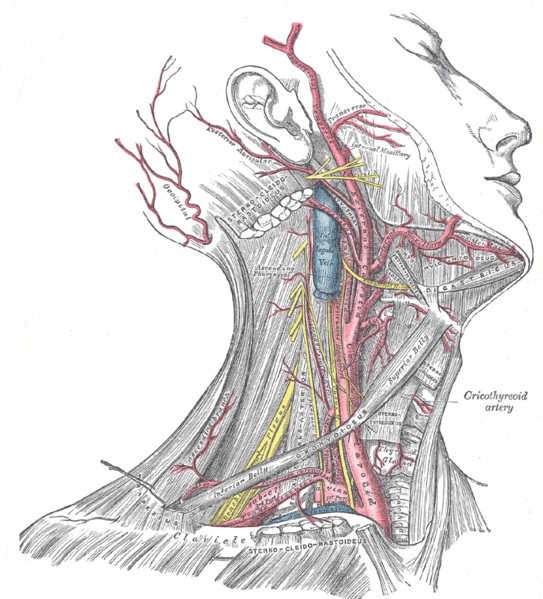 Superficial dissection of the right side of the neck, showing the carotid and subclavian arteries.
 -
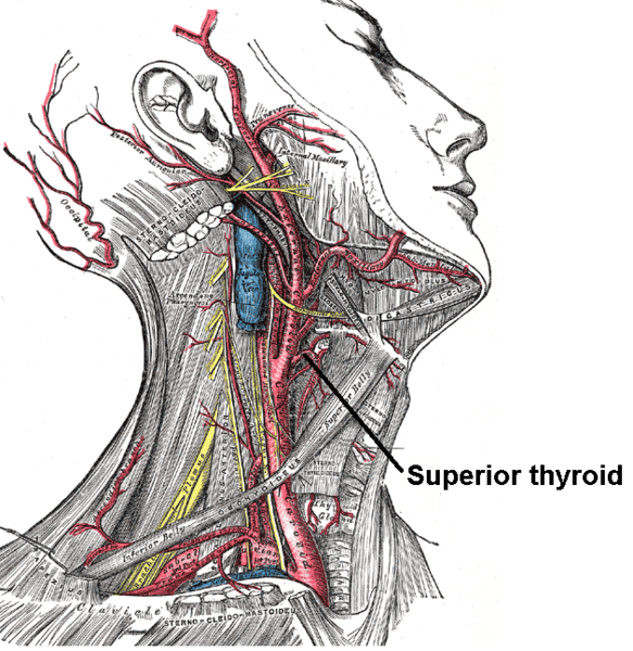 The internal carotid and vertebral arteries. Right side.
 -
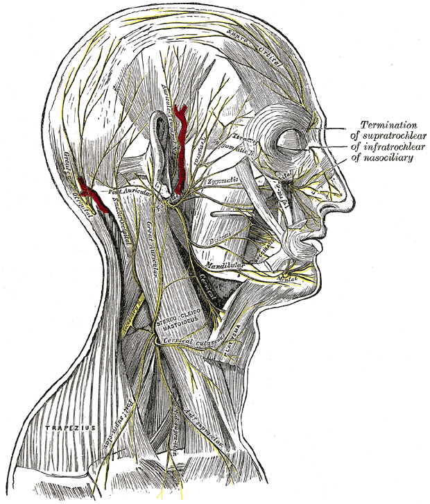 The nerves of the scalp, face, and side of neck.
 -
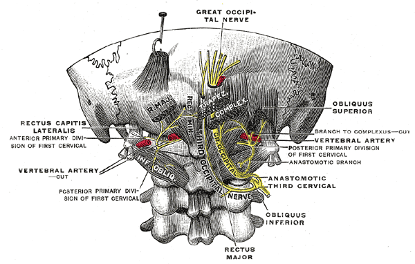 Posterior primary divisions of the upper three cervical nerves.
 -
 Side of neck, showing chief surface markings.


External links
- Template:EMedicineDictionary
- Template:MUNAnatomy
- Template:NormanAnatomy (Template:NormanAnatomyFig)
- Diagram at stchas.edu
- Description at okstate.edu