Dura mater
|
WikiDoc Resources for Dura mater |
|
Articles |
|---|
|
Most recent articles on Dura mater |
|
Media |
|
Evidence Based Medicine |
|
Clinical Trials |
|
Ongoing Trials on Dura mater at Clinical Trials.gov Clinical Trials on Dura mater at Google
|
|
Guidelines / Policies / Govt |
|
US National Guidelines Clearinghouse on Dura mater
|
|
Books |
|
News |
|
Commentary |
|
Definitions |
|
Patient Resources / Community |
|
Patient resources on Dura mater Discussion groups on Dura mater Patient Handouts on Dura mater Directions to Hospitals Treating Dura mater Risk calculators and risk factors for Dura mater
|
|
Healthcare Provider Resources |
|
Causes & Risk Factors for Dura mater |
|
Continuing Medical Education (CME) |
|
International |
|
|
|
Business |
|
Experimental / Informatics |
Overview
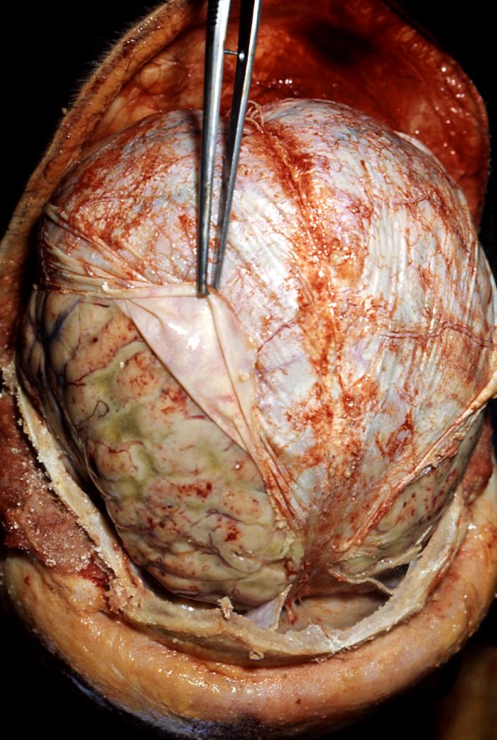
The dura mater (from the Latin "hard mother"), or pachymeninx, is the tough and inflexible outermost of the three layers of the meninges surrounding the brain and spinal cord. (The other two meningeal layers are the pia mater and the arachnoid mater.) The dura mater is not as tightly fitting around the spinal cord, extending past the spinal cord (at the second lumbar vertebra) to about the second sacral vertebra.
Layers and reflections
The dura mater has two layers:
- a superficial layer, which is actually the skull's inner periosteum
- a deep layer, the dura mater proper.
The dura separates into two layers at dural reflections, places where the inner dural layer is reflected as sheet-like protrusions into the cranial cavity. There are two main dural reflections:
- The tentorium cerebelli exists between and separates the cerebellum and brainstem from the occipital lobes of the cerebrum.[1]
- The falx cerebri, which separates the two hemispheres of the brain, is located in the longitudinal cerebral fissure between the hemispheres.[2]
Drainage
The two layers of dura mater run together throughout most of the skull. Where they separate, the gap between them is called a dural venous sinus. These sinuses drain blood and cerebrospinal fluid from the brain and empty into the internal jugular vein.
They drain via the arachnoid villi, which are outgrowths of the arachnoid mater (the middle meningeal layer) that extend into the venous sinuses. These villi act as one-way valves.
Meningeal veins, which course through the dura mater, and bridging veins, which drain the underlying neural tissue and puncture the dura mater, empty into these dural sinuses.
Clinical significance
A subdural hematoma occurs when there is an abnormal collection of blood between the dura and the arachnoid, usually as a result of torn bridging veins secondary to head trauma. An epidural hematoma is a collection of blood between the dura and the inner surface of the skull, and is usually due to arterial bleeding.
The American Red Cross and some other agencies accepting blood donations consider dura mater transplants, along with receipt of pituitary-derived growth hormone, a risk factor due to concerns about Creutzfeldt-Jakob disease.[3]
References
- ↑ Shepherd S. 2004. "Head Trauma." Emedicine.com.
- ↑ Vinas FC and Pilitsis J. 2004. "Penetrating Head Trauma." Emedicine.com.
- ↑ Red Cross: http://www.redcross.org/services/biomed/0,1082,0_553_,00.html
Additional images
-
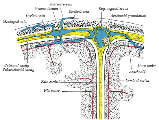 Diagrammatic representation of a section across the top of the skull, showing the membranes of the brain, etc.
 -
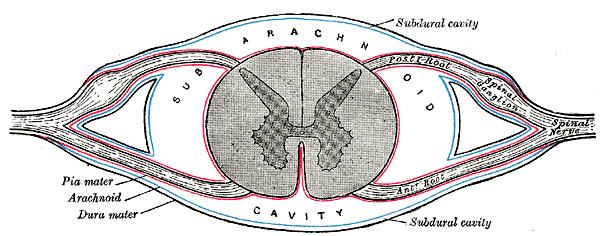 Diagrammatic transverse section of the medulla spinalis and its membranes.
 -
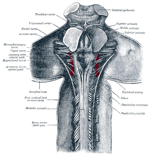 Upper part of medulla spinalis and hind- and mid-brains; posterior aspect, exposed in situ.
 -
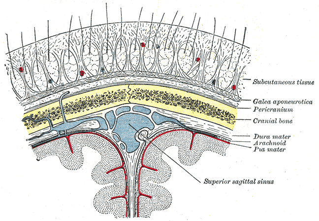 Diagrammatic section of scalp.
 -