Middle cerebral artery
Editor-In-Chief: C. Michael Gibson, M.S., M.D. [1]
Overview
The middle cerebral artery (MCA) is one of the three major paired arteries that supplies blood to the brain. The MCA arises from the internal carotid and continues into the lateral sulcus where it then branches and projects to many parts of the lateral cerebral cortex. It also supplies blood to the anterior temporal lobes and the insular cortices.
The MCAs rise from trifurcations of the internal carotid arteries and thus are connected to the anterior cerebral arteries and the posterior communicating arteries, which connect to the posterior cerebral arteries. The MCAs are not considered a part of the Circle of Willis.[1]
Areas supplied
Areas supplied by the middle cerebral artery include:
- The bulk of the lateral surface of the hemisphere. Exceptions are the superior inch of the frontal lobe and parietal lobe and the inferior part of the temporal lobe and the occipital pole, which are supplied by the posterior cerebral artery.
- Part of the internal capsule and basal ganglia.
Occlusion
Occlusion of the middle cerebral artery may result in the following defects:
- Paralysis of the contralateral face and arm.
- Sensory loss in the contralateral face and arm.
- Aphasia (e.g. Broca's, Wernicke's, conduction, and anomic types) when the dominant hemisphere (usually the left hemisphere for right handed individuals) is affected
- Contralateral neglect syndrome with damage to the right hemisphere
- Homonymous hemianopia or quadrantanopia.
Additional images
-
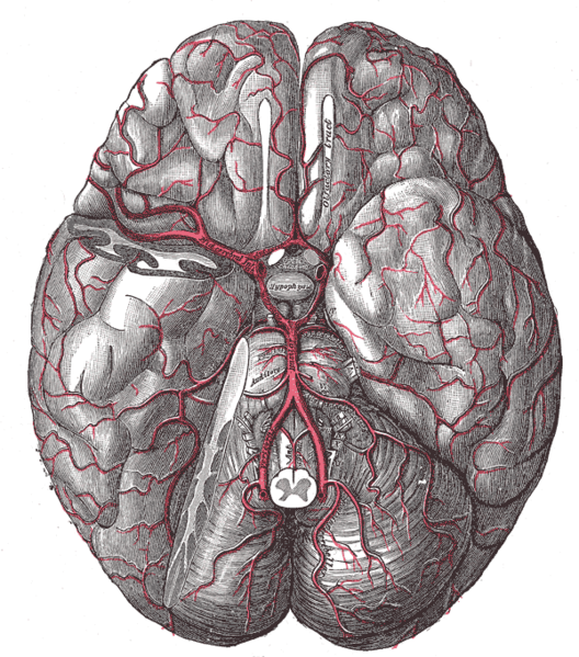 The arteries of the base of the brain.
 -
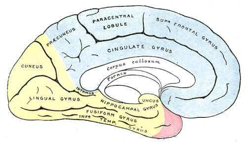 Medial surface of cerebral hemisphere, showing areas supplied by cerebral arteries.
 -
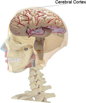 Location of the cerebral cortex


Reference
- ↑ Moore KL, Dalley AR. Clinically Oriented Anatomy, 4th Ed., Lippincott Williams & Wilkins, Toronto. Copyright 1999. ISBN 0-683-06141-0.
External link
- Template:LoyolaMedEd
- Template:SUNYAnatomyLabs
- Template:RocheLexicon
- Blood supply at neuropat.dote.hu
Template:Arteries of head and neck