General visceral efferent fibers
Jump to navigation
Jump to search
Template:Infobox Anatomy The general visceral efferent fibers (GVE or sympathetic efferent fibers), probably arise from cells in the lateral column or the base of the anterior column and emerge through the anterior roots and white rami communicantes.
These are preganglionic fibers which end in various sympathetic ganglia from which postganglionic fibers conduct the motor impulses to the smooth muscles of the viscera and vessels and secretory impulses to the glands.
The cell bodies of GVE fibers are present from the first thoracic to the second lumbar spinal levels (ie, T1-L2).
Examples of nerves containing GVE fibers include the oculomotor nerve, the facial nerve, the glossopharyngeal nerve, and the vagus nerve. [1]
Additional images
-
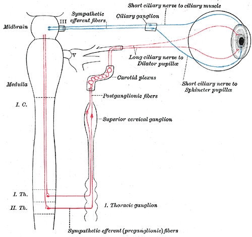 Sympathetic connections of the ciliary and superior cervical ganglia.

See also
References
- ↑ Mehta, Samir et al. Step-Up: A High-Yield, Systems-Based Review for the USMLE Step 1. Baltimore, MD: LWW, 2003.