Facial nerve
|
WikiDoc Resources for Facial nerve |
|
Articles |
|---|
|
Most recent articles on Facial nerve Most cited articles on Facial nerve |
|
Media |
|
Powerpoint slides on Facial nerve |
|
Evidence Based Medicine |
|
Clinical Trials |
|
Ongoing Trials on Facial nerve at Clinical Trials.gov Clinical Trials on Facial nerve at Google
|
|
Guidelines / Policies / Govt |
|
US National Guidelines Clearinghouse on Facial nerve
|
|
Books |
|
News |
|
Commentary |
|
Definitions |
|
Patient Resources / Community |
|
Patient resources on Facial nerve Discussion groups on Facial nerve Patient Handouts on Facial nerve Directions to Hospitals Treating Facial nerve Risk calculators and risk factors for Facial nerve
|
|
Healthcare Provider Resources |
|
Causes & Risk Factors for Facial nerve |
|
Continuing Medical Education (CME) |
|
International |
|
|
|
Business |
|
Experimental / Informatics |
Editor-In-Chief: C. Michael Gibson, M.S., M.D. [1]
The facial nerve is the seventh (VII) of twelve paired cranial nerves. It emerges from the brainstem between the pons and the medulla, and controls the muscles of facial expression, and taste to the anterior two-thirds of the tongue. It also supplies preganglionic parasympathetic fibers to several head and neck ganglia.
Structure
The motor part of the facial nerve arises from the facial nerve nucleus in the pons while the sensory part of the facial nerve arises from the nervus intermedius.
The motor part of the facial nerve enters the petrous temporal bone into the internal auditory meatus (intimately close to the inner ear) then runs a tortuous course (including two tight turns) through the facial canal, emerges from the stylomastoid foramen and passes through the parotid gland, where it divides into five major branches. Though it passes through the parotid gland, it does not innervate the gland. This action is the responsibility of cranial nerve IX, the glossopharyngeal nerve.
Inside one of the tight turns in the facial canal, the facial nerve forms the geniculate ganglion.
No other nerve in the body travels such a long distance through a bony canal.
Branches
Inside the facial canal
- Greater petrosal nerve - provides parasympathetic innervation to lacrimal gland, as well as special taste sensory fibers to the palate via the nerve of pterygoid canal(Vidian Nerve).
- Nerve to stapedius - provides motor innervation for stapedius muscle in middle ear
- Chorda tympani - provides parasympathetic innervation to submandibular and sublingual glands and special sensory taste fibers for the anterior 2/3 of the tongue.
Outside skull (distal to stylomastoid foramen)
- Posterior auricular nerve - controls movements of some of the scalp muscles around the ear
- Branch to Posterior belly of Digastric and Stylohyoid muscle
- Five major facial branches (in parotid gland) - from top to bottom:
A helpful mnemonic device for remembering the major branches are the phrases: "To Zanzibar By Motor Car", "Two Zebras Bit My Cat", "Tell Ziggy Bob Marley Called", or "Two Zulus Buggered My Cat"..
Function
Efferent
Its main function is motor control of most of the muscles of facial expression. It also innervates the posterior belly of the digastric muscle, the stylohyoid muscle, and the stapedius muscle of the middle ear. All of these muscles are striated muscles of branchiomeric origin developing from the 2nd pharyngeal arch.
The facial also supplies parasympathetic fibers to the submandibular gland and sublingual glands via chorda tympani and the submandibular ganglion. Parasympathetic innervation serves to increase the flow of saliva from these glands. It also supplies parasympathetic innervation to the nasal mucosa and the lacrimal gland via the pterygopalatine ganglion.
Afferent
In addition, it receives taste sensations from the anterior two-thirds of the tongue and sends them to the nucleus of solitary tract. The facial nerve also supplies a small amount of afferent innervation to the oropharynx above the palatine tonsil. There is also a small amount of cutaneous sensation carried by the nervus intermedius from the skin in and around the auricle (earlobe).
Location of Cell Bodies
The cell bodies for the facial nerve are grouped in anatomical areas called nuclei or ganglia. The cell bodies for the afferent nerves are found in the geniculate ganglion for both taste and general afferent sensation. The cell bodies for muscular efferent nerves are found in the facial motor nucleus whereas the cell bodies for the parasympathetic efferent nerves are found in the superior salivatory nucleus.
Pathology
People may suffer from acute facial nerve paralysis, which is usually manifested by facial paralysis. Bell's palsy is one type of idiopathic acute facial nerve paralysis, which is more accurately described as a multiple cranial nerve ganglionitis that involves the facial nerve, and most likely results from viral infection and also sometimes as a result of Lyme disease.
Testing the facial nerve
Voluntary facial movements, such as wrinkling the brow, showing teeth, frowning, closing the eyes tightly (lagophthalmos)[1] , pursing the lips and puffing out the cheeks, all test the facial nerve. There should be no noticeable asymmetry.
In an upper motor neuron lesion, called central seven, only the lower part of the face on the opposite side will be affected, due to the bilateral control to the upper facial muscles.
Lower motor neuron lesions can result in Bell's palsy, manifested as both upper and lower facial weakness on the same side of the lesion.
Taste can be tested on the anterior 2/3 of the tongue. This can be tested with a swab dipped in a flavoured solution, or with electronic stimulation (similar to putting your tongue on a battery).
Additional images
-
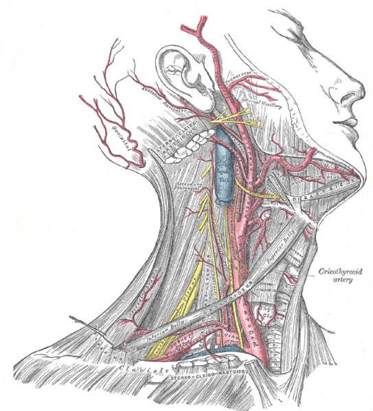
Superficial dissection of the right side of the neck, showing the carotid and subclavian arteries.
-
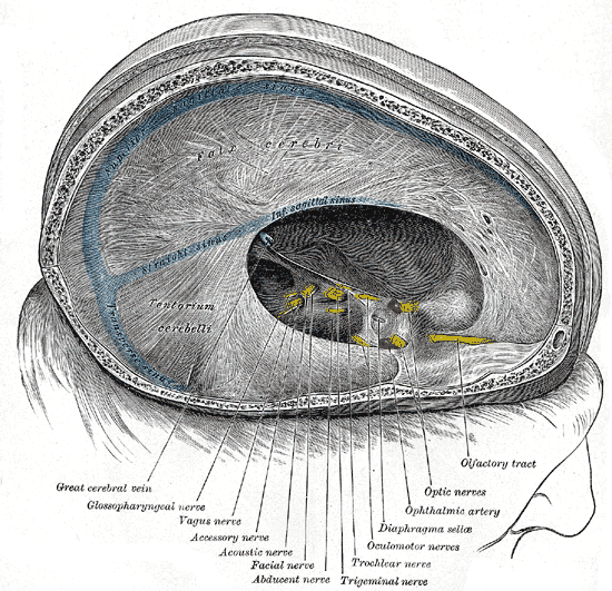
Dura mater and its processes exposed by removing part of the right half of the skull, and the brain.
-
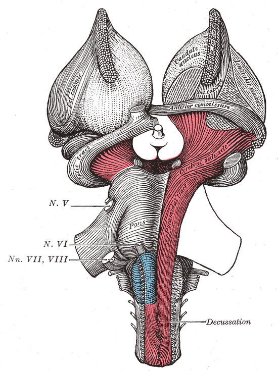
Superficial dissection of brain-stem. Ventral view.
-
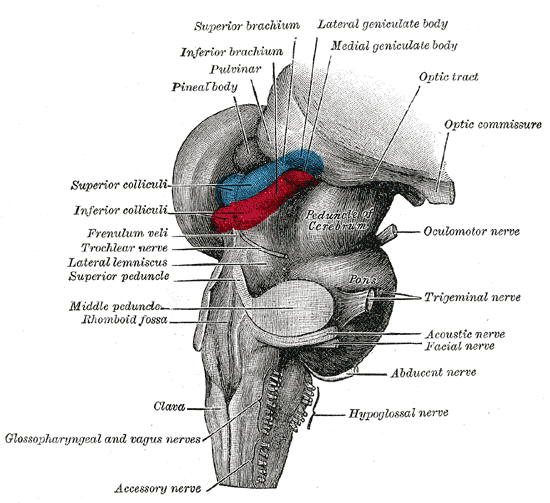
Hind- and mid-brains; postero-lateral view.
-
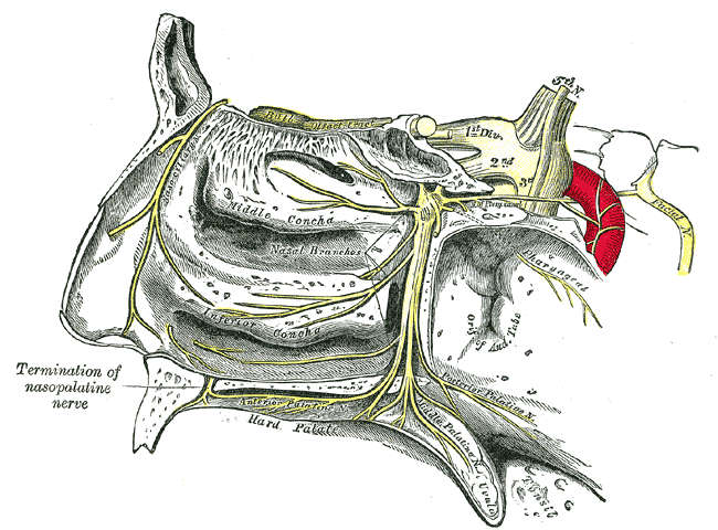
The sphenopalatine ganglion and its branches.
-
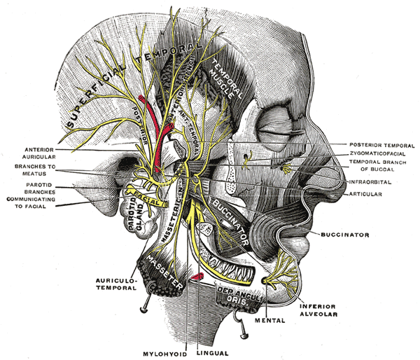
Mandibular division of the trifacial nerve.
-
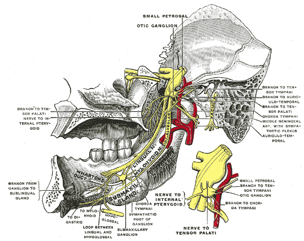
Mandibular division of trifacial nerve, seen from the middle line.
-
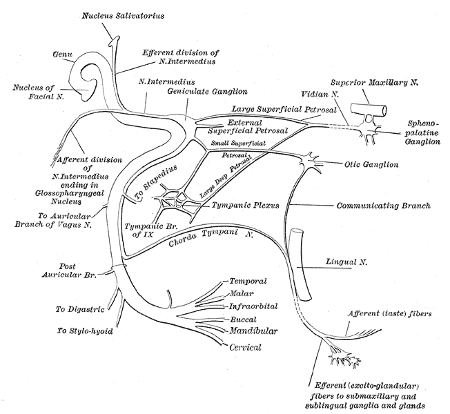
Plan of the facial and intermediate nerves and their communication with other nerves.
-
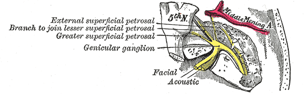
The course and connections of the facial nerve in the temporal bone.
-
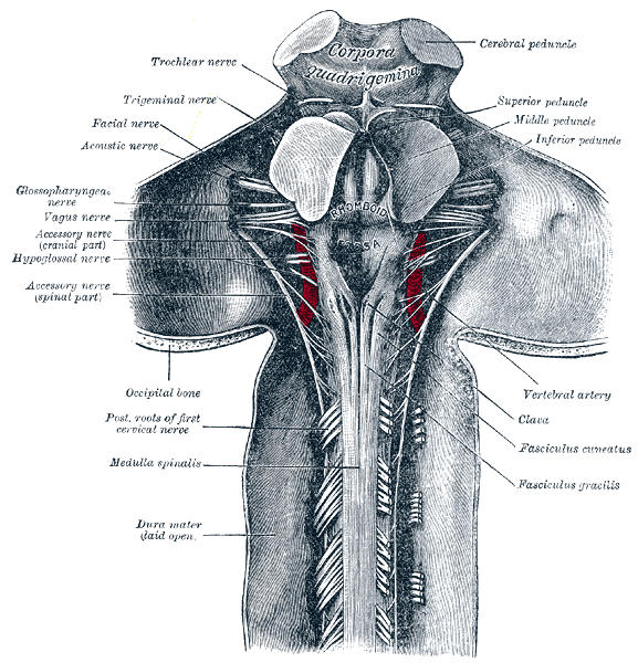
Upper part of medulla spinalis and hind- and mid-brains; posterior aspect, exposed in situ.
-
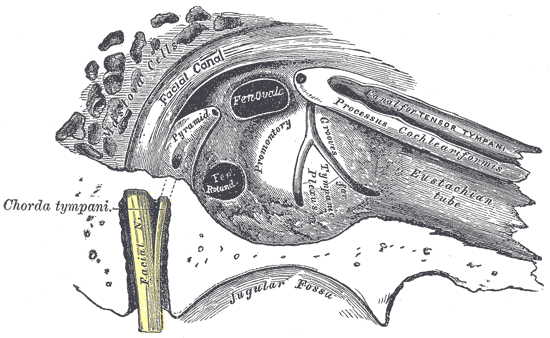
View of the inner wall of the tympanum (enlarged.)
-
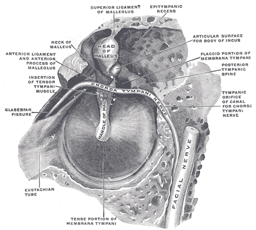
The right membrana tympani with the hammer and the chorda tympani, viewed from within, from behind, and from above.
-
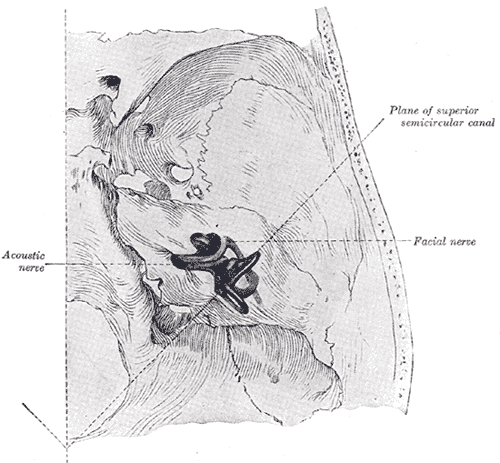
Position of the right bony labyrinth of the ear in the skull, viewed from above.
-

Left temporal bone showing surface markings for the tympanic antrum (red), transverse sinus (blue), and facial nerve (yellow).
-

Side of neck, showing chief surface markings.
-
Cranial nerves
-

Head facial nerve branches


References
- ↑ Kliniska Färdigheter: Informationsutbytet Mellan Patient Och Läkare, LINDGREN, STEFAN, ISBN 91-44-37271-X
External links
de:Nervus facialis lt:Veidinis nervas nl:Nervus facialis no:Nervus facialis fi:Kasvohermo sv:Ansiktsnerven