Meninges
|
WikiDoc Resources for Meninges |
|
Articles |
|---|
|
Most recent articles on Meninges |
|
Media |
|
Evidence Based Medicine |
|
Clinical Trials |
|
Ongoing Trials on Meninges at Clinical Trials.gov Clinical Trials on Meninges at Google
|
|
Guidelines / Policies / Govt |
|
US National Guidelines Clearinghouse on Meninges
|
|
Books |
|
News |
|
Commentary |
|
Definitions |
|
Patient Resources / Community |
|
Directions to Hospitals Treating Meninges Risk calculators and risk factors for Meninges
|
|
Healthcare Provider Resources |
|
Causes & Risk Factors for Meninges |
|
Continuing Medical Education (CME) |
|
International |
|
|
|
Business |
|
Experimental / Informatics |
Editor-In-Chief: C. Michael Gibson, M.S., M.D. [1]
Overview
The meninges (singular meninx) is the system of membranes which envelop the central nervous system. The meninges consist of three layers: the dura mater, the arachnoid mater, and the pia mater. The primary function of the meninges and of the cerebrospinal fluid is to protect the central nervous system.
Anatomy
Pia mater
The pia or pia mater is a very delicate membrane. It is attached to (nearest) the brain or the spinal cord. As such it follows all the minor contours of the brain (gyri and sulci). The pia mater is the meningeal envelope which firmly adheres to the surface of the brain and spinal cord. It is a very thin membrane composed of fibrous tissue covered on its outer surface by a sheet of flat cells thought to be impermeable to fluid. The pia mater is pierced by blood vessels which travel to the brain and spinal cord, and its capillaries are responsible for nourishing the brain.
Arachnoid mater
The middle element of the meninges is the arachnoid mater, so named because of its spider web-like appearance. It provides a cushioning effect for the central nervous system. The arachnoid mater exists as a thin, transparent membrane. It is composed of fibrous tissue and, like the pia mater, is covered by flat cells also thought to be impermeable to fluid. The arachnoid does not follow the convolutions of the surface of the brain and so looks like a loosely fitting sac. In the region of the brain, particularly, a large number of fine filaments called arachnoid trabeculae pass from the arachnoid through the subarachnoid space to blend with the tissue of the pia mater.
The arachnoid and pia mater are sometimes together called the leptomeninges.
Dura mater
The dura mater (also rarely called meninx fibrosa, or pachymeninx) is a thick, durable membrane, closest to the skull. It contains larger blood vessels which split into the capilliaries in the pia mater. It is composed of dense fibrous tissue, and its inner surface is covered by flattened cells like those present on the surfaces of the pia mater and arachnoid. The dura mater is a sac which envelops the arachnoid and has been modified to serve several functions. The dura mater surrounds and supports the large venous channels (dural sinuses) carrying blood from the brain toward the heart.
Spaces
The subarachnoid space is the space which normally exists between the arachnoid and the pia mater, which is filled with cerebrospinal fluid.
Normally, the dura mater is attached to the skull in the head, or to the bones of the vertebral canal in the spinal cord. The arachnoid is attached to the dura mater, and the pia mater is attached to the central nervous system tissue. When the dura mater and the arachnoid separate through injury or illness, the space between them is the subdural space.
Pathology
There are three types of hemorrhage involving the meninges:[1]
- A subarachnoid hemorrhage is acute bleeding under the arachnoid; it may occur spontaneously or as a result of trauma.
- A subdural hematoma is a hematoma (collection of blood) located in a separation of the arachnoid from the dura mater. The small veins which connect the dura mater and the arachnoid are torn, usually during an accident, and blood can leak into this area.
- An epidural hematoma similarly may arise after an accident or spontaneously.
Other medical conditions which affect the meninges include meningitis (usually from fungal, bacterial, or viral infection) and meningiomas arising from the meninges or from tumors formed elsewhere in the body which metastasize to the meninges.
Additional images
-
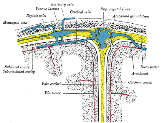 Diagrammatic representation of a section across the top of the skull
 -
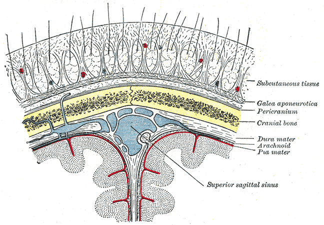 Diagrammatic section of scalp.
 -


References
- ↑ Orlando Regional Healthcare, Education and Development. 2004. "Overview of Adult Traumatic Brain Injuries." Retrieved on September 6, 2007.
de:Hirnhäute he:קרומי המוח it:Meningi la:Meninges no:Hjernehinne sv:Hjärnhinna