Muscle spindle
|
WikiDoc Resources for Muscle spindle |
|
Articles |
|---|
|
Most recent articles on Muscle spindle Most cited articles on Muscle spindle |
|
Media |
|
Powerpoint slides on Muscle spindle |
|
Evidence Based Medicine |
|
Clinical Trials |
|
Ongoing Trials on Muscle spindle at Clinical Trials.gov Trial results on Muscle spindle Clinical Trials on Muscle spindle at Google
|
|
Guidelines / Policies / Govt |
|
US National Guidelines Clearinghouse on Muscle spindle NICE Guidance on Muscle spindle
|
|
Books |
|
News |
|
Commentary |
|
Definitions |
|
Patient Resources / Community |
|
Patient resources on Muscle spindle Discussion groups on Muscle spindle Patient Handouts on Muscle spindle Directions to Hospitals Treating Muscle spindle Risk calculators and risk factors for Muscle spindle
|
|
Healthcare Provider Resources |
|
Causes & Risk Factors for Muscle spindle |
|
Continuing Medical Education (CME) |
|
International |
|
|
|
Business |
|
Experimental / Informatics |
Muscle structure is innervated by both sensory and motor neuron axons. The muscle spindle's functions are to send proprioceptive information about the muscle to the central nervous system, and to respond to muscle stretching.
Anatomy
Muscle spindles are found within the fleshy portions of muscles, embedded in so-called extrafusal muscle fibres. They are composed of 3-10 intrafusal muscle fibres, of which there are three types:
- dynamic nuclear bag fibres (bag1 fibres)
- static nuclear bag fibers (bag2 fibres)
- nuclear chain fibers and the axons of sensory neurons. Axons of motor neurons also terminate in muscle spindles; they make synapses at either or both of the ends of the intrafusal muscle fibers and regulate spindle sensitivity.
Muscle spindles are encapsulated by connective tissue, and are aligned parallel to extrafusal muscle fibers, unlike Golgi tendon organs, which are oriented in series.
The muscle spindle has both sensory and motor components.
- Primary and secondary sensory fibers spiral around and terminate on the central portions of intrafusal fibers, providing the sensory component of the structure via stretch-sensitive ion-channels of the axons.
- In humans, the motor component is provided by gamma motoneurons; in many other species, beta motoneurons innervate the spindles. They cause a slight contraction of the end portions of the intrafusal muscle fibers when activated. The gamma (fusimotor) axons only innervate the intrafusal muscle fibres, whereas the beta (skeletofusimotor) axons innervate both extrafusal and intrafusal muscle fibres.
These motorneurons are classified as static or dynamic according to their pattern of innervation and their physiological effects.
- The static axons innervate the chain or bag2 fibres.
- The dynamic axons innervate the bag1 fibres and increase the velocity sensitivity of the Ia afferents.
Sensitivity Modification
The function of the gamma motor neuron neuromuscular junction is not to supplement the general muscle contraction provided by extrafusal fibers, but to modify the sensitivity of the muscle spindle to stretch. Upon release of acetylcholine by the gamma neuron, the end portions of the intrafusal muscle fibers contract, thus deliberately elongating the non-contractile central portions of intrafusal muscle fibers. This opens stretch-sensitive ion channels of the centrally-positioned sensory axons, leading to an influx of sodium ions. This raises the resting potential of these axons, thereby increasing the probability of action potential firing, thus increasing the sensitivity of the muscle spindle.
Stretch reflex
When a muscle is stretched, primary sensory fibers (Group Ia afferent neurons) of the muscle spindle respond to both the velocity and the degree of stretch, and send this information to the spinal cord. Likewise, secondary sensory fibers (Group II afferent neurons) detect and send information about the degree of stretch (but not the velocity thereof) to the CNS. This information is transmitted monosynaptically to an alpha efferent motor fiber, which activates extrafusal fibers of the muscle to contract, thereby reducing stretch, and polysynaptically through an interneuron to another alpha motoneuron, which inhibits contraction in the antagonising muscles.
PNF stretching, or proprioceptive neuromuscular facilitation, is a method of flexibility training that reduces the automatic reflex action in order allow muscles to lengthen.
Development
It is also believed that muscle spindles play a critical role in sensorimotor development.
See also
Additional images
-
Muscle spindle
-
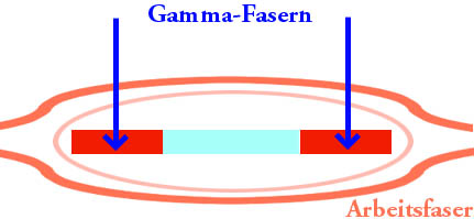
Gamma fiber
-
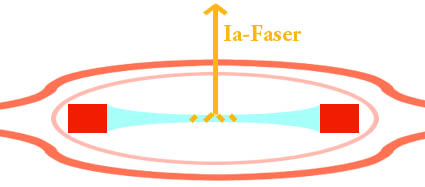
1A fiber
-
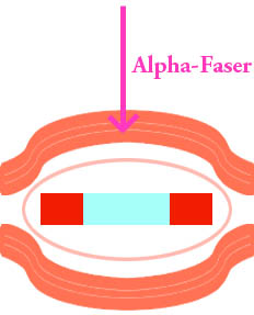
Alpha fiber


External links
- Muscle+Spindles at the US National Library of Medicine Medical Subject Headings (MeSH)