Oculomotor nerve
Template:Infobox Nerve Editor-In-Chief: C. Michael Gibson, M.S., M.D. [1]
Overview
The oculomotor nerve is the third of twelve paired cranial nerves. It controls most of the eye movements (cranial nerves IV and VI also do some), constriction of the pupil, and holding the eyelid open.
Pathophysiology
Nuclei
The oculomotor nerve arises from the anterior aspect of mesencephalon (midbrain). There are two nuclei for the oculomotor nerve:
- The oculomotor nucleus originates at the level of the superior colliculus. The muscles it controls are the ciliary muscle (affecting accommodation), and all extraocular muscles except for the superior oblique muscle and the lateral rectus muscle.
- The Edinger-Westphal nucleus supplies parasympathetic fibres to the eye via the ciliary ganglion, and thus controls pupil constriction.
Emergence from brain
On emerging from the brain, the nerve is invested with a sheath of pia mater, and enclosed in a prolongation from the arachnoid.
It passes between the superior cerebellar (below) and posterior cerebral arteries (above), and then pierces the dura mater in front of and lateral to the posterior clinoid process, passing between the free and attached borders of the tentorium cerebelli.
It runs along the lateral wall of the cavernous sinus, above the other orbital nerves, receiving in its course one or two filaments from the cavernous plexus of the sympathetic, and a communicating branch from the ophthalmic division of the trigeminal.
Superior and inferior rami
It then divides into two branches, which enter the orbit through the superior orbital fissure, between the two heads of the lateral rectus.
Here the nerve is placed below the trochlear nerve and the frontal and lacrimal branches of the ophthalmic nerve, while the nasociliary nerve is placed between its two rami:
Testing the oculomotor nerve
Cranial nerves III, IV and VI are usually tested together. The examiner typically instructs the patient to hold his head still and follow only with the eyes a finger or penlight that circumscribes a large "H" in front of the patient. By observing the eye movements and eyelids, the examiner is able to obtain more information about the extraocular muscles, the levator palpebrae superioris muscle, and cranial nerves III, IV, and VI.
Since the oculomotor nerve controls most of the eye muscles, it may be easier to detect damage to it. Damage to this nerve, termed oculomotor nerve palsy is also known by the down n' out symptoms, because of the position of the affected eye.
The oculomotor nerve also controls the constriction of the pupils. This can be tested in two main ways. By moving a finger towards a person's face to induce accommodation, as well as them going cross-eyed, their pupils should constrict.
Shining a light into their eyes should also make their pupils constrict. Both pupils should constrict at the same time, independent of what eye the light is actually shone on.
Additional images
-
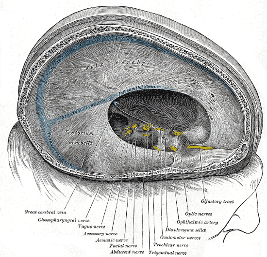 Dura mater and its processes exposed by removing part of the right half of the skull, and the brain.
 -
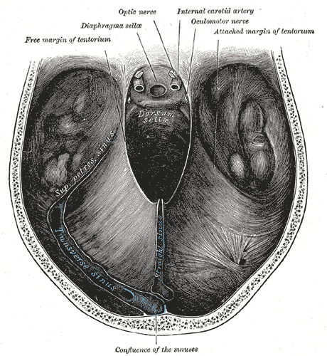 Tentorium cerebelli from above.
 -
 Coronal section through mid-brain.
 -
 Mesal aspect of a brain sectioned in the median sagittal plane.
 -
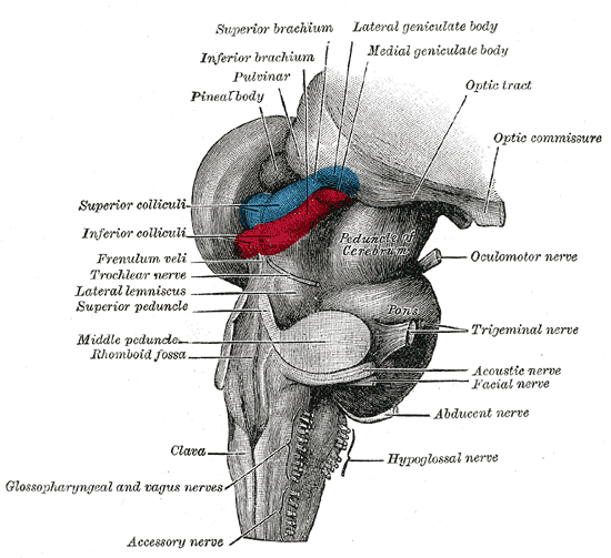 Hind- and mid-brains; postero-lateral view.
 -
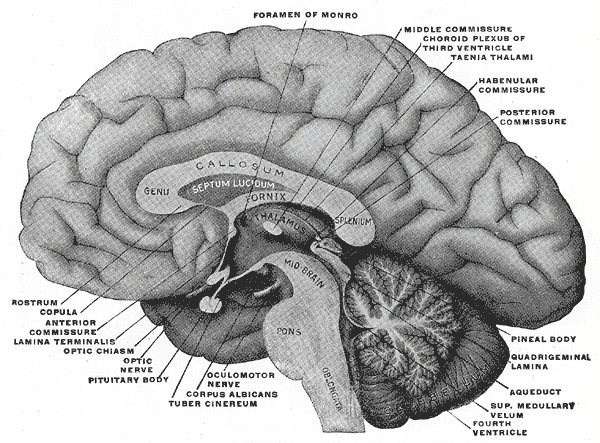
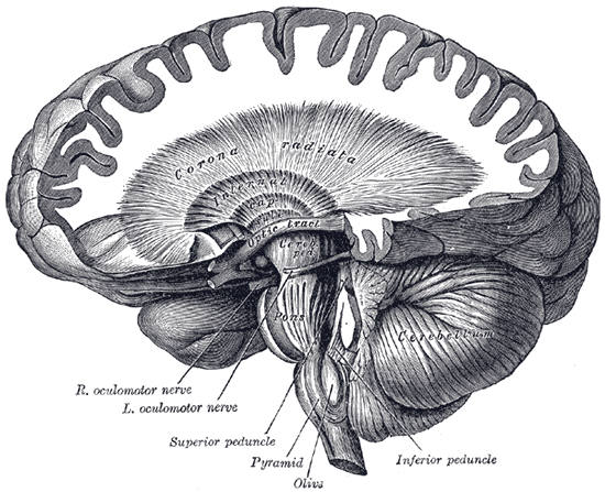
 Median sagittal section of brain.
 -
 Dissection showing the course of the cerebrospinal fibers.
 -
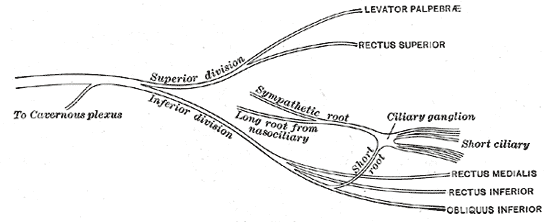 Plan of oculomotor nerve.
 -
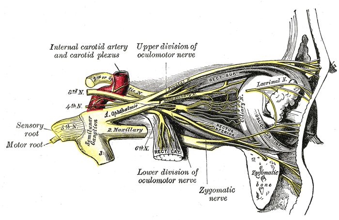 Nerves of the orbit, and the ciliary ganglion. Side view.
 -
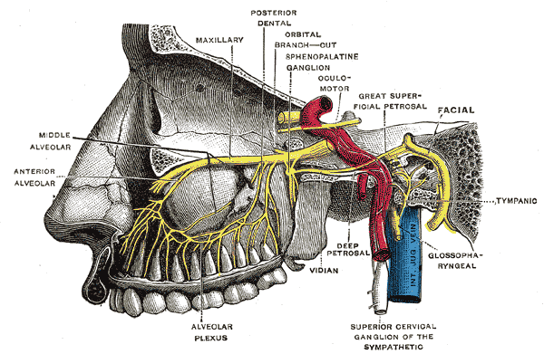 Alveolar branches of superior maxillary nerve and sphenopalatine ganglion.
 -
 Figure showing the mode of innervation of the Recti medialis and lateralis of the eye.
 -
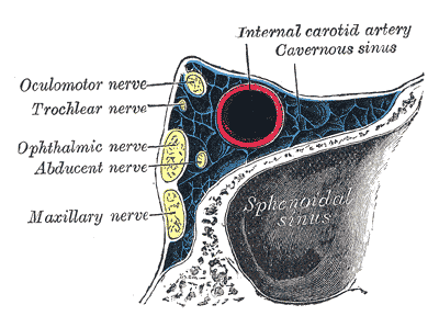 Oblique section through the right cavernous sinus.
 -
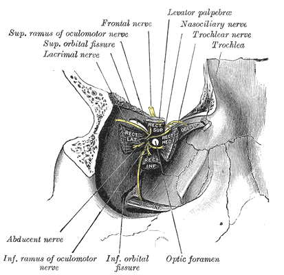 Dissection showing origins of right ocular muscles, and nerves entering by the superior orbital fissure.
 -
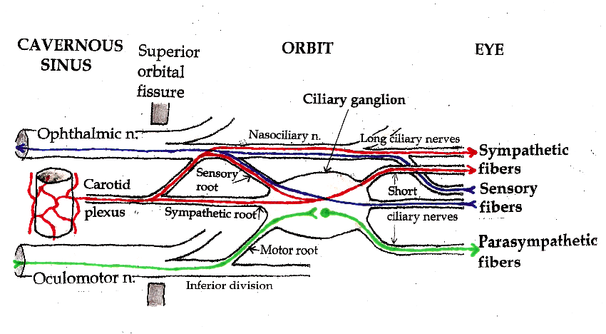 Pathways in the Ciliary Ganglion.


See also
External links
- Template:LoyolaMedEd
- oph/183 at eMedicine - "Oculomotor nerve palsy"
- Oculomotor+Nerve at the US National Library of Medicine Medical Subject Headings (MeSH)
- Template:BrainInfo
- Template:NormanAnatomy (Template:NormanAnatomyFig)
de:Nervus oculomotorius lt:Judinamasis akies nervas nl:Nervus oculomotorius no:Nervus oculomotorius fi:Silmän liikehermo