Parathyroid adenoma pathophysiology: Difference between revisions
| Line 148: | Line 148: | ||
==Microscopic Pathology== | ==Microscopic Pathology== | ||
*[[Chief cell|Chief cells]] are predominant in [[parathyroid adenoma]] on microcopy. | |||
*In majority of cases, a few nests of larger oxyphil cells are also present. | |||
*[[Adenoma]] is seperated from a rim of non-[[Neoplastic disease|neoplastic]] tissue on the edge by a [[fibrous]] [[capsule]]. | |||
*[[Chief cells]] present in [[adenoma]] larger than normal [[Chief cell|chief cells]] and shows greater variability on nuclear size. | |||
*[[Endocrine]] [[atypia]] (cells with bizarre and [[pleomorphic]] [[nuclei]]) is often seen in [[parathyroid adenoma]]. It should not be mistaken as a sign of [[malignancy]]. | |||
*Mitotic figures are rarely present. | |||
*[[Parathyroid adenoma]] has incospicuous [[adipose tissue]] when compared with normal [[parathyroid gland]]. | |||
<gallery> | |||
Image:Adenoma 01.jpg|<small>Intermediate/Low magnification micrograph of parathyroid adenoma. H&E stain. Features: Single cell population forming a single mass. Thin capsule. No adipose tissue. +/-Glandular architecture (which may lead to confusion with thyroid tissue). Normal parathyroid gland with prominent adipose tissue is seen on the right of the image. - [https://en.wikipedia.org/wiki/File:Parathyroid_adenoma_intermed_mag.jpg Source: Wikipedia]</small> | |||
Image:Parathyroid adenoma high mag 02.jpg|<small>High magnification micrograph of a parathyroid adenoma. H&E stain. Features: Single cell population forming a single mass. Thin capsule. No adipose tissue. +/-Glandular architecture (which may lead to confusion with thyroid tissue). Normal parathyroid gland with prominent adipose tissue is seen on the right of the image. - [https://en.wikipedia.org/wiki/File:Parathyroid_adenoma_high_mag.jpg Source:wikipeida]</small> | |||
Image:Parathyroid adenoma histopathology (03).jpg|<small>Histopatholgical image of parathyroid adenoma in a patient with primary hyperparathyroidism. Hematoxylin and eosin stain. - [https://en.wikipedia.org/wiki/File:Parathyroid_adenoma_histopathology_(1).jpg Source: Wikipedia]</small> | |||
Image:Parathyroid adenoma histopathology 04.jpg|<small>Histopatholgical image of parathyroid adenoma in a patient with primary hyperparathyroidism. Hematoxylin and eosin stain. - [https://en.wikipedia.org/wiki/File:Parathyroid_adenoma_histopathology_(2).jpg Source: Wikipedia]</small> | |||
Image:Parathyroid adenoma histopathology05.jpg|<small>Histopatholgical image of parathyroid adenoma in a patient with primary hyperparathyroidism. - [https://en.wikipedia.org/wiki/File:Parathyroid_adenoma_histopathology_(3).jpg Source: Wikipedia]</small> | |||
</gallery> | |||
==References== | ==References== | ||
Revision as of 13:46, 14 March 2019
|
Parathyroid adenoma Microchapters |
|
Diagnosis |
|---|
|
Treatment |
|
Case Studies |
|
Parathyroid adenoma pathophysiology On the Web |
|
American Roentgen Ray Society Images of Parathyroid adenoma pathophysiology |
|
Risk calculators and risk factors for Parathyroid adenoma pathophysiology |
Editor-In-Chief: C. Michael Gibson, M.S., M.D. [1]; Associate Editor(s)-in-Chief:
Overview
The exact pathogenesis of [disease name] is not fully understood.
OR
It is thought that [disease name] is the result of / is mediated by / is produced by / is caused by either [hypothesis 1], [hypothesis 2], or [hypothesis 3].
OR
[Pathogen name] is usually transmitted via the [transmission route] route to the human host.
OR
Following transmission/ingestion, the [pathogen] uses the [entry site] to invade the [cell name] cell.
OR
[Disease or malignancy name] arises from [cell name]s, which are [cell type] cells that are normally involved in [function of cells].
OR
The progression to [disease name] usually involves the [molecular pathway].
OR
The pathophysiology of [disease/malignancy] depends on the histological subtype.
Pathophysiology
Physiology
The effect of parathyroid hormone on mineral metabolism is as follows:[1][2]
- Effect of parathyroid hormone on inorganic phosphate metabolism:
- Increases excretion of inorganic phosphate from kidney resulting in decreased serum concentration of phosphate.
- Effect on parathyroid hormone on calcium metabolism:
- Direct effect:
- Increased resorption of bones.
- Decreases excretion from kidney.
- Indirect effect:
- Increases conversion of inactive 25-hydroxy vitamin D to the active 1,25-dihydroxy vitamin D which increases absorption of calcium from gut. Decreased phosphate concentration also increases this conversion process. Vitamin D shows synergism with parathyroid hormone action on bone.
- Decreased serum inorganic phosphate concentration prevents precipitation of calcium phosphate in bones.
- Both these direct and indirect mechanism results in an increased serum calcium concentration.
- Direct effect:
- Effect of parathyroid hormone on magnesium concentration:
Effect of minerals and vitamin D on parathyroid hormone:
- Decrease in serum calcium concentration stimulates parathyroid hormone.
- Calcium provides negative feedback on parathyroid hormone.
- Magnesium provides negative feedback on parathyroid hormone.
- Vitamin D decreases the concentration of parathyroid hormone.
| Parathyroid hormone | |||||||||||||||||||||||||||||||||||||||||||||||||||||||||||||||||||
| Kidney | Bone | ||||||||||||||||||||||||||||||||||||||||||||||||||||||||||||||||||
| Decreased excretion of magnesium | Increasead conversion of inactive 25-hydroxy vitamin D to the active 1,25-dihydroxy vitamin D | Increase excretion of inorganic phosphate | Decrease excretion of calcium | Increased resorption of bone | |||||||||||||||||||||||||||||||||||||||||||||||||||||||||||||||
| Increased serum concentration of magnesium | Increased absorption of calcium from gut | Decreased serum concentration of inorganic phosphate | |||||||||||||||||||||||||||||||||||||||||||||||||||||||||||||||||
| Prevents precipitation of calcium phosphate in bones | |||||||||||||||||||||||||||||||||||||||||||||||||||||||||||||||||||
| Increased serum concentration of calcium | |||||||||||||||||||||||||||||||||||||||||||||||||||||||||||||||||||
Pathogenesis
- Parathyroid adenoma results in an increase in parathyroid hormone secretion.[3]
- Calcium-sensing receptor expression in reduced in parathyroid adenoma resulting in an increase in calcium sensing set point.[4][5]
Calcium-sensing receptors
- Calcium-sensing receptors are present on parathyroid glands. They are a type of 7-transmembrane receptors in G-protein coupled receptors superfamily of receptors.[6]
- Calcium-sensing receptors sense change in extracellular concentration of ionized calcium.[7]
- Calcium-sensing receptor expression in reduced in primary hyperparathyroidism (parathyroid adenoma) and secondary hyperparathyroidism.[4]
- This reduced expression of receptor causes an increases in calcium sensing set point.[5]
- This in turn leads to increase in secretion of parathyroid hormone in presence on normal serum concentration of extracellular ionized calcium.
Genetics
The development of parathyroid adenoma is the result of multiple genetic mutations in minority of cases. Genes involved in the pathogenesis of parathyroid adenoma include calcium-sensing receptor gene, HRPT2 gene (CDC73 gene), Cyclin D1 gene (CCND1)/PRAD1 gene, MEN1 gene, and RET gene.
- Calcium-sensing receptor gene mutation:[8]
- Calcium-sensing receptor (CSR) gene is present on chromosome 3q.
- Few individuals carries an inherited mutation in the extracellular calcium-sensing receptor gene.
- The first identified mutation in CSR gene is a point mutation in which phenylalanine is replaced with leucine at codon 881 of CSR gene.[9]
- This mutation reduces the activity of calcium-sensing receptor.
- This mutation can be heterozygous or homozygous.
- Individuals carrying heterozygous mutation have familial hypocalciuric hypercalcemia (FHH) or familial benign hypercalcemia. FHH is characterized by parathyroid dependent hypercalcemia and decreased responsiveness of parathyroid and kidney to hypercalcemia.
- Individuals carrying homozygous mutation have neonatal severe hyperparathyroidism. Neonatal severe hyperparathyroidism is characterized by marked parathyroid hyperplasia.
- Familial hypocalciuric hypercalcemia (FHH) and neonatal severe hyperparathyroidism are transmitted in autosomal dominant pattern.
- HRPT2 gene(CDC73 gene) mutations:[10]
- HRPT2 gene code for parafibromin protein.
- HRPT2 gene mutations are found in a type of familial hyperparathyroidism, hyperparathyroidism-jaw tumor (HPT-JT) syndrome.
- HRTP2 gene mutations increases risk of parathyroid carcinoma.
- Cyclin D1 gene (CCND1)/PRAD1 gene:[11][12]
- PRAD1 (parathyroid adenoma 1) is a protooncogene located on chromosome 11q13.
- Cyclin D1 gene translocation and oncogene action observed in 8% of adenomas.
- Cyclin D1 gene overexpression is observed in 20% to 40% of parathyroid adenomas.
- MEN1 gene:[11][13]
- MEN 1 ics a tumor supressor gene on chromosome 11q13.
- Somatic loss of single MEN1 allele is observed in 25% to 40% of sporadic parathyroid adenomas.
- RET gene:[14]
- RET gene is a proto-oncogene.
- RET proto-oncogene is associated with multiple endocrine neoplasia type 2 (MEN 2).
- MEN2A caries increased risk of parathyroid adenoma and/or parathyroid hyperplasia.
- CDNK1B gene:[15]
- CDNK1B mutation causes Multiple endocrine neoplasia type 4 (MEN 4).
- Parathyroid tumors are found along with anterior pituitary, gonadal, adrenal, and renal tumors in MEN 4 syndrome.
- CDNK1B encodes for the cyclin-dependent kinase inhibitor p27kip1.
Associated Conditions
The conditions associated with parathyroid adenoma include:[16][17][18][8][10][13][19][20][21][22][23][24]
- Brown tumor
- Chronic renal failure
- Depression
- Familial hypocalciuric hypercalcemia
- Hyperparathyroid-jaw tumor syndrome
- Hypertension
- Multiple endocrine neoplasia type 1
- Multiple endocrine neoplasia type 2A
- Multiple endocrine neoplasia type 4
- Neonatal severe hyperparathyroidism
- Osteitis fibrosa cystica
- Osteoporosis
- Osteomalacia
- Osteoarthritis
- Pancreatitis
Gross Pathology
- On gross pathology, parathyroid adenoma is a soft, tan nodule which is well-circumscribed by a delicate capsule.[25]
- Most commonly, parathyroid adenoma is present in single gland. Some times multiple glands are involved.
- If single gland is involved, the other glands may shrink due to negative feedback.
- Majority of times, parathyroid adenoma weight ranges between 0.5 gram to 5 gram.
- Typically, cut surface of parathyroid adenoma is smooth, soft, and reddish brown in color. It should be differentiated from normal parathyroid gland tissue which is yellow-brown color.[26]
- The tissue of parathyroid gland that is not involved in parathyroid adenoma is typically atrophied and compressed. Fat component of normal parathyroid tissue is also observed.
- On rare occasion, parathyroid adenoma may be cystic.
 |
 |
 |
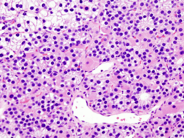
Microscopic Pathology
- Chief cells are predominant in parathyroid adenoma on microcopy.
- In majority of cases, a few nests of larger oxyphil cells are also present.
- Adenoma is seperated from a rim of non-neoplastic tissue on the edge by a fibrous capsule.
- Chief cells present in adenoma larger than normal chief cells and shows greater variability on nuclear size.
- Endocrine atypia (cells with bizarre and pleomorphic nuclei) is often seen in parathyroid adenoma. It should not be mistaken as a sign of malignancy.
- Mitotic figures are rarely present.
- Parathyroid adenoma has incospicuous adipose tissue when compared with normal parathyroid gland.
-
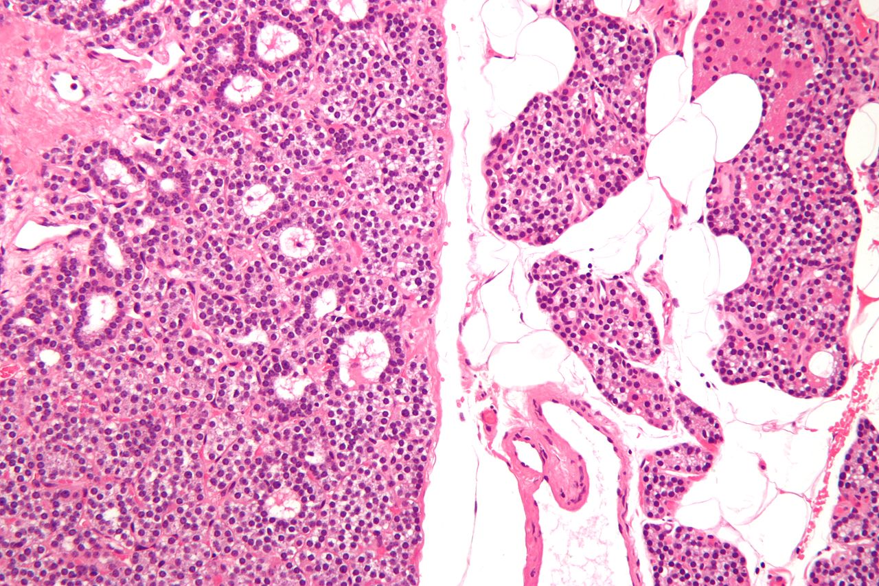
Intermediate/Low magnification micrograph of parathyroid adenoma. H&E stain. Features: Single cell population forming a single mass. Thin capsule. No adipose tissue. +/-Glandular architecture (which may lead to confusion with thyroid tissue). Normal parathyroid gland with prominent adipose tissue is seen on the right of the image. - Source: Wikipedia
-
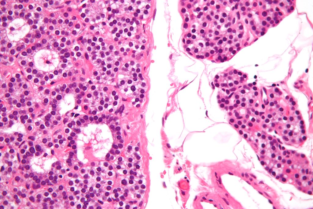
High magnification micrograph of a parathyroid adenoma. H&E stain. Features: Single cell population forming a single mass. Thin capsule. No adipose tissue. +/-Glandular architecture (which may lead to confusion with thyroid tissue). Normal parathyroid gland with prominent adipose tissue is seen on the right of the image. - Source:wikipeida
-
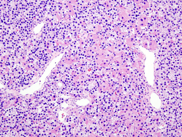
Histopatholgical image of parathyroid adenoma in a patient with primary hyperparathyroidism. Hematoxylin and eosin stain. - Source: Wikipedia
-
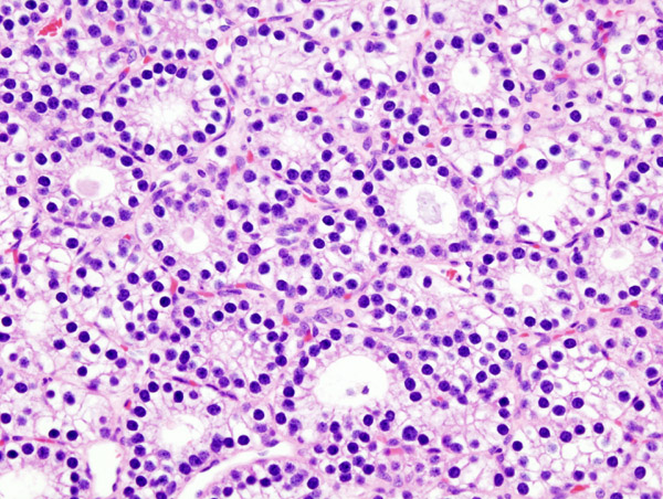
Histopatholgical image of parathyroid adenoma in a patient with primary hyperparathyroidism. Hematoxylin and eosin stain. - Source: Wikipedia
-
Histopatholgical image of parathyroid adenoma in a patient with primary hyperparathyroidism. - Source: Wikipedia


.jpg)


.jpg)
.jpg)
.jpg)
References
- ↑ HARRISON MT (1964). "INTERRELATIONSHIPS OF VITAMIN D AND PARATHYROID HORMONE IN CALCIUM HOMEOSTASIS". Postgrad Med J. 40: 497–505. PMC 2482768. PMID 14184232.
- ↑ Nussey, Stephen (2001). Endocrinology : an integrated approach. Oxford, UK Bethesda, Md: Bios NCBI. ISBN 1-85996-252-1.
- ↑ Wieneke JA, Smith A (2008). "Parathyroid adenoma". Head Neck Pathol. 2 (4): 305–8. doi:10.1007/s12105-008-0088-8. PMC 2807581. PMID 20614300.
- ↑ 4.0 4.1 Gogusev J, Duchambon P, Hory B, Giovannini M, Goureau Y, Sarfati E; et al. (1997). "Depressed expression of calcium receptor in parathyroid gland tissue of patients with hyperparathyroidism". Kidney Int. 51 (1): 328–36. PMID 8995751.
- ↑ 5.0 5.1 Kifor O, Moore FD, Wang P, Goldstein M, Vassilev P, Kifor I; et al. (1996). "Reduced immunostaining for the extracellular Ca2+-sensing receptor in primary and uremic secondary hyperparathyroidism". J Clin Endocrinol Metab. 81 (4): 1598–606. doi:10.1210/jcem.81.4.8636374. PMID 8636374.
- ↑ Brown EM, Gamba G, Riccardi D, Lombardi M, Butters R, Kifor O; et al. (1993). "Cloning and characterization of an extracellular Ca(2+)-sensing receptor from bovine parathyroid". Nature. 366 (6455): 575–80. doi:10.1038/366575a0. PMID 8255296.
- ↑ Brown EM, Pollak M, Seidman CE, Seidman JG, Chou YH, Riccardi D; et al. (1995). "Calcium-ion-sensing cell-surface receptors". N Engl J Med. 333 (4): 234–40. doi:10.1056/NEJM199507273330407. PMID 7791841.
- ↑ 8.0 8.1 Hosokawa Y, Pollak MR, Brown EM, Arnold A (1995). "Mutational analysis of the extracellular Ca(2+)-sensing receptor gene in human parathyroid tumors". J. Clin. Endocrinol. Metab. 80 (11): 3107–10. doi:10.1210/jcem.80.11.7593409. PMID 7593409.
- ↑ Carling T, Szabo E, Bai M, Ridefelt P, Westin G, Gustavsson P, Trivedi S, Hellman P, Brown EM, Dahl N, Rastad J (2000). "Familial hypercalcemia and hypercalciuria caused by a novel mutation in the cytoplasmic tail of the calcium receptor". J. Clin. Endocrinol. Metab. 85 (5): 2042–7. doi:10.1210/jcem.85.5.6477. PMID 10843194.
- ↑ 10.0 10.1 Shattuck TM, Välimäki S, Obara T, Gaz RD, Clark OH, Shoback D; et al. (2003). "Somatic and germ-line mutations of the HRPT2 gene in sporadic parathyroid carcinoma". N Engl J Med. 349 (18): 1722–9. doi:10.1056/NEJMoa031237. PMID 14585940.
- ↑ 11.0 11.1 Westin G, Björklund P, Akerström G (2009). "Molecular genetics of parathyroid disease". World J Surg. 33 (11): 2224–33. doi:10.1007/s00268-009-0022-6. PMID 19373510.
- ↑ Hsi ED, Zukerberg LR, Yang WI, Arnold A (1996). "Cyclin D1/PRAD1 expression in parathyroid adenomas: an immunohistochemical study". J Clin Endocrinol Metab. 81 (5): 1736–9. doi:10.1210/jcem.81.5.8626826. PMID 8626826.
- ↑ 13.0 13.1 Agarwal SK, Kester MB, Debelenko LV, Heppner C, Emmert-Buck MR, Skarulis MC; et al. (1997). "Germline mutations of the MEN1 gene in familial multiple endocrine neoplasia type 1 and related states". Hum Mol Genet. 6 (7): 1169–75. PMID 9215689.
- ↑ Marquard, Jessica; Eng, Charis (September 27, 1999). "Multiple Endocrine Neoplasia Type 2". GeneReviews® [Internet].
- ↑ Bilezikian JP (January 15, 2017). De Groot LJ, Chrousos G, Dungan K, et al., eds. Primary Hyperparathyroidism. Endotext [Internet]: South Dartmouth (MA): MDText.com, Inc.
- ↑ Bandeira F, Cusano NE, Silva BC, Cassibba S, Almeida CB, Machado VC, Bilezikian JP (2014). "Bone disease in primary hyperparathyroidism". Arq Bras Endocrinol Metabol. 58 (5): 553–61. PMC 4315357. PMID 25166047.
- ↑ Rodriguez M, Nemeth E, Martin D (2005). "The calcium-sensing receptor: a key factor in the pathogenesis of secondary hyperparathyroidism". Am J Physiol Renal Physiol. 288 (2): F253–64. doi:10.1152/ajprenal.00302.2004. PMID 15507543.
- ↑ Espiritu RP, Kearns AE, Vickers KS, Grant C, Ryu E, Wermers RA (2011). "Depression in primary hyperparathyroidism: prevalence and benefit of surgery". J. Clin. Endocrinol. Metab. 96 (11): E1737–45. doi:10.1210/jc.2011-1486. PMID 21917870.
- ↑ Marquard, Jessica; Eng, Charis (September 27, 1999). "Multiple Endocrine Neoplasia Type 2". GeneReviews® [Internet].
- ↑ Bilezikian JP (January 15, 2017). De Groot LJ, Chrousos G, Dungan K, et al., eds. Primary Hyperparathyroidism. Endotext [Internet]: South Dartmouth (MA): MDText.com, Inc.
- ↑ Mazzuoli GF, D'Erasmo E, Pisani D (1998). "Primary hyperparathyroidism and osteoporosis". Aging (Milano). 10 (3): 225–31. PMID 9801732.
- ↑ Lips P (2001). "Vitamin D deficiency and secondary hyperparathyroidism in the elderly: consequences for bone loss and fractures and therapeutic implications". Endocr Rev. 22 (4): 477–501. doi:10.1210/edrv.22.4.0437. PMID 11493580.
- ↑ Michael JW, Schlüter-Brust KU, Eysel P (2010). "The epidemiology, etiology, diagnosis, and treatment of osteoarthritis of the knee". Dtsch Arztebl Int. 107 (9): 152–62. doi:10.3238/arztebl.2010.0152. PMC 2841860. PMID 20305774.
- ↑ Bai HX, Giefer M, Patel M, Orabi AI, Husain SZ (2012). "The association of primary hyperparathyroidism with pancreatitis". J. Clin. Gastroenterol. 46 (8): 656–61. doi:10.1097/MCG.0b013e31825c446c. PMC 4428665. PMID 22874807.
- ↑ Kumar, Vinay (2013). Robbins basic pathology. Philadelphia, PA: Elsevier/Saunders. p. 736-737. ISBN 9781437717815.
- ↑ Wieneke JA, Smith A (2008). "Parathyroid adenoma". Head Neck Pathol. 2 (4): 305–8. doi:10.1007/s12105-008-0088-8. PMC 2807581. PMID 20614300.