Cyanosis physical examination
Jump to navigation
Jump to search
|
Cyanosis Microchapters |
|
Diagnosis |
|---|
|
Treatment |
|
Case Studies |
|
Cyanosis physical examination On the Web |
|
American Roentgen Ray Society Images of Cyanosis physical examination |
|
Risk calculators and risk factors for Cyanosis physical examination |
Editor-In-Chief: C. Michael Gibson, M.S., M.D. [1]; Associate Editor(s)-in-Chief: Chandrakala Yannam, MD [2]
Overview
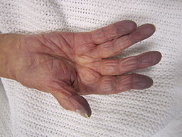
Patients with cyanosis show bluish discolration of skin and mucous membranes. Common locations to look for cyanosis include tongue, buccal mucosa, lips, hands and feet.
Physical Examination
- Physical examination of patients with cyanosis will show bluish discoration of lips, tongue, oral mucosa, nose tip, ear lobules, hands and feet. Because cyanosis is a symptom of disease process careful physical examination for associated symptoms include tachypnea, tachycardia, abnormal heart sounds or murmurs, wheezing, crackles, fever, clubbing, edema of extremities will be necessary to identify underlying disease process.
Appearance of the Patient
- Appearance of patients with cyanosis will vary depending to the underlying condition.
Vital Signs
Skin
- Skin usually appears blue in patients with cyanosis.
- It is very difficult to find bluish discoration in dark-skinned individuals and in poor lighting conditions.
- Sites to look for central cyanosis: Tongue, inner aspect of lips, gums, soft palate, buccal mucosa, and sites of peripheral cyanosis.
- Sites to look for peripheral cyanosis: Nose tip, ear lobules, outer aspect of lips, fingertips, nail bed, extremities.
- Central cyanosis with gray appearance to the skin is characteristic of methemoglobinemia.
- Certain skin conditions and exposure to dyes may mimic cyanosis have to be ruled out(eg, Mongolian spot, tattoo, blue clothing dye, finger paints).

HEENT
- Evidence of head trauma
Neck
Heart
- Cardiovascular examination of patients with cyanosis will show:[2][3][4]
- Tachycardia/ Bradycardia
- Bradycardia is an ominous sign for imminent cardiovascular collapse.
- Paradoxic pulse: Acute airway obstruction, Pulmonary embolism, cardiac tamponade, and asthma.
- Auscultate for abnormal (loud, single or widely split S2) and additional heart sounds and murmurs (Grade, timing, location, radiation, intensity).
- Tachycardia/ Bradycardia
| S2 | Murmur | |
|---|---|---|
| TOF | single | systolic |
| Tricuspid atresia | single | with or with out systolic |
| Ebstein's anomaly | split | systolic |
| TGA | single | none |
| Truncus arteriosus | single | systolic murmur/ with or with out diastolic murmur |
| Pulmonary stenosis | single | systolic |
| Pulmonary atresia | single | systolic |
| TAPVC | split | systolic |
| HLHS | single | with or with out systolic |
| Tricuspid atresia | single | with or with out systolic |
- Measure blood pressure in both upper and lower extremities
Lungs
- Patients with cyanosis will show:[3][5][6]
- Tachypnea is seen in patients with respiratory and cardiac diseases presenting with cyanosis.
- Nasal flaring, grunting, intercostal and substernal retractions, and prolonged breathing may indicate respiratory distress.
- Traumatic injury involving chest wall or lung will show following abnormalities:
- Chest wall movement abnormalities
- Chest wound (eg, open or sucking)
- Abrasions
- Ecchymosis
- Focal tenderness can be elicited on palpation over ribs, sternum, or scapula.
- Deviation of Trachea
- Subcutaneous air with crepitus
- Stridor, voice changes, suprasternal retractions, drooling and prolonged inspiration can be found in patients with upper airway obstruction.
- Wheezing, rales / crackles and assymmetric breath sounds will suggest Intrinsic lung diseases.
- Tactile fremitus:
- Increased: Consolidation by pneumonia ,atelactasis
- Decresed: Pleural effusion, pneumothorax
- Lung sounds may be clear on auscultation in patients with
- Cyanotic congenital heart disease
- Methemoglobinemia
- Neurologic conditions associated with hypoventilation (eg, muscle weakness, coma, and seizures)
- Pulmonary embolism
Extremities
- Clubbing is seen in some patients presenting with cyanosis.[7][8]
- Congenital heart diseases
- Pulmonary diseases: COPD, bronchiectasis, cystic fibrosis, pulmonary fibrosis, pulmonary arteriovenous malformations.
- Edema of lower extremities due to congestive heart failure and pulmonary embolism, pulmonary edema, pulmonary hypertension.
References
- ↑ https://commons.wikimedia.org/w/index.php?curid=17978808
- ↑ Berg A, Greve G, Hirth A, Rosland GA, Norgård G (April 2005). "[Evaluation of cardiac murmurs in children]". Tidsskr. Nor. Laegeforen. (in Norwegian). 125 (8): 1000–3. PMID 15852070.
- ↑ 3.0 3.1 Sasidharan P (August 2004). "An approach to diagnosis and management of cyanosis and tachypnea in term infants". Pediatr. Clin. North Am. 51 (4): 999–1021, ix. doi:10.1016/j.pcl.2004.03.010. PMID 15275985.
- ↑ Ammash N, Warnes CA (September 1996). "Cerebrovascular events in adult patients with cyanotic congenital heart disease". J. Am. Coll. Cardiol. 28 (3): 768–72. PMID 8772770.
- ↑ Maitre B, Similowski T, Derenne JP (September 1995). "Physical examination of the adult patient with respiratory diseases: inspection and palpation". Eur. Respir. J. 8 (9): 1584–93. PMID 8575588.
- ↑ Kosacka M, Brzecka A, Jankowska R, Lewczuk J, Mroczek E, Weryńska B (2009). "[Combined pulmonary fibrosis and emphysema - case report and literature review]". Pneumonol Alergol Pol (in Polish). 77 (2): 205–10. PMID 19462358.
- ↑ Srinivas SK, Manjunath CN (September 2013). "Differential clubbing and cyanosis: classic signs of patent ductus arteriosus with Eisenmenger syndrome". Mayo Clin. Proc. 88 (9): e105–6. doi:10.1016/j.mayocp.2013.02.016. PMID 24001503.
- ↑ Wald R, Crean A (June 2010). "Differential clubbing and cyanosis in a patient with pulmonary hypertension". CMAJ. 182 (9): E380. doi:10.1503/cmaj.091003. PMC 2882471. PMID 20421356.