Gallstone disease differential diagnosis

Jump to navigation Jump to search

Gallstone disease Microchapters

Home

Patient Information

Overview

Historical Perspective

Classification

Pathophysiology

Causes

Differentiating Gallstone disease from other Diseases

Epidemiology and Demographics

Risk Factors

Screening

Natural History, Complications and Prognosis

Diagnosis

Diagnostic Study of Choice

History and Symptoms

Physical Examination

Laboratory Findings

Electrocardiogram

X Ray

CT

MRI

Ultrasound

Other Imaging Findings

Other Diagnostic Studies

Treatment

Medical Therapy

Surgery

Lithotripsy
Surgical management

Primary Prevention

Secondary Prevention

Cost-Effectiveness of Therapy

Future or Investigational Therapies

Case Studies

Case #1

Gallstone disease differential diagnosis On the Web

Most recent articles

Most cited articles

Review articles

CME Programs

Powerpoint slides

Images

American Roentgen Ray Society Images of Gallstone disease differential diagnosis

All Images
X-rays
Echo & Ultrasound
CT Images
MRI

Ongoing Trials at Clinical Trials.gov

US National Guidelines Clearinghouse

NICE Guidance

FDA on Gallstone disease differential diagnosis

CDC on Gallstone disease differential diagnosis

Gallstone disease differential diagnosis in the news

Blogs on Gallstone disease differential diagnosis

Directions to Hospitals Treating Gallstone disease

Risk calculators and risk factors for Gallstone disease differential diagnosis

Editor-In-Chief: C. Michael Gibson, M.S., M.D. [1]; Associate Editor(s)-in-Chief: Hadeel Maksoud M.D.[2]

Overview

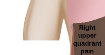
Gallstone disease must be differentiated from other diseases that cause epigastric, and left and right hypochondriac pain (right upper quadrant) such as: Abdominal pain, esophageal chest pain,gastroesophageal reflux disorder, peptic ulcer disease, non-ulcer dyspepsia,hepatitis, functional gallbladder disorder, sphincter of Oddi dysfunction,appendicitis, bile duct stricture, chronic pancreatitis, irritable bowel syndrome, ischemic heart disease, pyelonephritis, ureteral calculi and complications of gallstone disease include: acute cholecystitis, choledocholithiasis, acute pancreatitis, and acute cholangitis.[1][2]

Differentiating Gallstone disease from other Diseases

As Gallstone disease manifests in a variety of clinical forms, differentiation must be established in accordance with the particular subtype. The presence of biliary colic is an important diagnostic feature to distinguish between gallstones and non-biliary disorders. It has been shown that this feature is predictive of finding stones on imaging. [3] However, it is important to note that biliary colic concomitant in patients with other biliary disorders such as acute cholecystitis, choledocholithiasis, sphincter of Oddi dysfunction, and functional gallbladder disorder.

Laboratory studies can be helpful, along side clinical presentation in making a preliminary diagnosis:

Other tests that may be indicated depending upon the patient's symptoms and history including:

To review a table of differential diagnoses for disease symptoms including jaundice, abdominal pain and fever, please click here:

Differential diagnosis

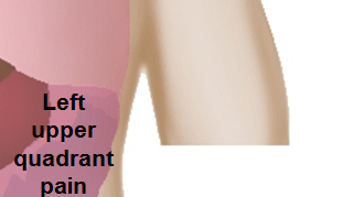
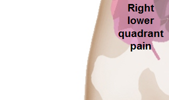
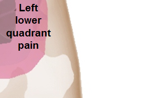
Abbreviations: RUQ= Right upper quadrant of the abdomen, LUQ= Left upper quadrant, LLQ= Left lower quadrant, RLQ= Right lower quadrant, LFT= Liver function test, SIRS= Systemic inflammatory response syndrome, ERCP= Endoscopic retrograde cholangiopancreatography, IV= Intravenous, N= Normal, AMA= Anti mitochondrial antibodies, LDH= Lactate dehydrogenase, GI= Gastrointestinal, CXR= Chest X ray, IgA= Immunoglobulin A, IgG= Immunoglobulin G, IgM= Immunoglobulin M, CT= Computed tomography, PMN= Polymorphonuclear cells, ESR= Erythrocyte sedimentation rate, CRP= C-reactive protein, TS= Transferrin saturation, SF= Serum Ferritin, SMA= Superior mesenteric artery, SMV= Superior mesenteric vein, ECG= Electrocardiogram

Classification of pain in the abdomen based on etiology Disease Clinical manifestations Diagnosis Comments
Symptoms Signs
Abdominal Pain Fever Rigors and chills Jaundice Diarrhea GI Bleed Hypo-

tension

Guarding Rebound Tenderness Bowel sounds Lab Findings Imaging
Abdominal causes Inflammatory causes Pancreato-biliary disorders Acute suppurative cholangitis RUQ + + + + + + N
  • Abnormal LFT
  • WBC >10,000
Ultrasound shows biliary dilatation/stents/tumor Septic shock occurs with features of SIRS
Acute cholangitis RUQ + + N Ultrasound shows biliary dilatation/stents/tumor Biliary drainage (ERCP) + IV antibiotics
Acute cholecystitis RUQ + + Hypoactive Ultrasound shows gallstone and evidence of inflammation Murphy’s sign
Acute pancreatitis Epigastric + ± ± + + N
  • Ultrasound shows evidence of inflammation
  • CT scan shows severity of pancreatitis
Pain radiation to back
Chronic pancreatitis Epigastric ± + N
  • Pancreatic function test
CT scan
  • Calcification
  • Pseudocyst
  • Dilation of main pancreatic duct
Predisposes to pancreatic cancer
Pancreatic carcinoma Epigastric - - + + - - - - N

Skin manifestations may include:

Primary biliary cirrhosis RUQ/Epigastric + + in late presentation N
  • Increased AMA level, abnormal LFTs
Primary sclerosing cholangitis RUQ + + N ERCP and MRCP shows
  • Multiple segmental strictures
  • Mural irregularities
  • Biliary dilatation and diverticula
  • Distortion of biliary tree
The risk of cholangiocarcinoma in patients with primary sclerosing cholangitis is 400 times higher than the risk in the general population.
Cholelithiasis RUQ/Epigastric ± ± N to hyperactive for dislodged stone Ultrasound shows gallstone Fatty food intolerance
Gastric causes Peptic ulcer disease Diffuse ± + in perforated + + N
  • Ascitic fluid
    • LDH > serum LDH
    • Glucose < 50mg/dl
    • Total protein > 1g/dl
Air under diaphragm in upright CXR Upper GI endoscopy for diagnosis
Gastritis Epigastric ± + in chronic gastritis N H.pylori infection diagnostic tests Endoscopy H.pylori gastritis guideline recommendation
Gastroesophageal reflux disease Epigastric N Esophageal manometry Gastric emptying studies Endoscopy for alarm signs
Gastric outlet obstruction Epigastric Hyperactive Succussion splash
Gastroparesis Epigastric - - - - - ± - - Hyperactive/hypoactive
  • Hemoglobin
  • Fasting plasma glucose
  • Serum total protein, albumin, thyrotropin (TSH), and an antinuclear antibody (ANA) titer
  • HbA1c
  • Scintigraphic gastric emptying
  • Succussion splash
  • Single photon emission computed tomography (SPECT)
  • Full thickness gastric and small intestinal biopsy
Gastrointestinal perforation Diffuse + ± ± +, depends on site + + ± Hyperactive/hypoactive
  • WBC> 10,000
Air under diaphragm in upright CXR Hamman's sign
Dumping syndrome Lower and then diffuse + + Hyperactive
  • Glucose challenge test
  • Hydrogen breath test
  • Upper GI series
  • Gastric emptying study
Postgastrectomy
Intestinal causes Acute appendicitis Starts in epigastrium, migrates to RLQ + +in pyogenic appendicitis + in perforated appendicitis + + Hypoactive Ultrasound shows evidence of inflammation Nausea & vomiting, decreased appetite
Acute diverticulitis LLQ + ± ± Hematochezia + in perforated diverticulitis + + Hypoactive CT scan and ultrasound shows evidence of inflammation History of constipation
Inflammatory bowel disease Diffuse ± ± ± Hematochezia N/ Hyperactive String sign on abdominal x-ray in Crohn's disease

Extra intestinal findings:

Irritable bowel syndrome Diffuse ± + N Tests done to exclude other diseases as it diagnosis of exclusion Tests done to exclude other diseases as it diagnosis of exclusion Symptomatic treatment
Whipple's disease Diffuse ± ± + ± N Endoscopy is used to confirm diagnosis.

Images used to find complications

Extra intestinal findings:
Toxic megacolon Diffuse + + + ± Hypoactive CT scan shows:

Ultrasound shows:

  • Loss of haustra coli of the colon
  • Hypoechoic and thickened bowel walls with irregular internal margins in the sigmoid and descending colon
  • Prominent dilation of the transverse colon (>6 cm)
  • Insignificant dilation of ileal bowel loops (diameter >18 mm) with increased intraluminal gas and fluid
Tropical sprue Diffuse + + N Barium studies show dilation and edema of mucosal folds Steatorrhea- 10-40 g/day (Normal=5 g/day)
Celiac disease Diffuse + Hyperactive USG
  • Bull’s eye or target pattern
  • Pseudokidney sign
Gluten allergy
Infective colitis Diffuse + + Hematochezia + in fulminant colitis ± ± Hyperactive CT scan
  • Bowel wall thickening
  • Edema
Colon carcinoma Diffuse/localized - - - ± + ± - -
  • Normal
  • Hyperactive if obstruction present
  • CBC
  • Carcinoembryonic antigen (CEA)
  • Colonoscopy
  • Flexible sigmoidoscopy
  • Barium enema
  • CT colonography 
PILLCAM 2: A colon capsule for CRC screening may be used in patients with an incomplete colonoscopy who lacks obstruction
Hepatic causes Viral hepatitis RUQ + + + in Hep A and E + in fulminant hepatitis +in acute + N
  • Abnormal LFTs
  • Viral serology
USG Hep A and E have fecoral route of transmission and Hep B and C transmits via blood transfusion and sexual contact.
Liver masses RUQ + + in Liver abscess ± + in Hepatocellular carcinoma + in sepsis + in Liver abscess + in Liver abscess N
  • CBC
  • LFTs
USG
Liver abscess RUQ + + + ± - + + ± Normal/hypoactive
  • US
  • CT
Hepatocellular carcinoma/Metastasis RUQ + - + - - - - -
  • Normal
  • Hyperactive if obstruction present
  • US
  • CT
  • Liver biopsy

Other symptoms:

Budd-Chiari syndrome RUQ ± ± + in liver failure leading to varices N
Findings on CT scan suggestive of Budd-Chiari syndrome include:
Ascitic fluid examination shows:
Hemochromatosis RUQ + in cirrhotic patients N
  • >60% TS
  • >240 μg/L SF
  • Raised LFT
    Hyperglycemia
Ultrasound shows evidence of cirrhosis Extra intestinal findings:
  • Hyperpigmentation
  • Diabetes mellitus
  • Arthralgia
  • Impotence in males
  • Cardiomyopathy
  • Atherosclerosis
  • Hypopituitarism
  • Hypothyroidism
  • Extrahepatic cancer
  • Prone to specific infections
Cirrhosis RUQ + varices + N USG
  • Stigmata of liver disease
  • Cruveilhier- Baumgarten murmur
Peritoneal causes Spontaneous bacterial peritonitis Diffuse + + + in cirrhotic patients + ± + + Hypoactive
  • Ascitic fluid PMN>250 cells/mm³
  • Culture: Positive for single organism
Ultrasound for evaluation of liver cirrhosis
Renal causes Pyelonephritis Lumbar region + ± - - - + ± ± Hypoactive
  • Urinalysis
  • Urine culture
  • Blood culture
  • CT
  • MRI
  • Renal punch positive
Renal colic Flank pain N Hematuria CT scan and ultrasound
Hollow Viscous Obstruction Small intestine obstruction Diffuse + + + ± Hyperactive then absent Leukocytosis with left shift indicates complications Abdominal X ray
  • dilated loops of bowel with air fluid levels
  • gasless abdonen
  • "Target sign"– , indicative of intussusception
  • Venous cut-off sign" – suggests thrombosis
Volvulus Diffuse +in perforated cases +in perforated cases + + Hyperactive then absent Leukocytosis CT scan and abdominal X ray
  • U shaped sigmoid colon
"Whirl sign"
Biliary colic RUQ + N Increased bilirubin and alkaline phosphatase Ultrasound Nausea & vomiting
Vascular Disorders Ischemic causes Mesenteric ischemia Periumbilical + if bowel becomes gangrenous + Hematochezia + if bowel becomes gangrenous + if bowel becomes gangrenous Hyperactive to absent CT angiography
  • SMA or SMV thrombosis
Also known as abdominal angina, worsens with eating
Acute ischemic colitis Diffuse + ± + Massive + + + Hyperactive then absent Leukocytosis Abdominal x-ray
  • Distension and pneumatosis

CT scan

  • Double halo appearance, thumbprinting
  • Thickening of bowel
Can lead to shock
Hemorrhagic causes Ruptured abdominal aortic aneurysm Diffuse ± Herald to massive + N Focused Assessment with Sonography in Trauma (FAST)  Unstable hemodynamics
Intra-abdominal or retroperitoneal hemorrhage Diffuse ± Massive + N Anemia CT scan History of trauma
Gynaecological Causes Tubal causes Torsion of the cyst RLQ / LLQ ± ± N Increased ESR and CRP Ultrasound Sudden onset & severe pain with nausea and vomiting
Acute salpingitis RLQ / LLQ + ± ± ± N Leukocytosis Pelvic ultrasound Vaginal discharge
Cyst rupture RLQ / LLQ + ± ± N Increased ESR and CRP Ultrasound Sudden onset sever pain with nausea and vomiting
Pregnancy Ruptured ectopic pregnancy RLQ / LLQ + + + N Positive pregnancy test Ultrasound History of missed period and vaginal bleeding
Extra-abdominal causes Pulmonary disorders Pleural empyema RUQ/Epigastric + ± N Thoracentesis Chest X-ray
  • Pleural opacity

USG

  • Localization of effusion
Physical examination
Pulmonary embolism RUQ/LUQ ± - - - - ± - - N
  • ABGs
  • D-dimer
Pneumonia RUQ/LUQ + + - - - + - - N/Hypoactive
  • ABGs
  • Eosinophilia
  • Pancytopenia
  • CXR
  • CT chest
  • Bronchoscopy
Cardiovascular disorders Myocardial Infarction Epigastric + in cardiogenic shock N Echocardiogram
  • Wall motion abnormality
  • Wall rupture
  • Septal rupture
Complications:

References

  1. Portincasa P, Moschetta A, Palasciano G (2006). "Cholesterol gallstone disease". Lancet. 368 (9531): 230–9. doi:10.1016/S0140-6736(06)69044-2. PMID 16844493.
  2. Center SA (2009). "Diseases of the gallbladder and biliary tree". Vet. Clin. North Am. Small Anim. Pract. 39 (3): 543–98. doi:10.1016/j.cvsm.2009.01.004. PMID 19524793.
  3. Kraag N, Thijs C, Knipschild P (1995). "Dyspepsia--how noisy are gallstones? A meta-analysis of epidemiologic studies of biliary pain, dyspeptic symptoms, and food intolerance". Scand. J. Gastroenterol. 30 (5): 411–21. PMID 7638565.
  4. Poupon R, Rosmorduc O, Boëlle PY, Chrétien Y, Corpechot C, Chazouillères O, Housset C, Barbu V (2013). "Genotype-phenotype relationships in the low-phospholipid-associated cholelithiasis syndrome: a study of 156 consecutive patients". Hepatology. 58 (3): 1105–10. doi:10.1002/hep.26424. PMID 23533021.
  5. Shaffer EA (2005). "Epidemiology and risk factors for gallstone disease: has the paradigm changed in the 21st century?". Curr Gastroenterol Rep. 7 (2): 132–40. PMID 15802102.
  6. Julliard O, Hauters P, Possoz J, Malvaux P, Landenne J, Gherardi D (2016). "Incisional hernia after single-incision laparoscopic cholecystectomy: incidence and predictive factors". Surg Endosc. 30 (10): 4539–43. doi:10.1007/s00464-016-4790-4. PMID 26895902.

Template:WH Template:WS |}