White blood cells

|
WikiDoc Resources for White blood cells |
|
Articles |
|---|
|
Most recent articles on White blood cells Most cited articles on White blood cells |
|
Media |
|
Powerpoint slides on White blood cells |
|
Evidence Based Medicine |
|
Cochrane Collaboration on White blood cells |
|
Clinical Trials |
|
Ongoing Trials on White blood cells at Clinical Trials.gov Trial results on White blood cells Clinical Trials on White blood cells at Google
|
|
Guidelines / Policies / Govt |
|
US National Guidelines Clearinghouse on White blood cells NICE Guidance on White blood cells
|
|
Books |
|
News |
|
Commentary |
|
Definitions |
|
Patient Resources / Community |
|
Patient resources on White blood cells Discussion groups on White blood cells Patient Handouts on White blood cells Directions to Hospitals Treating White blood cells Risk calculators and risk factors for White blood cells
|
|
Healthcare Provider Resources |
|
Causes & Risk Factors for White blood cells |
|
Continuing Medical Education (CME) |
|
International |
|
|
|
Business |
|
Experimental / Informatics |
Editor-In-Chief: C. Michael Gibson, M.S., M.D. [1]
Overview
White blood cells or leukocytes are cells of the immune system which defend the body against both infectious disease and foreign materials. Several different and diverse types of leukocytes exist, but they are all produced and derived from a multipotent cell in the bone marrow known as a hematopoietic stem cell. Leukocytes are found throughout the body, including the blood and lymphatic system.
The number of leukocytes in the blood is often an indicator of disease. There are normally between 4×10⁹ and 11×109 white blood cells in a litre of blood, making up approximately 1% of blood in a healthy adult.[1] In conditions such as leukemia the number of leukocytes is higher than normal, and in leukopenia this number is much lower. The physical properties of leukocytes, such as volume, conductivity, and granularity, may change due to activation, the presence of immature cells, or the presence of malignant leukocytes in leukemia.
Etymology
The name "white cells" derives from the fact that after centrifugation of a blood sample, the white cells are found in the Buffy coat, a thin layer of nucleated cells between the sedimented red blood cells and the blood plasma, which is typically white in color. The scientific term leukocyte directly reflects this description, derived from Greek leukos - white, and kytos - cell. Blood plasma may sometimes be green if there are large amounts of neutrophils in the sample, due to the heme-containing enzyme myeloperoxidase that they produce.
Types
There are several different types of white blood cells. One primary technique to classify them is to look for the presence of granules, which allows the differentiation of cells into the categories granulocytes and agranulocytes:
- Granulocytes: leukocytes characterised by the presence of differently staining granules in their cytoplasm when viewed under light microscopy. These granules are membrane-bound enzymes which primarily act in the digestion of endocytosed particles. There are three types of granulocytes: neutrophils, basophils, and eosinophils, which are named according to their staining properties.
- Agranulocytes: leukocytes characterized by the absence of granules in their cytoplasm. These include lymphocytes, monocytes, and macrophages.
The functions and morphology of these cells are as follows:[1]
| Type | Image | Diagram | Approx. % in humans | Description |
|---|---|---|---|---|
| Neutrophil |  |
 |
65% | Neutrophils deal with defense against bacterial or fungal infection and other very small inflammatory processes and are usually first responders to microbial infection; their activity and death in large numbers forms pus. |
| Eosinophil |  |
 |
4% | Eosinophils primarily deal with parasitic infections and an increase in them may indicate such. Eosinophils are also the predominant inflammatory cells in allergic reactions. The most important causes of eosinophilia include allergies such as asthma, hay fever, and hives; and also parasitic infections. |
| Basophil |  |
 |
<1% | Basophils are chiefly responsible for allergic and antigen response by releasing the chemical histamine causing inflammation. |
| Lymphocyte |  |
 |
25% | Lymphocytes are much more common in the lymphatic system. The blood has three types of lymphocytes:
|
| Monocyte |  |
6% | Monocytes share the "vacuum cleaner" (phagocytosis) function of neutrophils, but are much longer lived as they have an additional role: they present pieces of pathogens to T cells so that the pathogens may be recognized again and killed, or so that an antibody response may be mounted. | |
| Macrophage |  |
 |
(see above) | Monocytes are able to develop into the professional phagocytosing macrophage cell after they migrate from the bloodstream into the tissue and undergo differentiation. |
Medications causing leukopenia
Some medications can have an impact on the number and function of white blood cells. Leukopenia is the reduction in the number of white blood cells, which may affect the overall white cell count or one of the specific populations of white blood cells. For example, if the number of neutrophils is low, the condition is known as neutropenia. Likewise, low lymphocyte levels are termed lymphopenia. Medications which can cause leukopenia include clozapine, an antipsychotic medication with a rare adverse effect leading to the total absence of all granulocytes (neutrophils, basophils, eosinophils). Other medications include immunosuppressive drugs, such as sirolimus, mycophenolate mofetil, tacrolimus, and cyclosporine.
Fixed leukocytes
Some leukocytes migrate into the tissues of the body to take up a permanent residence at that location rather than remaining in the blood. Often these cells have specific names depending upon which tissue they settle in, such as fixed macrophages in the liver which become known as Kupffer cells. These cells still serve a role in the immune system.
Additional image(s)
-
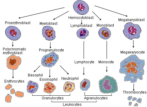 Blood cell lineage

See also
References
- ↑ 1.0 1.1 Alberts, Bruce (2005). "Leukocyte functions and percentage breakdown". Molecular Biology of the Cell. NCBI Bookshelf. Retrieved 2007-04-14.
External links
- Template:Dorlands
- Template:EMedicineDictionary
- Leukocytes at the US National Library of Medicine Medical Subject Headings (MeSH)
Suggested Reading and Key General References
Suggested Links and Web Resources
For Patients
Template:Hematology Template:Blood
bn:শ্বেত রক্তকণিকা
bs:Bijele krvne ćelije
bg:Левкоцит
ca:Leucòcit
cs:Bílá krvinka
da:Leukocyt
de:Leukozyt
eo:Leŭkocito
eu:Leukozito
fa:گویچه سفید
ko:백혈구
ia:Leucocyto
it:Leucocita
he:תא דם לבן
hr:Leukociti
id:Sel darah putih
lt:Baltasis kraujo kūnelis
mk:Леукоцит
nl:Witte bloedcel
nn:Kvite blodceller
simple:White blood cell
sk:Biela krvinka
sl:Bele krvničke
sq:Leukociti
sr:Бела крвна зрнца
su:Leukosit
fi:Valkosolu
sv:Vita blodkroppar
ta:இரத்த வெள்ளையணு
th:เม็ดเลือดขาว
yi:ווייסע בלוט צעל