Atrial fibrillation EKG examples
Editor-In-Chief: C. Michael Gibson, M.S., M.D. [1] Associate Editor: Cafer Zorkun, M.D., Ph.D. [2]
For the main page on atrial fibrillation, click here.
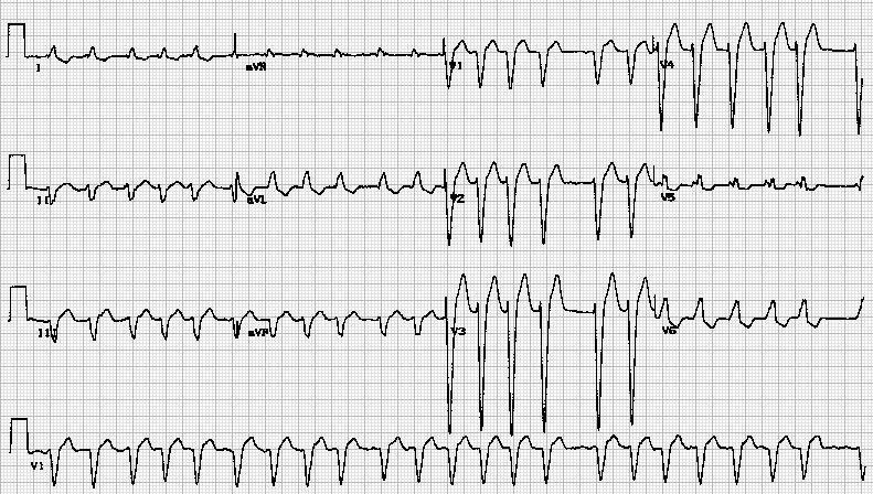
Shown below is an example of an EKG showing atrial fibrillation with rapid ventricular rate response and left bundle branch block.

Shown below is an example of an EKG showing atrial fibrillation with rapid ventricular rate response and left bundle branch block.

Shown below is an example of an EKG showing atrial fibrillation with rapid ventricular rate response and left bundle branch block.

Shown below is an example of an EKG showing atrial fibrillation with rapid ventricular rate response and left bundle branch block.

Shown below is an example of an EKG showing atrial fibrillation with rapid ventricular rate response and left bundle branch block.

Shown below is an example of an EKG showing atrial fibrillation with rapid ventricular rate response and left bundle branch block.

Shown below is an example of an EKG showing atrial fibrillation with rapid ventricular rate response and left bundle branch block.

Shown below is an example of an EKG showing atrial fibrillation with rapid ventricular rate response and left bundle branch block.

Shown below is an example of an EKG showing atrial fibrillation with rapid ventricular rate response and left bundle branch block.

Shown below is an example of an EKG showing atrial fibrillation with rapid ventricular rate response and left bundle branch block.

Shown below is an example of an EKG showing atrial fibrillation with rapid ventricular rate response and left bundle branch block.

Shown below is an example of an EKG showing atrial fibrillation with rapid ventricular rate response and left bundle branch block.

Shown below is an example of an EKG showing atrial fibrillation with rapid ventricular rate response and left bundle branch block.

Shown below is an example of an EKG showing atrial fibrillation with rapid ventricular rate response and left bundle branch block.

Shown below is an example of an EKG showing atrial fibrillation with rapid ventricular rate response and left bundle branch block.

Shown below is an example of an EKG showing atrial fibrillation with rapid ventricular rate response and left bundle branch block.

Shown below is an example of an EKG showing atrial fibrillation with rapid ventricular rate response and left bundle branch block.

Shown below is an example of an EKG showing atrial fibrillation with rapid ventricular rate response and left bundle branch block.

Shown below is an example of an EKG showing atrial fibrillation with rapid ventricular rate response and left bundle branch block.

Shown below is an example of an EKG showing atrial fibrillation with rapid ventricular rate response and left bundle branch block.

Shown below is an example of an EKG showing atrial fibrillation with rapid ventricular rate response and left bundle branch block.

Shown below is an example of an ECG showing atrial fibrillation with rapid ventricular response. Left axis deviation.

Shown below is an example of an ECG showing atrial fibrillation with rapid ventricular response. Left axis deviation.

Shown below is an example of an ECG showing Atrial fibrillation with slow ventricular response. Bifascicular block [Right Bundle Branch Block + Left anterior fascicular block (LAFB)].

Shown below is an example of an ECG showing Atrial fibrillation. Septal myocardial infarction (age indetermined)

Shown below is an example of an ECG showing Atrial fibrillation.

Shown below is an example of an ECG showing Atrial fibrillation.

Shown below is an example of an ECG showing Atrial fibrillation.

Shown below is an example of an ECG showing Atrial fibrillation.

Shown below is an example of an ECG showing Atrial fibrillation.

Shown below is an example of an ECG showing Atrial fibrillation.

Shown below is an example of an ECG showing Atrial fibrillation.

Shown below is an example of an ECG showing Atrial fibrillation.

Shown below is an example of an ECG showing Atrial fibrillation.

Shown below is an example of an ECG showing Atrial fibrillation.

Shown below is an example of an ECG showing Atrial fibrillation.

Shown below is an example of an ECG showing Atrial fibrillation.

Shown below is an example of an ECG showing Atrial fibrillation.

Shown below is an example of an ECG showing Atrial fibrillation.

Shown below is an example of an ECG showing Atrial fibrillation.

Shown below is an example of an ECG showing Atrial fibrillation.

Shown below is an example of an ECG showing Atrial fibrillation.


-

-
Atrial Fibrillation with old LBBB


Shown below is an example of an ECG showing Atrial Fibrillation with LVH and Strain.

Shown below is an example of a low quality ECG showing atrial fibrillation. The R wave in leads V5 and V6 are greater than 35mm and there are ST/T wave changes. This is consistent with left ventricular hypertrophy.

Shown below is an EKG that shows atrial fibrillation with wide spread ST depressions suggestive of ischemia or possibly non-Q myocardial infarction. In this case there were no enzyme changes suggestive of a myocardial infarction and the changes probably represent ischemia secondary to the ventricular tachycardia terminated by the lidocaine. It is also possible that the ST changes are purely a result of the tachycardia but in this case this would seem unlikely.

Sources
Copyleft image obtained courtesy of ECGpedia, http://en.ecgpedia.org/index.php?title=Special:NewFiles&dir=prev&offset=20080806182927&limit=500
References