Occipital bone
|
WikiDoc Resources for Occipital bone |
|
Articles |
|---|
|
Most recent articles on Occipital bone Most cited articles on Occipital bone |
|
Media |
|
Powerpoint slides on Occipital bone |
|
Evidence Based Medicine |
|
Clinical Trials |
|
Ongoing Trials on Occipital bone at Clinical Trials.gov Trial results on Occipital bone Clinical Trials on Occipital bone at Google
|
|
Guidelines / Policies / Govt |
|
US National Guidelines Clearinghouse on Occipital bone NICE Guidance on Occipital bone
|
|
Books |
|
News |
|
Commentary |
|
Definitions |
|
Patient Resources / Community |
|
Patient resources on Occipital bone Discussion groups on Occipital bone Patient Handouts on Occipital bone Directions to Hospitals Treating Occipital bone Risk calculators and risk factors for Occipital bone
|
|
Healthcare Provider Resources |
|
Causes & Risk Factors for Occipital bone |
|
Continuing Medical Education (CME) |
|
International |
|
|
|
Business |
|
Experimental / Informatics |
Editor-In-Chief: C. Michael Gibson, M.S., M.D. [1]
Overview
The occipital bone, a saucer-shaped membrane bone situated at the back and lower part of the cranium, is trapezoid in shape and curved on itself. It is pierced by a large oval aperture, the foramen magnum, through which the cranial cavity communicates with the vertebral canal.
- The curved, expanded plate behind the foramen magnum is named the squama occipitalis.
- The thick, somewhat quadrilateral piece in front of the foramen is called the basilar part of occipital bone.
- On either side of the foramen are the lateral parts of occipital bone.
Foramen magnum
- See also Foramen magnum
The foramen magnum is a large oval aperture with its long diameter antero-posterior; it is wider behind than in front where it is encroached upon by the condyles.
It transmits the medulla oblongata and its membranes, the accessory nerves, the vertebral arteries, the anterior and posterior spinal arteries, and the membrana tectoria and alar ligaments.
Angles
The superior angle of the occipital bone articulates with the occipital angles of the parietal bones and, in the fetal skull, corresponds in position with the posterior fontanelle.
The inferior angle is fused with the body of the sphenoid. The lateral angles are situated at the extremities of the grooves for the transverse sinuses: each is received into the interval between the mastoid angle of the parietal and the mastoid part of the temporal.
Borders
The superior borders extend from the superior to the lateral angles: they are deeply serrated for articulation with the occipital borders of the parietals, and form by this union the lambdoidal suture.
The inferior borders extend from the lateral angles to the inferior angle; the upper half of each articulates with the mastoid portion of the corresponding temporal, the lower half with the petrous part of the same bone.
These two portions of the inferior border are separated from one another by the jugular process, the notch on the anterior surface of which forms the posterior part of the jugular foramen.
Structure
The occipital, like the other cranial the outer and inner tables, between which is the cancellous tissue or diploë; the bone is especially thick at the ridges, protuberances, condyles, and anterior part of the basilar part; in the inferior fossæ it is thin, semitransparent, and destitute of diploë.
Additional images
-
 Cranial bones
 -
 Base of skull. Inferior surface.
 -
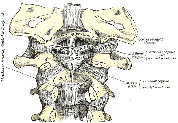 Membrana tectoria, transverse, and alar ligaments.
 -
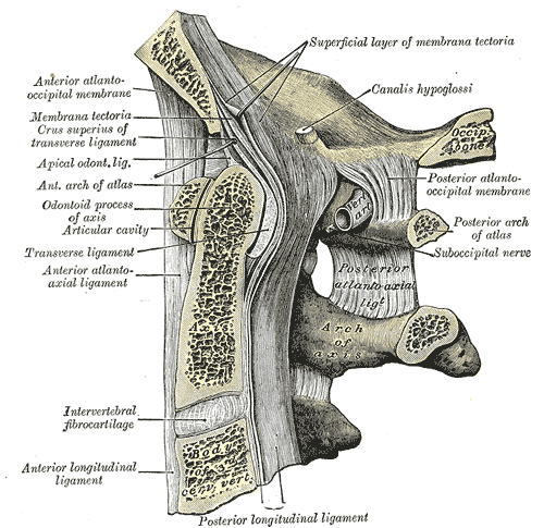 Median sagittal section through the occipital bone and first three cervical vertebræ.
 -
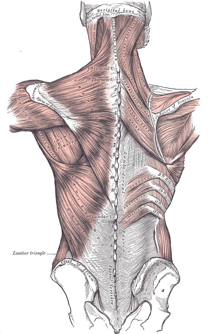 Muscles connecting the upper extremity to the vertebral column.
 -
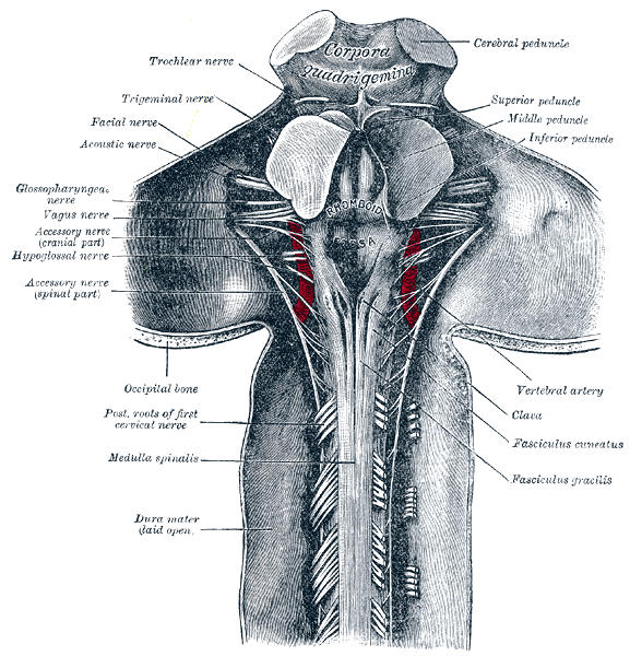 Upper part of medulla spinalis and hind- and mid-brains; posterior aspect, exposed in situ.
 -
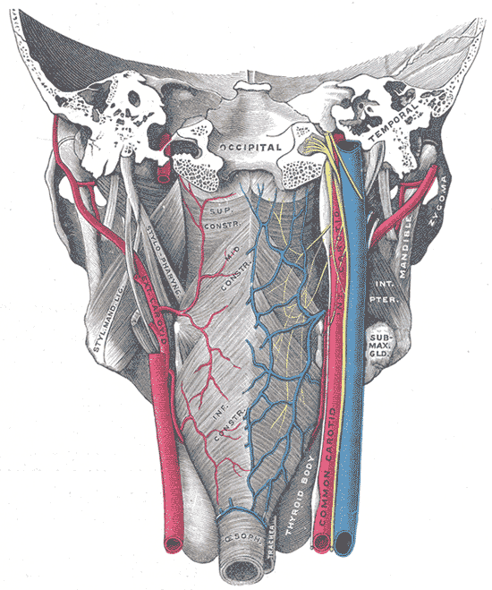 Muscles of the pharynx, viewed from behind, together with the associated vessels and nerves.


See also
- Bone terminology
- Comparative anatomy
- Neanderthal
- Occipital bun
- Terms for anatomical location
- Ossification of occipital bone
ca:Occipital
da:Nakkeben
de:Hinterhauptsbein
gl:Occipital
ko:뒤통수뼈
it:Osso occipitale
he:עצם העורף
la:Os occipitale
lt:Pakauškaulis
sk:Záhlavná kosť
sl:Zatilnica
sr:Потиљачна кост
fi:Takaraivoluu
sv:Nackben
uk:Потилична кістка