Neural crest
|
WikiDoc Resources for Neural crest |
|
Articles |
|---|
|
Most recent articles on Neural crest Most cited articles on Neural crest |
|
Media |
|
Powerpoint slides on Neural crest |
|
Evidence Based Medicine |
|
Clinical Trials |
|
Ongoing Trials on Neural crest at Clinical Trials.gov Clinical Trials on Neural crest at Google
|
|
Guidelines / Policies / Govt |
|
US National Guidelines Clearinghouse on Neural crest
|
|
Books |
|
News |
|
Commentary |
|
Definitions |
|
Patient Resources / Community |
|
Patient resources on Neural crest Discussion groups on Neural crest Patient Handouts on Neural crest Directions to Hospitals Treating Neural crest Risk calculators and risk factors for Neural crest
|
|
Healthcare Provider Resources |
|
Causes & Risk Factors for Neural crest |
|
Continuing Medical Education (CME) |
|
International |
|
|
|
Business |
|
Experimental / Informatics |
Editor-In-Chief: C. Michael Gibson, M.S., M.D. [1]
Overview
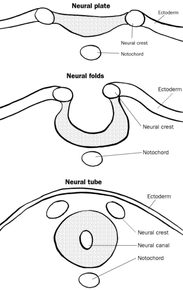
The neural crest, a transient component of the ectoderm, is located in between the neural tube and the epidermis (or the free margins of the neural folds) of an embryo during neural tube formation. Neural crest cells quickly migrate during or shortly after neurulation, an embryological event marked by neural tube closure.
It has been referred to as the fourth germ layer, due to its great importance. The neural crest can give rise to neurons and glia of the peripheral nervous system (PNS); some skeletal elements, tendons and smooth muscle; chondrocytes, osteocytes, melanocytes, chromaffin cells, and supporting cells and hormone producing cells in certain organs.
Clinical significance
Diseases due to defects in the neural crest induction, formation or migration are referred to as neurocristopathies, and genes that cause some of these like piebaldism and Hirschprung's disease have been cloned in mice models.
History and Nomenclature
In 1868 His described Neural Crest as "zwischenstrang"- a strip of cells lying between the dorsal ectoderm and the neural tube.[1]
From this time till almost 1950s most of the work on this structure was done on amphibian embryos, eg a 1950 comprehensive review in a monograph by the Swedish embryologist Sven Hörstadius.[2] Newth (who also studied it in fishes)[3] in 1951 described it as such by "a remarkable embryonic structure" and till another decade its origin still remained an enigma!
In 1960s with the invent of cell labeling with tritiated thymidine by Chibon[4] and Weston[5] gave rise to a major breakthrough in this field through amphibian and avian studies. But this was a transient method of cell labeling and the field had to wait till the chick-quail transfer studies were devised for a definitive confirmation of those results. These extensive works in 1970s was reviewed extensively in "the Neural Crest" by Nicole Le Douarin first published in 1982 (and second ed in 1999).[6]
The nomenclature of these cells derives from amphibian and avian studies which demonstrate migration from the neural crest which forms on the rostral region of the neurulating ectoderm in the trilaminar disc. In humans, the cells actually migrate from the lateral margins of the neural tube however the use of 'crest cells' in this regard is retained.
Induction
Cells fated to become neural crest tissue are induced by BMP, Wnt and FGF signaling to express the proteins Fox3D, RhoB and Slug, and to lose expression of E-cadherin.
- RhoB is likely to signal cytoskeletal changes required for migration. [7]
- Slug is a repressor[8] that leads to an activation of factors that dissociate tight junctions.
Categories
There are several main categories of neural crest based upon function:[9]
Cranial neural crest
- The cranial neural crest arises in the anterior and populates the face and the pharyngeal arches giving rise to bones, cartilage, nerves and connective tissue.
- Other Migration Locations:
- Into the pharyngeal arches and play an inductive in thymus development.
- Into the pharyngeal arches and form the parafollicular cell or ultimobranchial bodies of the thyroid gland.
- Into the pharyngeal arches and play an inductive role in parathyroid gland development.
- Facial ectomesenchyme of the pharyngeal arches forming skeletal muscle, bone, and cartilage in the face.
- Odontoblasts (dentin-producing cells) of the teeth.
- Into the optic vesicle and the developing eye and contributes to many anterior eye elements such the cornea, sclera, and ciliary muscle. It also contributes to the attaching skeletal muscles of the eye.
- Into the otic placode and participates in the inner ear development.
- Sensory ganglia of the fifth, seventh, ninth and tenth cranial nerves.
Vagal and sacral neural crest
- The vagal and sacral neural crest arises in the neck and tail and populates the gut, forming the parasympathetic neurons that regulates peristalsis and control blood vessel dilation.
- Other Migration Locations:
- Walls of the viscera to become enteric ganglia.
Trunk neural crest
- The trunk neural crest lies between the vagal and sacral neural crest and gives rise to two groups of cells. One group migrates dorsolateral and populates the skin, forming pigment cells and the other migrates ventrolateral through the anterior sclerotome to become the epinephrine-producing cells of the adrenal gland and the neurons of the sympathetic nervous system. Some cells remain in the sclerotome to form the dorsal root ganglia
- Other Migration Locations:
- Proximal to the spinal cord and line up symmetrically to form the dorsal root ganglia.
- Into the skin to form melanocytes and Merkel cells.
- Chromaffin cells of the adrenal medulla.
- Near the vertebral column and become sympathetic chain ganglia.
Cardiac neural crest
- The cardiac neural crest overlaps the vagal neural crest and migrates to populate the pharyngeal arches 3, 4 and 6 (producing structures in the head) and to the heart, forming connective tissue that separates the great vessels of the heart.
- Other Migration Locations:
- Into the pharyngeal arches and Truncus arteriosus (embryology), forming the aorticopulmonary septum and the smooth muscle of great arteries.
- Anterior of the aorta to become the four pre-aortic ganglia (celiac ganglion, superior mesenteric ganglion, inferior mesenteric ganglion and aortical renal ganglia)
Migration
Neural crest cells require extracellular matrix to migrate through interactions between integrins and fibronectin and laminin. Migration is directed by inhibitory and attractive signals from cells. Ephrin is an inhibitory ligand in posterior sclerotome that affects ventral pathway trunk neural crest cells and causes them to migrate through the anterior sclerotome instead. Thrombospondin promotes migration through the anterior sclerotome. Another signal, stem cell factor is involved in specifying the destination of migration. If expressed in the wrong locations, pigment cells migrate to that site and proliferate there.
Plasticity
Neural crest cells show varying degrees of plasticity. Some trunk neural crest cells are pluripotent. Cranial neural crest cells can give rise to trunk neural crest cells if transplanted. However, heart neural crest cells are committed before migration. Individual neural crest cells can take on a new fate, however groups of neural crest cells cannot.
See also
References
- ↑ "Neural Crest Introduction". Retrieved 2007-09-18.
- ↑ "Neural Crest and the Origins of Craniofacial Pattern". Retrieved 2007-09-18.
- ↑ Newth DR (1950). "Fate of the neural crest in lampreys". Nature. 165 (4190): 284. PMID 15405801.
- ↑ Chibon P (1967). "[Nuclear labelling by tritiated thymidine of neural crest derivatives in the amphibian Urodele Pleurodeles waltlii Michah]". Journal of embryology and experimental morphology (in French). 18 (3): 343–58. PMID 5590717.
- ↑ Weston JA (1963). "A radioautographic analysis of the migration and localization of trunk neural crest cells in the chick". Dev. Biol. 6: 279–310. PMID 14000137.
- ↑ Kalcheim, Chaya; Le Douarin, N. (1999). The neural crest. Cambridge, UK: Cambridge University Press. ISBN 0-521-62010-4.
- ↑ Liu JP, Jessell TM (1998). "A role for rhoB in the delamination of neural crest cells from the dorsal neural tube". Development. 125 (24): 5055–67. PMID 9811589.
- ↑ Vernon AE, LaBonne C (2006). "Slug stability is dynamically regulated during neural crest development by the F-box protein Ppa". Development. 133 (17): 3359–70. doi:10.1242/dev.02504. PMID 16887825.
- ↑ "Neural Crest Migration". Retrieved 2007-09-18.
External links
Template:Developmental biology Template:Development of nervous system de:Neuralleiste