Eustachian tube
| https://https://www.youtube.com/watch?v=H29571Ex-kY%7C350}} |
Template:Infobox Anatomy Editor-In-Chief: C. Michael Gibson, M.S., M.D. [1]
The Eustachian tube (or auditory tube) is a tube that links the pharynx to the middle ear. In adults the Eustachian tube is approximately 35 mm long. It is named after the 16th century anatomist Eustachius.[1] Some modern medical books call this the pharyngotympanic tube.[2]
Location
The Eustachian tube extends from the anterior wall of the middle ear to the lateral wall of the nasopharynx, approximately at the level of the inferior nasal concha. A portion of the tube (~1/3) proximal to the middle ear is made of bone; the rest is composed of cartilage [3] and raises a tubal elevation, the torus tubarius, in the nasopharynx where it opens.
In the equids (horses) and some rodent-like species such as the desert hyrax, an evagination of the eustachian tube is known as the gutteral pouch and is divided into medial and lateral compartments by the stylohyoid bone of the hyoid apparatus. This is of great importance in equine medicine as the pouches are prone to infections, and due to their intimate relationship to the cranial nerves (VII,IX,X,XI) and the internal and external carotid artery, various syndromes may arise relating to which is damaged. Epistaxis (nosebleed) is a very common presentation to veterinary surgeons and this may often be fatal unless a balloon catheter can be placed in time to suppress bleeding.
Functions
Pressure Equalization
Normally the Eustachian tube is closed, but it can open to let a small amount of air through to equalize the pressure between the middle ear and the atmosphere. When this happens we hear a small pop, an event familiar to aircraft passengers or drivers in mountainous regions. Yawning or swallowing can pull on muscles in the neck, causing the tube to open. Some people are born with the ability to contract just these muscles voluntarily, similar to people who can wiggle their ears. Doing so will make one's voice sound louder to oneself. Without this airway, air would be unable to escape from one's ear, the middle ear would be isolated from the atmosphere, and could be easily damaged by pressure changes.
Some who have the ability to voluntarily contract these muscles can hear a "popclickity" sound in the middle ear when actuating these muscles, and are able to hold the muscle contraction (some refer to this as 'clicking your ears to equalize the pressure'). This allows such people to voluntarily equalize pressures at will when making rapid ascents or descents, typically in aircraft flights or large elevation changes in either tall buildings or mountainous treks. When the breath (inhale or exhale) is controlled, air pressure can be intentionally increased or decreased in the middle ear (breathing through the nose only or mouth), where the feeling of a cool air breeze can be felt inside the eustachian tube.
Occasionally, if the voluntary contraction timing is missed during a rapid pressure change, a slight yawning (opening of the jaw) action combines to assist in pressure equalization.
Mucus Drainage
The Eustachian tube also drains mucus from the middle ear. Upper airway infections or allergies can cause the Eustachian tube to become swollen, trapping bacteria and causing ear infections. This swelling can be reduced through the use of pseudoephedrine. Earaches are more common in children because the tube is more horizontal and thinner, making the movement of fluid harder.
Embryologic Development
The Eustachian tube is derived from the first pharyngeal pouch, which during embryogenesis forms a recess called the tubotympanic sulcus. The sulcus deepens to meet the first pharyngeal cleft forming the tympanic membrane. The distal part of the tubotympanic sulcus gives rise to the tympanic cavity, while the proximal tubular structure becomes the Eustachian tube.
Muscles
There are four muscles associated with the function of the Eustachian tube:
- levator veli palatini (innervated by the vagus nerve)
- salpingopharyngeus (innervated by the vagus nerve)
- tensor tympani (innervated by the mandibular nerve of CN V)
- tensor veli palatini (innervated by the mandibular nerve of CN V)
Disorders of the Eustachian tube
Some people are born with a dysfunctional Eustachian tube, which is much slimmer than the usual human Eustachian tube. This can either be genetically, or has been suggested to be a phenomenon occurring as a result of a hard birth, in which the baby's head was subject to an extraordinary amount of pressure, thus reshaping the Eustachian tube, making it much slimmer. The result of this disorder is a huge amount of mucus gathering in the middle ear, often impairing hearing to a degree. This condition is known as otitis media or glue ear. The disorder is treated by inserting small tubes (often known as Grommets) through the tympanic membrane (ear drum). This allows any accumulation of mucus to flow directly out through the ear, as well as continually equalizing the pressure of the ear, throughout a subject's day. In some cases a person may be able to blow small amounts of air through the Eustachian tube and out through the ear, particularly if the Eustachian tubes are patulous.
Some people suffer from the rare Patulous Eustachian tube condition, in which the Eustachian tube remains intermittently open, causing an echoing sound of their own heartbeat, breathing and speech, which may be temporarily relieved by flipping the head upside down.
Additional images
-
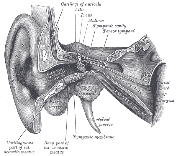 External and middle ear, opened from the front. Right side.
 -
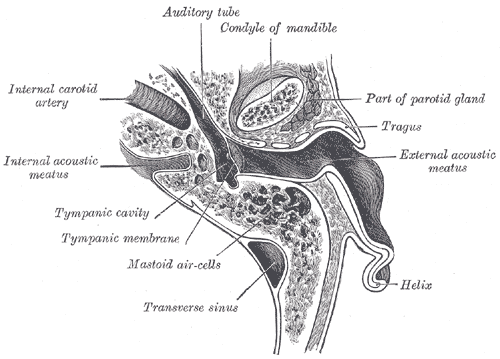 Horizontal section through left ear; upper half of section.
 -
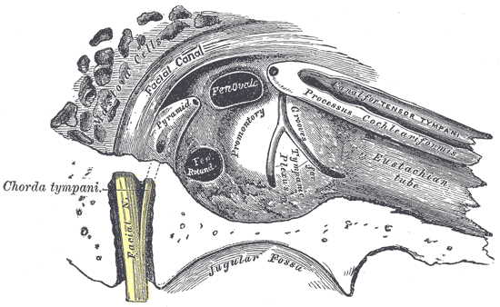 View of the inner wall of the tympanum (enlarged.)
 -
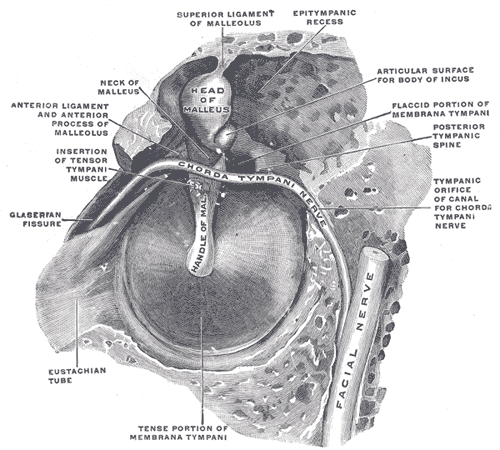 The right membrana tympani with the hammer and the chorda tympani, viewed from within, from behind, and from above.


References
de:Eustachi-Röhre hr:Eustahijeva cijev it:Tromba di Eustachio lt:Ausies trimitas nl:Buis van Eustachius sk:Eustachova trubica fi:Korvatorvi sv:Örontrumpet