Subclavian artery
|
WikiDoc Resources for Subclavian artery |
|
Articles |
|---|
|
Most recent articles on Subclavian artery Most cited articles on Subclavian artery |
|
Media |
|
Powerpoint slides on Subclavian artery |
|
Evidence Based Medicine |
|
Cochrane Collaboration on Subclavian artery |
|
Clinical Trials |
|
Ongoing Trials on Subclavian artery at Clinical Trials.gov Trial results on Subclavian artery Clinical Trials on Subclavian artery at Google
|
|
Guidelines / Policies / Govt |
|
US National Guidelines Clearinghouse on Subclavian artery NICE Guidance on Subclavian artery
|
|
Books |
|
News |
|
Commentary |
|
Definitions |
|
Patient Resources / Community |
|
Patient resources on Subclavian artery Discussion groups on Subclavian artery Patient Handouts on Subclavian artery Directions to Hospitals Treating Subclavian artery Risk calculators and risk factors for Subclavian artery
|
|
Healthcare Provider Resources |
|
Causes & Risk Factors for Subclavian artery |
|
Continuing Medical Education (CME) |
|
International |
|
|
|
Business |
|
Experimental / Informatics |
Editor-In-Chief: C. Michael Gibson, M.S., M.D. [1]
In human anatomy, the subclavian artery is a major artery of the upper thorax that mainly supplies blood to the head and arms. It is located below the clavicle, hence the name. There is a left subclavian and a right subclavian.
- On the left side of the body, the subclavian comes directly off the arch of aorta.
- On the right side of the body, the subclavian arises from the relatively short brachiocephalic artery (trunk) when it bifurcates into the subclavian and the right common carotid artery.
The usual branches of the subclavian on both sides of the body are the vertebral artery, the internal thoracic artery, the thyrocervical trunk the costocervical trunk and the dorsal scapular artery. The subclavian becomes the axillary artery at the lateral border of the first rib.
Course
From its origin, the subclavian artery travels laterally, passing between anterior and middle scalene muscles, with the anterior scalene (scalenus anterior) on its anterior side and the middle scalene (scalenus medius) on its posterior. This is in contrast to the subclavian vein, which travels anterior to the scalenus anterior. As the subclavian artery crosses the border of the first rib, it becomes the axillary artery.
Branches
- vertebral artery -From 1st part of Subclavian
- internal thoracic artery-From 1st part of Subclavian
- thyrocervical trunk -From 1st part of Subclavian
- costocervical trunk -From 2nd part of the Subclavian
- dorsal scapular artery- This can emerge from the 3rd part (rarely from the 2nd part) of the Subclavian as its own branch or from the Thyrocervical Artery as a branch of the Transverse Cervical artery.

These may be remembered by the mnemonic VITamin C and D.
The subclavian artery continues as the axillary artery.
 |
 |
 |
Embryology
Embryologically, the left subclavian simply arises from the left 7th intersegmental artery, while the right subclavian arises, proximal to distal:
- aortic arch IV
- right dorsal aorta (between the 4th and the 7th intersegmental arteries)
- right 7th intersegmental artery
Anatomical details
On the right side the subclavian artery arises from the innominate artery behind the right sternoclavicular articulation; on the left side it springs from the arch of the aorta. The two vessels, therefore, in the first part of their course, differ in length, direction, and relation with neighboring structures.
In order to facilitate the description, each subclavian artery is divided into three parts:
- The first portion extends from the origin of the vessel to the medial border of the Scalenus anterior.
- The second lies behind this muscle.
- The third extends from the lateral margin of the muscle to the outer border of the first rib, where it becomes the axillary artery.
The first portions of the two vessels require separate descriptions; the second and third parts of the two arteries are practically alike.
First Part of the Right Subclavian Artery
The first part of the right subclavian artery arises from the innominate artery, behind the upper part of the right sternoclavicular articulation, and passes upward and lateralward to the medial margin of the Scalenus anterior. It ascends a little above the clavicle, the extent to which it does so varying in different cases.
Relations
It is covered, in front, by the integument, superficial fascia, Platysma, deep fascia, the clavicular origin of the Sternocleidomastoideus, the Sternohyoideus, and Sternothyreoideus, and another layer of the deep fascia. It is crossed by the internal jugular and vertebral veins, by the vagus nerve and the cardiac branches of the vagus and sympathetic, and by the subclavian loop of the sympathetic trunk which forms a ring around the vessel. The anterior jugular vein is directed lateralward in front of the artery, but is separated from it by the Sternohyoideus and Sternothyreoideus. Below and behind the artery is the pleura, which separates it from the apex of the lung; behind is the sympathetic trunk, the Longus collie and the first thoracic vertebra. The right recurrent nerve winds around the lower and back part of the vessel.
First Part of the Left Subclavian Artery
The first part of the left subclavian artery arises from the arch of the aorta, behind the left common carotid, and at the level of the fourth thoracic vertebra; it ascends in the superior mediastinal cavity to the root of the neck and then arches lateralward to the medial border of the Scalenus anterior.
Relations
It is in relation, in front, with the vagus, cardiac, and phrenic nerves, which lie parallel with it, the left common carotid artery, left internal jugular and vertebral veins, and the commencement of the left innominate vein, and is covered by the Sternothyreoideus, Sternohyoideus, and Sternocleidomastoideus; behind, it is in relation with the esophagus, thoracic duct, left recurrent nerve, inferior cervical ganglion of the sympathetic trunk, and Longus colli; higher up, however, the esophagus and thoracic duct lie to its right side; the latter ultimately arching over the vessel to join the angle of union between the subclavian and internal jugular veins. Medial to it are the esophagus, trachea, thoracic duct, and left recurrent nerve; lateral to it, the left pleura and lung.
Second Part of the Subclavian Artery
The second portion of the subclavian artery lies behind the Scalenus anterior; it is very short, and forms the highest part of the arch described by the vessel.
Relations
It is covered, in front, by the skin, superficial fascia, Platysma, deep cervical fascia, Sternocleidomastoideus, and Scalenus anterior. On the right side of the neck the phrenic nerve is separated from the second part of the artery by the Scalenus anterior, while on the left side it crosses the first part of the artery close to the medial edge of the muscle. Behind the vessel are the pleura and the Scalenus medius; above, the brachial plexus of nerves; below, the pleura. The subclavian vein lies below and in front of the artery, separated from it by the Scalenus anterior.
Third Part of the Subclavian Artery
The third portion of the subclavian artery runs downward and lateralward from the lateral margin of the Scalenus anterior to the outer border of the first rib, where it becomes the axillary artery. This is the most superficial portion of the vessel, and is contained in the subclavian triangle.
Relations
It is covered, in front, by the skin, the superficial fascia, the Platysma, the supraclavicular nerves, and the deep cervical fascia. The external jugular vein crosses its medial part and receives the transverse scapular, transverse cervical, and anterior jugular veins, which frequently form a plexus in front of the artery. Behind the veins, the nerve to the Subclavius descends in front of the artery. The terminal part of the artery lies behind the clavicle and the Subclavius and is crossed by the transverse scapular vessels. The subclavian vein is in front of and at a slightly lower level than the artery. Behind, it lies on the lowest trunk of the brachial plexus, which intervenes between it and the Scalenus medius. Above and to its lateral side are the upper trunks of the brachial plexus and the Omohyoideus. Below, it rests on the upper surface of the first rib.
Peculiarities
The subclavian arteries vary in their origin, their course, and the height to which they rise in the neck.
The origin of the right subclavian from the innominate takes place, in some cases, above the sternoclavicular articulation, and occasionally, but less frequently, below that joint. The artery may arise as a separate trunk from the arch of the aorta, and in such cases it may be either the first, second, third, or even the last branch derived from that vessel; in the majority, however, it is the first or last, rarely the second or third. When it is the first branch, it occupies the ordinary position of the innominate artery; when the second or third, it gains its usual position by passing behind the right carotid; and when the last branch, it arises from the left extremity of the arch, and passes obliquely toward the right side, usually behind the trachea, esophagus, and right carotid, sometimes between the esophagus and trachea, to the upper border of the first rib, whence it follows its ordinary course. In very rare instances, this vessel arises from the thoracic aorta, as low down as the fourth thoracic vertebra. Occasionally, it perforates the Scalenus anterior; more rarely it passes in front of that muscle. Sometimes the subclavian vein passes with the artery behind the Scalenus anterior. The artery may ascend as high as 4 cm. above the clavicle, or any intermediate point between this and the upper border of the bone, the right subclavian usually ascending higher than the left.
The left subclavian is occasionally joined at its origin with the left carotid.
The left subclavian artery is more deeply placed than the right in the first part of its course, and, as a rule, does not reach quite as high a level in the neck. The posterior border of the Sternocleidomastoideus corresponds pretty closely to the lateral border of the Scalenus anterior, so that the third portion of the artery, the part most accessible for operation, lies immediately lateral to the posterior border of the Sternocleidomastoideus.
Additional images
-
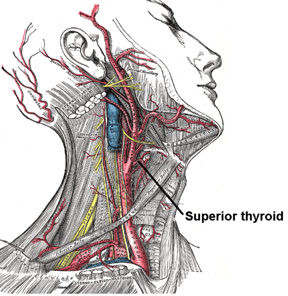 The internal carotid and vertebral arteries. Right side.
 -
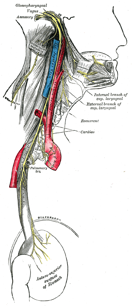 Course and distribution of the glossopharyngeal, vagus, and accessory nerves.
 -
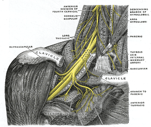 The right brachial plexus with its short branches, viewed from in front.
 -
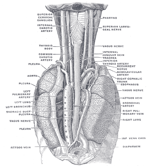 The position and relation of the esophagus in the cervical region and in the posterior mediastinum. Seen from behind.
 -
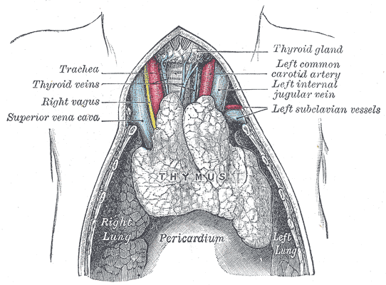 The thymus of a full-time fetus, exposed in situ.
 -
 Side of neck, showing chief surface markings.


See also
External links
- Template:DukeOrtho
- Template:GPnotebook
- Template:SUNYAnatomyFigs
- Diagram of branches at informatics.jax.org
- Template:LoyolaMedEd
Template:Gray's Template:Arteries of head and neck Template:Arteries of chest
de:Arteria subclavia nn:Arteria subclavia no:Arteria subclavia