Appendix cancer CT scan: Difference between revisions
Jump to navigation
Jump to search
No edit summary |
No edit summary |
||
| Line 5: | Line 5: | ||
==Overview== | ==Overview== | ||
Abdominal [[Computed tomography|CT scan]] is | Abdominal [[Computed tomography|CT scan]] is helpful in the [[diagnosis]] and management of appendix cancer. Findings on [[Computed tomography|CT scan]] suggestive of [[Vermiform appendix|appendix]] [[cancer]] include soft tissue thickening, wall irregularity, [[Calcification|calcification,]] internal septations, preappendiceal fat stranding as well as [[Peritoneum|intraperitoneal]] free fluid. [[Computed tomography|CT scan]] is also one of the best [[Imaging studies|imaging modalities]] to assess disease burden, [[Metastasis CT|metastatic lesions]] as well as disease [[Cancer staging|stage]]. | ||
==CT scan== | ==CT scan== | ||
* | * Experts believe that [[Computed tomography|enhanced CT]] is the [[Imaging studies|imaging modality]] of choice for [[Vermiform appendix|appendix]] [[cancer]].<ref name="pmid26648795">Kelly KJ (2015) [https://www.ncbi.nlm.nih.gov/entrez/eutils/elink.fcgi?dbfrom=pubmed&retmode=ref&cmd=prlinks&id=26648795 Management of Appendix Cancer.] ''Clin Colon Rectal Surg'' 28 (4):247-55. [http://dx.doi.org/10.1055/s-0035-1564433 DOI:10.1055/s-0035-1564433] PMID: [https://pubmed.gov/26648795 26648795]</ref> | ||
* On [[Computed tomography|CT,]] [[appendix cancer]] is characterized by the following findings: | * On [[Computed tomography|CT,]] [[appendix cancer]] is characterized by the following findings: | ||
** [[Soft tissue]] thickening | ** [[Soft tissue]] thickening | ||
| Line 31: | Line 31: | ||
:*'''''Table and figure below demonstrate abdominal regions as well as scoring system for [[Peritoneal carcinomatosis|PCI]].''''' | :*'''''Table and figure below demonstrate abdominal regions as well as scoring system for [[Peritoneal carcinomatosis|PCI]].''''' | ||
[[Image:Peritoneal Carcinomatosis Index (PCI) Regions.jpg|thumb|left|'''Peritoneal Carcinomatosis Index (PCI) Regions'''| | [[Image:Peritoneal Carcinomatosis Index (PCI) Regions.jpg|thumb|left|'''Peritoneal Carcinomatosis Index (PCI) Regions'''|850px|right|Peritoneal Carcinomatosis Index (PCI) Regions]] | ||
{| class="wikitable" | {| class="wikitable" | ||
|+PCI Scoring System | |+PCI Scoring System | ||
| Line 101: | Line 101: | ||
* | * | ||
[[Image:Liver-metastases-from-carcinoid.jpg|thumb|centre|'''Liver metastases from gastrointestinal carcinoid'''. Case courtesy of Dr Natalie Yang, <a | [[Image:Liver-metastases-from-carcinoid.jpg|thumb|centre|'''Liver metastases from gastrointestinal carcinoid'''. Case courtesy of Dr Natalie Yang, <a | ||
<nowiki><nowiki>&lt;nowiki&gt;&amp;lt;nowiki&amp;gt;&amp;amp;lt;nowiki&amp;amp;gt;&amp;amp;amp;lt;nowiki&amp;amp;amp;gt; &amp;amp;amp;lt;/nowiki&amp;amp;amp;gt;&amp;amp;lt;/nowiki&amp;amp;gt;&amp;lt;/nowiki&amp;gt;&lt;/nowiki&gt;</nowiki></nowiki>ref="<nowiki><nowiki>&lt;nowiki&gt;&amp;lt;nowiki&amp;gt;&amp;amp;lt;nowiki&amp;amp;gt;&amp;amp;amp;lt;nowiki&amp;amp;amp;gt;https://radiopaedia.org/&amp;amp;amp;lt;/nowiki&amp;amp;amp;gt;&amp;amp;lt;/nowiki&amp;amp;gt;&amp;lt;/nowiki&amp;gt;&lt;/nowiki&gt;</nowiki></nowiki>">Radiopaedia.org<nowiki></a></nowiki>. From the case <nowiki><a href="https://radiopaedia.org/cases/7010">rID: 7010</a></nowiki>]] | <nowiki><nowiki>&lt;nowiki&gt;&amp;lt;nowiki&amp;gt;&amp;amp;lt;nowiki&amp;amp;gt;&amp;amp;amp;lt;nowiki&amp;amp;amp;gt;&amp;amp;amp;amp;lt;nowiki&amp;amp;amp;amp;gt; &amp;amp;amp;amp;lt;/nowiki&amp;amp;amp;amp;gt;&amp;amp;amp;lt;/nowiki&amp;amp;amp;gt;&amp;amp;lt;/nowiki&amp;amp;gt;&amp;lt;/nowiki&amp;gt;&lt;/nowiki&gt;</nowiki></nowiki>ref="<nowiki><nowiki>&lt;nowiki&gt;&amp;lt;nowiki&amp;gt;&amp;amp;lt;nowiki&amp;amp;gt;&amp;amp;amp;lt;nowiki&amp;amp;amp;gt;&amp;amp;amp;amp;lt;nowiki&amp;amp;amp;amp;gt;https://radiopaedia.org/&amp;amp;amp;amp;lt;/nowiki&amp;amp;amp;amp;gt;&amp;amp;amp;lt;/nowiki&amp;amp;amp;gt;&amp;amp;lt;/nowiki&amp;amp;gt;&amp;lt;/nowiki&amp;gt;&lt;/nowiki&gt;</nowiki></nowiki>">Radiopaedia.org<nowiki></a></nowiki>. From the case <nowiki><a href="https://radiopaedia.org/cases/7010">rID: 7010</a></nowiki>]] | ||
* [[Bone metastasis|Metastatic bone lesions]] of both [[adenocarcinoma]] and [[Carcinoid|carcinoid tumor]]<nowiki/>s of [[Vermiform appendix|appendix]] are extremely rare but might present with [[Osteolysis|osteolitic (adenocarcinom)]] and a mixture of [[Osteosclerosis|osteosclerot]]<nowiki/>ic and [[Osteolysis|osteolytic changes (carcinoid tumors)]].<ref name="pmid22783400">Hori T, Yasuda T, Suzuki K, Kanamori M, Kimura T (2012) [https://www.ncbi.nlm.nih.gov/entrez/eutils/elink.fcgi?dbfrom=pubmed&retmode=ref&cmd=prlinks&id=22783400 Skeletal metastasis of carcinoid tumors: Two case reports and review of the literature.] ''Oncol Lett'' 3 (5):1105-1108. [http://dx.doi.org/10.3892/ol.2012.622 DOI:10.3892/ol.2012.622] PMID: [https://pubmed.gov/22783400 22783400]</ref> <ref name="pmid27121744">Ganesh V, Probyn L, Vuong S, Caskenette S, Chow E (2016) [https://www.ncbi.nlm.nih.gov/entrez/eutils/elink.fcgi?dbfrom=pubmed&retmode=ref&cmd=prlinks&id=27121744 A case report of bone metastases from appendiceal adenocarcinoma and a review of literature.] ''Ann Palliat Med'' 5 (2):149-52. [http://dx.doi.org/10.21037/apm.2016.01.03 DOI:10.21037/apm.2016.01.03] PMID: [https://pubmed.gov/27121744 27121744]</ref> | * [[Bone metastasis|Metastatic bone lesions]] of both [[adenocarcinoma]] and [[Carcinoid|carcinoid tumor]]<nowiki/>s of [[Vermiform appendix|appendix]] are extremely rare but might present with [[Osteolysis|osteolitic (adenocarcinom)]] and a mixture of [[Osteosclerosis|osteosclerot]]<nowiki/>ic and [[Osteolysis|osteolytic changes (carcinoid tumors)]].<ref name="pmid22783400">Hori T, Yasuda T, Suzuki K, Kanamori M, Kimura T (2012) [https://www.ncbi.nlm.nih.gov/entrez/eutils/elink.fcgi?dbfrom=pubmed&retmode=ref&cmd=prlinks&id=22783400 Skeletal metastasis of carcinoid tumors: Two case reports and review of the literature.] ''Oncol Lett'' 3 (5):1105-1108. [http://dx.doi.org/10.3892/ol.2012.622 DOI:10.3892/ol.2012.622] PMID: [https://pubmed.gov/22783400 22783400]</ref> <ref name="pmid27121744">Ganesh V, Probyn L, Vuong S, Caskenette S, Chow E (2016) [https://www.ncbi.nlm.nih.gov/entrez/eutils/elink.fcgi?dbfrom=pubmed&retmode=ref&cmd=prlinks&id=27121744 A case report of bone metastases from appendiceal adenocarcinoma and a review of literature.] ''Ann Palliat Med'' 5 (2):149-52. [http://dx.doi.org/10.21037/apm.2016.01.03 DOI:10.21037/apm.2016.01.03] PMID: [https://pubmed.gov/27121744 27121744]</ref> | ||
Revision as of 16:37, 22 February 2019
|
Appendix cancer Microchapters |
|
Diagnosis |
|---|
|
Treatment |
|
Appendix cancer CT scan On the Web |
|
American Roentgen Ray Society Images of Appendix cancer CT scan |
|
Risk calculators and risk factors for Appendix cancer CT scan |
Editor-In-Chief: C. Michael Gibson, M.S., M.D. [1]; Associate Editor(s)-in-Chief: Soroush Seifirad, M.D.[2]
Overview
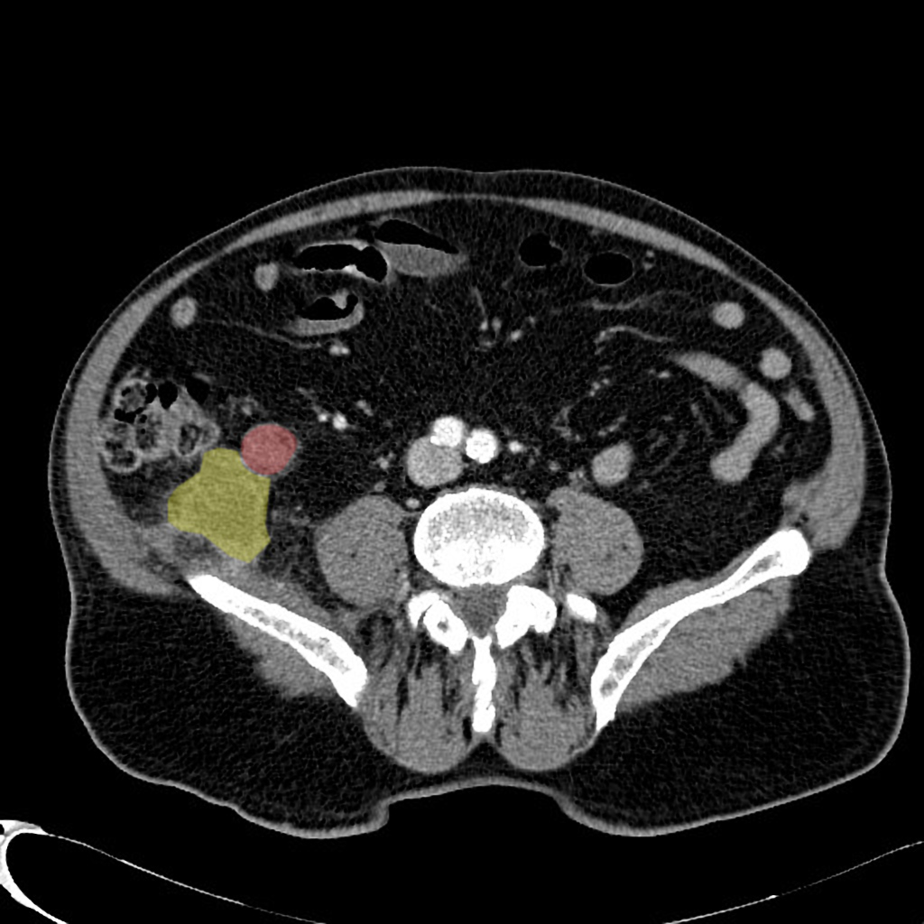
Abdominal CT scan is helpful in the diagnosis and management of appendix cancer. Findings on CT scan suggestive of appendix cancer include soft tissue thickening, wall irregularity, calcification, internal septations, preappendiceal fat stranding as well as intraperitoneal free fluid. CT scan is also one of the best imaging modalities to assess disease burden, metastatic lesions as well as disease stage.
CT scan
- Experts believe that enhanced CT is the imaging modality of choice for appendix cancer.[1]
- On CT, appendix cancer is characterized by the following findings:
- Soft tissue thickening
- Wall irregularity
- Presence of pseudomyxoma peritonei
- Calcification
- Internal septations
- Periappendiceal fat stranding and intraperitoneal free fluid which is a nonspecific finding
- Cystic lesion


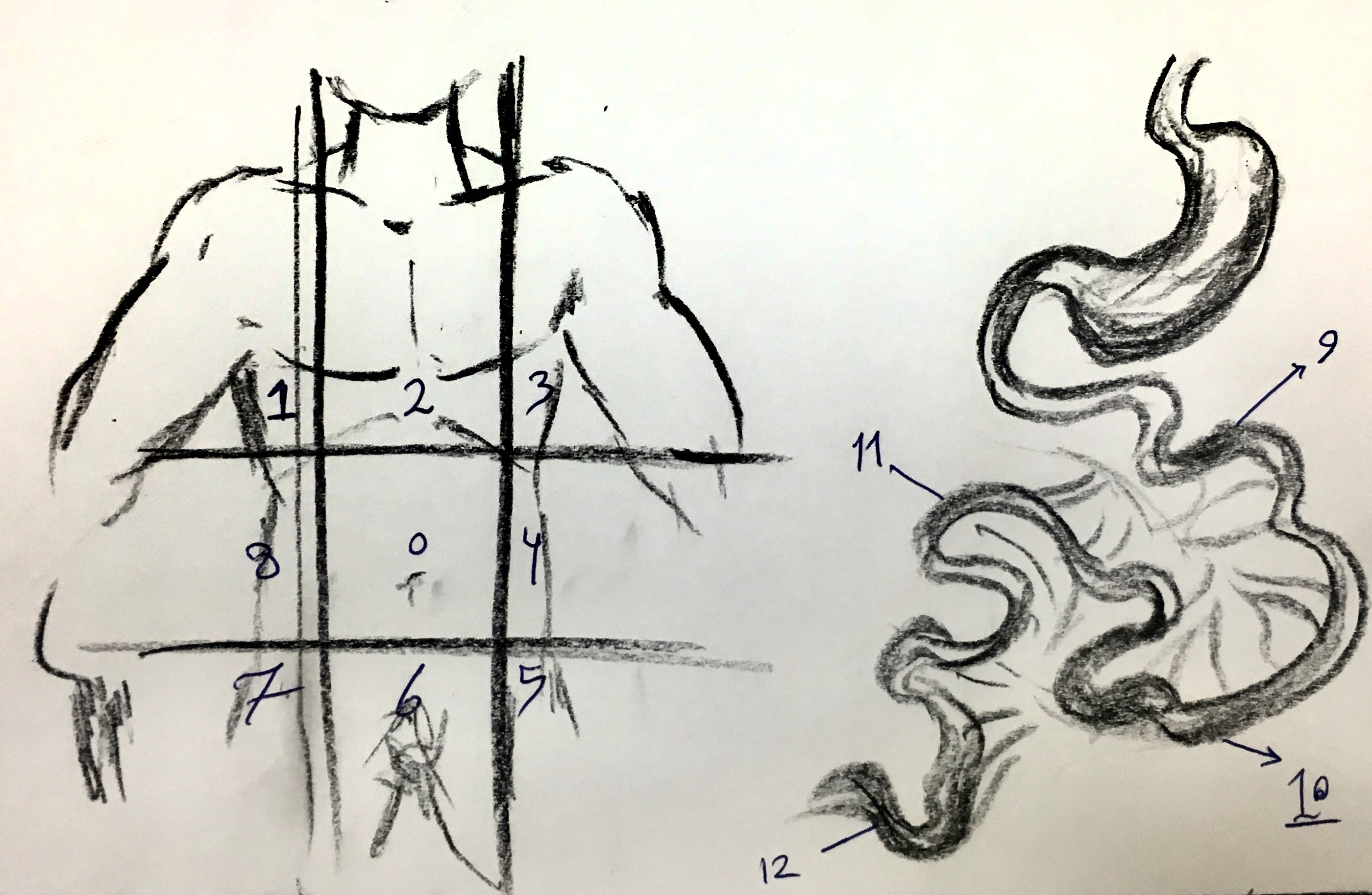
- Peritoneal carcinomatosis index (PCI): a widely accepted metric for assessment of disease border in appendix cancer[2]
- Estimated by contrast enhanced cross sectional imaging.
- Both MRI and CT scan has been used and are globally accepted imaging modalities.
- Small peritoneal seeding might be difficult to appreciate on CT.
- Sometimes it is challenging to distinguish between tumor and mucin.
- There are reports in favor of diffusion weighted MRI superiority compared to CT in evaluating extent of peritoneal involvement.[3]
- Table and figure below demonstrate abdominal regions as well as scoring system for PCI.

| Lesion Size Score | |
|---|---|
| LS0 | No tumor seen |
| LS1 | Tumor up to 0.5 cm |
| LS2 | Tumor up to 5 cm cm |
| LS3 | Tumor > 5 cm or confluence |
| Maximum Score = 3 | |
| Regions (0-3) | |
| 0 | Central |
| 1 | Right Upper |
| 2 | Epigasterium |
| 3 | Left Upper |
| 4 | Left Flank |
| 5 | Left Lower |
| 6 | Pelvis |
| 7 | Right Upper |
| 8 | Right Flank |
| 9 | Upper Jejunum |
| 10 | Lower Jejunum |
| 11 | Upper Illeum |
| 12 | lower Illeum |
| Maximum Score = 36 | |
| Total Maximum Score = 39 | |
- CT scan also helps in discovering distant metastatic lesions in the other organs like bone, lungs, brain and particularly liver.
- Carcinoid tumors that metastases to liver presents with carcinoid syndrome.
- Helical, contrast enhanced thriplephase CT scan is the best Ct scan method to assess liver involvement.

- Metastatic bone lesions of both adenocarcinoma and carcinoid tumors of appendix are extremely rare but might present with osteolitic (adenocarcinom) and a mixture of osteosclerotic and osteolytic changes (carcinoid tumors).[4] [5]
- Generally there is no need for imaging studies in carcinoid tumors.
- Radiographic investigation indications in carcinoid tumors are as follows:
- Tumor size > 2 cm
- Incomplete tumor resection
- Evidence of mesentric or intraabdominal involvement
- Carcinoid syndrome
- Low attenuated, well defined mass in right lower quadrant, near cecum without inflammation points to appendiceal mucocele.
- Wall thinkness does not differenciate between benign and malignant lesions.
- Intramural nodule raise suspension for cystadenocarcinoma.
References
- ↑ Kelly KJ (2015) Management of Appendix Cancer. Clin Colon Rectal Surg 28 (4):247-55. DOI:10.1055/s-0035-1564433 PMID: 26648795
- ↑ Jacquet P, Sugarbaker PH (1996) Clinical research methodologies in diagnosis and staging of patients with peritoneal carcinomatosis. Cancer Treat Res 82 ():359-74. PMID: 8849962
- ↑ Low RN, Barone RM (2012) Combined diffusion-weighted and gadolinium-enhanced MRI can accurately predict the peritoneal cancer index preoperatively in patients being considered for cytoreductive surgical procedures. Ann Surg Oncol 19 (5):1394-1401. DOI:10.1245/s10434-012-2236-3 PMID: 22302265
- ↑ Hori T, Yasuda T, Suzuki K, Kanamori M, Kimura T (2012) Skeletal metastasis of carcinoid tumors: Two case reports and review of the literature. Oncol Lett 3 (5):1105-1108. DOI:10.3892/ol.2012.622 PMID: 22783400
- ↑ Ganesh V, Probyn L, Vuong S, Caskenette S, Chow E (2016) A case report of bone metastases from appendiceal adenocarcinoma and a review of literature. Ann Palliat Med 5 (2):149-52. DOI:10.21037/apm.2016.01.03 PMID: 27121744