Acoustic neuroma MRI: Difference between revisions
Jump to navigation
Jump to search
(→MRI) |
No edit summary |
||
| Line 3: | Line 3: | ||
{{CMG}}{{AE}}{{Simrat}} | {{CMG}}{{AE}}{{Simrat}} | ||
==Overview== | ==Overview== | ||
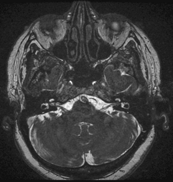
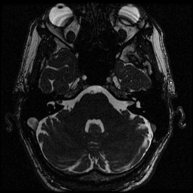
[[Gadolinium]]-enhanced [[MRI]] | [[Gadolinium]]-enhanced [[Magnetic resonance imaging|MRI scan]] is the definitive [[diagnostic test]] for acoustic neuroma and can identify [[tumors]] as small as 1-2 millimeter in [[diameter]]. On [[brain]] [[Magnetic resonance imaging|MRI]], acoustic neuroma characterized by hypointense mass on T1-weighted [[Magnetic resonance imaging|MRI]], and hyperintense mass on T2-weighted [[Magnetic resonance imaging|MRI]]. | ||
==MRI== | ==MRI== | ||
*[[Gadolinium]]-enhanced [[magnetic resonance imaging | *[[Gadolinium]]-enhanced [[magnetic resonance imaging|magnetic resonance imaging (MRI)]] is the preferred [[diagnostic test]] for identifying acoustic neuromas and can identify [[tumors]] as small as 1-2 millimeter in [[diameter]].<ref>{{Cite journal | ||
| author = [[E. P. Lin]] & [[B. T. Crane]] | | author = [[E. P. Lin]] & [[B. T. Crane]] | ||
| title = The Management and Imaging of Vestibular Schwannomas | | title = The Management and Imaging of Vestibular Schwannomas | ||
| Line 28: | Line 28: | ||
| pmid = 1518359 | | pmid = 1518359 | ||
}}</ref> | }}</ref> | ||
{| style="border: 0px; font-size: 90%; margin: 3px; width: 600px" align="center" | {| style="border: 0px; font-size: 90%; margin: 3px; width: 600px" align="center" | ||
Revision as of 17:46, 26 April 2019
|
Acoustic neuroma Microchapters | |
|
Diagnosis | |
|---|---|
|
Treatment | |
|
Case Studies | |
|
Acoustic neuroma MRI On the Web | |
|
American Roentgen Ray Society Images of Acoustic neuroma MRI | |
Editor-In-Chief: C. Michael Gibson, M.S., M.D. [1]Associate Editor(s)-in-Chief: Simrat Sarai, M.D. [2]
Overview
Gadolinium-enhanced MRI scan is the definitive diagnostic test for acoustic neuroma and can identify tumors as small as 1-2 millimeter in diameter. On brain MRI, acoustic neuroma characterized by hypointense mass on T1-weighted MRI, and hyperintense mass on T2-weighted MRI.
MRI
- Gadolinium-enhanced magnetic resonance imaging (MRI) is the preferred diagnostic test for identifying acoustic neuromas and can identify tumors as small as 1-2 millimeter in diameter.[1][2]
| MRI component | Features |
|---|---|
|
|
|
|
|
|
Post-op MRI: Linear enhancement may not indicate tumor, but if there is nodular enhancement suspect tumor recurrence (needs follow up MRI).[3]


References
- ↑ E. P. Lin & B. T. Crane (2017). "The Management and Imaging of Vestibular Schwannomas". AJNR. American journal of neuroradiology. 38 (11): 2034–2043. doi:10.3174/ajnr.A5213. PMID 28546250. Unknown parameter
|month=ignored (help) - ↑ D. F. Wilson, R. S. Hodgson, M. F. Gustafson, S. Hogue & L. Mills (1992). "The sensitivity of auditory brainstem response testing in small acoustic neuromas". The Laryngoscope. 102 (9): 961–964. doi:10.1288/00005537-199209000-00001. PMID 1518359. Unknown parameter
|month=ignored (help) - ↑ Acoustic Schwannoma. Radiopedia(2015) http://radiopaedia.org/articles/acoustic-schwannoma Accessed on October 2 2015
- ↑ Image courtesy of Dr Frank Gaillard. Radiopaedia (original file here).[http://radiopaedia.org/licence Creative Commons BY-SA-NC
- ↑ Image courtesy of Dr. Roberto Schubert Radiopaedia (original file here).[http://radiopaedia.org/licence Creative Commons BY-SA-NC