Sandbox SSW: Difference between revisions
Sargun Walia (talk | contribs) No edit summary |
Aditya Ganti (talk | contribs) |
||
| Line 18: | Line 18: | ||
| | | | ||
{| style="border: 0px; font-size: 90%; margin: 3px;" align="center" | {| style="border: 0px; font-size: 90%; margin: 3px;" align="center" | ||
! rowspan="3" style="background:#4479BA; color: #FFFFFF;" align="center" |Disease | ! rowspan="3" style="background:#4479BA; color: #FFFFFF;" align="center" |Disease | ||
| colspan="13" rowspan="1" style="background:#4479BA; color: #FFFFFF;" align="center" |'''Clinical manifestations''' | | colspan="13" rowspan="1" style="background:#4479BA; color: #FFFFFF;" align="center" |'''Clinical manifestations''' | ||
| Line 44: | Line 43: | ||
! style="background:#4479BA; color: #FFFFFF;" align="center" |Imaging | ! style="background:#4479BA; color: #FFFFFF;" align="center" |Imaging | ||
|- | |- | ||
| colspan="1" rowspan="1" style="padding: 5px 5px; background: #DCDCDC;" align="center" |Acute suppurative cholangitis | | colspan="1" rowspan="1" style="padding: 5px 5px; background: #DCDCDC;" align="center" |Acute suppurative cholangitis | ||
| style="padding: 5px 5px; background: #F5F5F5;" align="center" |[[RUQ]] | | style="padding: 5px 5px; background: #F5F5F5;" align="center" |[[RUQ]] | ||
| Line 280: | Line 276: | ||
* Fatty food intolerance | * Fatty food intolerance | ||
|- | |- | ||
| colspan="1" rowspan="1" style="padding: 5px 5px; background: #DCDCDC;" align="center" | [[Peptic Ulcer Disease|Peptic ulcer disease]] | | colspan="1" rowspan="1" style="padding: 5px 5px; background: #DCDCDC;" align="center" | [[Peptic Ulcer Disease|Peptic ulcer disease]] | ||
| style="padding: 5px 5px; background: #F5F5F5;" align="center" |Diffuse | | style="padding: 5px 5px; background: #F5F5F5;" align="center" |Diffuse | ||
| Line 461: | Line 456: | ||
* Postgastrectomy | * Postgastrectomy | ||
|- | |- | ||
! style="background:#4479BA; color: #FFFFFF;" align="center" |Disease | ! style="background:#4479BA; color: #FFFFFF;" align="center" |Disease | ||
! style="background:#4479BA; color: #FFFFFF;" align="center" |Abdominal Pain | ! style="background:#4479BA; color: #FFFFFF;" align="center" |Abdominal Pain | ||
| Line 769: | Line 763: | ||
* PILLCAM 2: A colon capsule for CRC screening may be used in patients with an incomplete colonoscopy who lacks obstruction | * PILLCAM 2: A colon capsule for CRC screening may be used in patients with an incomplete colonoscopy who lacks obstruction | ||
|- | |- | ||
| style="padding: 5px 5px; background: #DCDCDC;" align="center" |[[Hepatitis|Viral hepatitis]] | | style="padding: 5px 5px; background: #DCDCDC;" align="center" |[[Hepatitis|Viral hepatitis]] | ||
| style="padding: 5px 5px; background: #F5F5F5;" align="center" |[[RUQ]] | | style="padding: 5px 5px; background: #F5F5F5;" align="center" |[[RUQ]] | ||
| Line 974: | Line 967: | ||
! style="background:#4479BA; color: #FFFFFF;" align="center" |Comments | ! style="background:#4479BA; color: #FFFFFF;" align="center" |Comments | ||
|- | |- | ||
| colspan="1" rowspan="1" style="padding: 5px 5px; background: #DCDCDC;" align="center" |[[Spontaneous bacterial peritonitis]] | | colspan="1" rowspan="1" style="padding: 5px 5px; background: #DCDCDC;" align="center" |[[Spontaneous bacterial peritonitis]] | ||
| style="padding: 5px 5px; background: #F5F5F5;" align="center" |Diffuse | | style="padding: 5px 5px; background: #F5F5F5;" align="center" |Diffuse | ||
| Line 996: | Line 988: | ||
| style="padding: 5px 5px; background: #F5F5F5;" align="left" | | | style="padding: 5px 5px; background: #F5F5F5;" align="left" | | ||
|- | |- | ||
| style="padding: 5px 5px; background: #DCDCDC;" align="center" |Pyelonephritis | | style="padding: 5px 5px; background: #DCDCDC;" align="center" |Pyelonephritis | ||
| style="padding: 5px 5px; background: #F5F5F5;" align="center" |Unilateral | | style="padding: 5px 5px; background: #F5F5F5;" align="center" |Unilateral | ||
| Line 1,044: | Line 1,035: | ||
* [[Dysuria]] | * [[Dysuria]] | ||
|- | |- | ||
| colspan="1" rowspan="1" style="padding: 5px 5px; background: #DCDCDC;" align="center" |[[Small bowel obstruction]] | | colspan="1" rowspan="1" style="padding: 5px 5px; background: #DCDCDC;" align="center" |[[Small bowel obstruction]] | ||
| style="padding: 5px 5px; background: #F5F5F5;" align="center" |Diffuse | | style="padding: 5px 5px; background: #F5F5F5;" align="center" |Diffuse | ||
| Line 1,146: | Line 1,136: | ||
! style="background:#4479BA; color: #FFFFFF;" align="center" |Comments | ! style="background:#4479BA; color: #FFFFFF;" align="center" |Comments | ||
|- | |- | ||
| style="padding: 5px 5px; background: #DCDCDC;" align="center" |[[Mesenteric ischemia]] | | style="padding: 5px 5px; background: #DCDCDC;" align="center" |[[Mesenteric ischemia]] | ||
| style="padding: 5px 5px; background: #F5F5F5;" align="center" |Periumbilical | | style="padding: 5px 5px; background: #F5F5F5;" align="center" |Periumbilical | ||
| Line 1,195: | Line 1,183: | ||
* May lead to shock | * May lead to shock | ||
|- | |- | ||
| style="padding: 5px 5px; background: #DCDCDC;" align="center" |[[Ruptured abdominal aortic aneurysm]] | | style="padding: 5px 5px; background: #DCDCDC;" align="center" |[[Ruptured abdominal aortic aneurysm]] | ||
| style="padding: 5px 5px; background: #F5F5F5;" align="center" | Diffuse | | style="padding: 5px 5px; background: #F5F5F5;" align="center" | Diffuse | ||
| Line 1,259: | Line 1,246: | ||
! style="background:#4479BA; color: #FFFFFF;" align="center" |Comments | ! style="background:#4479BA; color: #FFFFFF;" align="center" |Comments | ||
|- | |- | ||
| style="padding: 5px 5px; background: #DCDCDC;" align="center" |Torsion of the cyst/ovary | | style="padding: 5px 5px; background: #DCDCDC;" align="center" |Torsion of the cyst/ovary | ||
| style="padding: 5px 5px; background: #F5F5F5;" align="center" |RLQ / LLQ | | style="padding: 5px 5px; background: #F5F5F5;" align="center" |RLQ / LLQ | ||
| Line 1,325: | Line 1,310: | ||
| style="padding: 5px 5px; background: #F5F5F5;" align="left" | | | style="padding: 5px 5px; background: #F5F5F5;" align="left" | | ||
|- | |- | ||
| style="padding: 5px 5px; background: #DCDCDC;" align="center" |Ruptured [[ectopic pregnancy]] | | style="padding: 5px 5px; background: #DCDCDC;" align="center" |Ruptured [[ectopic pregnancy]] | ||
| style="padding: 5px 5px; background: #F5F5F5;" align="center" |RLQ / LLQ | | style="padding: 5px 5px; background: #F5F5F5;" align="center" |RLQ / LLQ | ||
| Line 1,348: | Line 1,332: | ||
* Vaginal bleeding | * Vaginal bleeding | ||
|- | |- | ||
|[[Pleural empyema]] | |||
| style="padding: 5px 5px; background: #F5F5F5;" align="center" |[[RUQ]]/[[Epigastric]] | | style="padding: 5px 5px; background: #F5F5F5;" align="center" |[[RUQ]]/[[Epigastric]] | ||
| style="padding: 5px 5px; background: #F5F5F5;" align="center" | + | | style="padding: 5px 5px; background: #F5F5F5;" align="center" | + | ||
| Line 1,375: | Line 1,357: | ||
* Increased [[tactile fremitus]] | * Increased [[tactile fremitus]] | ||
|- | |- | ||
|[[Pulmonary embolism]] | |||
| style="padding: 5px 5px; background: #F5F5F5;" align="center" |RUQ/LUQ | | style="padding: 5px 5px; background: #F5F5F5;" align="center" |RUQ/LUQ | ||
| style="padding: 5px 5px; background: #F5F5F5;" align="center" | ± | | style="padding: 5px 5px; background: #F5F5F5;" align="center" | ± | ||
| Line 1,401: | Line 1,383: | ||
* Pleuretic chest pain | * Pleuretic chest pain | ||
|- | |- | ||
|[[Pneumonia]] | |||
| style="padding: 5px 5px; background: #F5F5F5;" align="center" |RUQ/LUQ | | style="padding: 5px 5px; background: #F5F5F5;" align="center" |RUQ/LUQ | ||
| style="padding: 5px 5px; background: #F5F5F5;" align="center" | + | | style="padding: 5px 5px; background: #F5F5F5;" align="center" | + | ||
| Line 1,427: | Line 1,409: | ||
* Cough | * Cough | ||
|- | |- | ||
|[[Myocardial Infarction]] | |||
| style="padding: 5px 5px; background: #F5F5F5;" align="center" |[[Epigastric]] | | style="padding: 5px 5px; background: #F5F5F5;" align="center" |[[Epigastric]] | ||
| style="padding: 5px 5px; background: #F5F5F5;" align="center" | ± | | style="padding: 5px 5px; background: #F5F5F5;" align="center" | ± | ||
| Line 1,462: | Line 1,443: | ||
{| | {| | ||
|- | |- | ||
| <figure-inline class="mw-default-size">[[Image:Right_upper_quadrant.PNG|link=Right upper quadrant abdominal pain resident survival guide|339x339px]]</figure-inline>||<figure-inline class="mw-default-size">[[Image:Epigastric_quadrant_pain.PNG|link=Epigastric pain resident survival guide|179x179px]]</figure-inline>||<figure-inline class="mw-default-size">[[Image:Left_upper_quadrant.PNG|link=Left upper quadrant abdominal pain resident survival guide|329x329px]]</figure-inline> | | <figure-inline class="mw-default-size"><figure-inline>[[Image:Right_upper_quadrant.PNG|link=Right upper quadrant abdominal pain resident survival guide|339x339px]]</figure-inline></figure-inline>||<figure-inline class="mw-default-size"><figure-inline>[[Image:Epigastric_quadrant_pain.PNG|link=Epigastric pain resident survival guide|179x179px]]</figure-inline></figure-inline>||<figure-inline class="mw-default-size"><figure-inline>[[Image:Left_upper_quadrant.PNG|link=Left upper quadrant abdominal pain resident survival guide|329x329px]]</figure-inline></figure-inline> | ||
|- | |- | ||
| <figure-inline class="mw-default-size">[[Image:Right_flank_quadrant.PNG|link=Right flank pain resident survival guide|338x338px]]</figure-inline>||<figure-inline class="mw-default-size">[[Image:Umbilical_pain.PNG|link=Umbilical region pain resident survival guide|165x165px]]</figure-inline>||<figure-inline class="mw-default-size">[[Image:Left_flank_quadrant.PNG|link=Left flank quadrant abdominal pain resident survival guide|335x335px]]</figure-inline> | | <figure-inline class="mw-default-size"><figure-inline>[[Image:Right_flank_quadrant.PNG|link=Right flank pain resident survival guide|338x338px]]</figure-inline></figure-inline>||<figure-inline class="mw-default-size"><figure-inline>[[Image:Umbilical_pain.PNG|link=Umbilical region pain resident survival guide|165x165px]]</figure-inline></figure-inline>||<figure-inline class="mw-default-size"><figure-inline>[[Image:Left_flank_quadrant.PNG|link=Left flank quadrant abdominal pain resident survival guide|335x335px]]</figure-inline></figure-inline> | ||
|- | |- | ||
| <figure-inline class="mw-default-size">[[Image:Right_lower_quadrant.PNG|link=Right lower quadrant abdominal pain resident survival guide|338x338px]]</figure-inline>||<figure-inline class="mw-default-size">[[Image:Hypogastric.PNG|link=Hypogastric pain resident survival guide|199x199px]]</figure-inline>||<figure-inline class="mw-default-size">[[Image:Left_lower_quadrant.PNG|link=Left lower quadrant abdominal pain resident survival guide|335x335px]]</figure-inline> | | <figure-inline class="mw-default-size"><figure-inline>[[Image:Right_lower_quadrant.PNG|link=Right lower quadrant abdominal pain resident survival guide|338x338px]]</figure-inline></figure-inline>||<figure-inline class="mw-default-size"><figure-inline>[[Image:Hypogastric.PNG|link=Hypogastric pain resident survival guide|199x199px]]</figure-inline></figure-inline>||<figure-inline class="mw-default-size"><figure-inline>[[Image:Left_lower_quadrant.PNG|link=Left lower quadrant abdominal pain resident survival guide|335x335px]]</figure-inline></figure-inline> | ||
|} | |} | ||
Revision as of 19:46, 8 January 2018
Editor-In-Chief: C. Michael Gibson, M.S., M.D. [1]; Associate Editor(s)-in-Chief: Sargun Singh Walia M.B.B.S.[2]
Overview
Editor-In-Chief: C. Michael Gibson, M.S., M.D. [3]; Associate Editor(s)-in-Chief: Amandeep Singh M.D.[4]
Differential diagnosis
Abbreviations: RUQ= Right upper quadrant of the abdomen, LUQ= Left upper quadrant, LLQ= Left lower quadrant, RLQ= Right lower quadrant, LFT= Liver function test, SIRS= Systemic inflammatory response syndrome, ERCP= Endoscopic retrograde cholangiopancreatography, IV= Intravenous, N= Normal, AMA= Anti mitochondrial antibodies, LDH= Lactate dehydrogenase, GI= Gastrointestinal, CXR= Chest X ray, IgA= Immunoglobulin A, IgG= Immunoglobulin G, IgM= Immunoglobulin M, CT= Computed tomography, PMN= Polymorphonuclear cells, ESR= Erythrocyte sedimentation rate, CRP= C-reactive protein, TS= Transferrin saturation, SF= Serum Ferritin, SMA= Superior mesenteric artery, SMV= Superior mesenteric vein, ECG= Electrocardiogram, US = Ultrasound
| |||||||||||||||||||||||||||||||||||||||||||||||||||||||||||||||||||||||||||||||||||||||||||||||||||||||||||||||||||||||||||||||||||||||||||||||||||||||||||||||||||||||||||||||||||||||||||||||||||||||||||||||||||||||||||||||||||||||||||||||||||||||||||||||||||||||||||||||||||||||||||||||||||||||||||||||||||||||||||||||||||||||||||||||||||||||||||||||||||||||||||||||||||||||||||||||||||||||||||||||||||||||||||||||||||||||||||||||||||||||||||||||||||||||||||||||||||||||||||||||||||||||||||||||||||||||||||||||||||||||||||||||||||||||||||||||||||||||||||||||||||||||||||||||||||||||||||||||||||||||||||||||||||||||||||||||||||||||||||||||||||||||||||||||||||||||||||||||||||||||||||||||||||||||||||||||||||||||||||||||||||||||||||||||||||||||||||||||||||||||||||||||||||||||||||||||||||||||||||||||||||||||||||||||||||||||||||||||||||||||||||||||||||||||||||||||||||||||||||||||||||||||||||||||||||||||||||||||||||||||||||||||||||||||||||||||||||||||||||||||||||||||||||||||||||||||||||||||||||||||
<figure-inline class="mw-default-size"><figure-inline> </figure-inline></figure-inline> </figure-inline></figure-inline> |
<figure-inline class="mw-default-size"><figure-inline> </figure-inline></figure-inline> </figure-inline></figure-inline> |
<figure-inline class="mw-default-size"><figure-inline>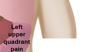 </figure-inline></figure-inline> </figure-inline></figure-inline>
|
<figure-inline class="mw-default-size"><figure-inline> </figure-inline></figure-inline> </figure-inline></figure-inline> |
<figure-inline class="mw-default-size"><figure-inline> </figure-inline></figure-inline> </figure-inline></figure-inline> |
<figure-inline class="mw-default-size"><figure-inline> </figure-inline></figure-inline> </figure-inline></figure-inline>
|
<figure-inline class="mw-default-size"><figure-inline>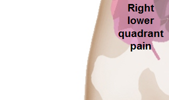 </figure-inline></figure-inline> </figure-inline></figure-inline> |
<figure-inline class="mw-default-size"><figure-inline>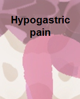 </figure-inline></figure-inline> </figure-inline></figure-inline> |
<figure-inline class="mw-default-size"><figure-inline>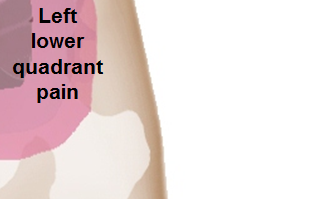 </figure-inline></figure-inline> </figure-inline></figure-inline>
|