Thoracic aorta
Editor-In-Chief: C. Michael Gibson, M.S., M.D. [1]
Overview
The thoracic aorta is contained in the posterior mediastinal cavity.
It begins at the lower border of the fourth thoracic vertebra where it is continuous with the aortic arch, and ends in front of the lower border of the twelfth thoracic vertebra, at the aortic hiatus in the diaphragm where it becomes the abdominal aorta.
At its commencement, it is situated on the left of the vertebral column; it approaches the median line as it descends; and, at its termination, lies directly in front of the column.
The vessel describes a curve which is concave forward; as the branches given off from it are small, its diminution in size is insignificant.
Relations
It is in relation, anteriorly, from above downward, with the root of the left lung, the pericardium, the esophagus, and the diaphragm; posteriorly, with the vertebral column and the hemiazygos veins; on the right side, with the azygos vein and thoracic duct; on the left side, with the left pleura and lung.
The esophagus, with its accompanying plexus of nerves, lies on the right side of the aorta above; but at the lower part of the thorax it is placed in front of the aorta, and, close to the diaphragm, is situated on its left side.
Branches
Branches before thoracic aorta
The initial part of the aorta, the ascending aorta, rises out of the left ventricle, from which it is separated by the aortic valve. The two coronary arteries of the heart arise from the aortic root, just above the cusps of the aortic valve.
The aorta then arches back over the right pulmonary artery. Three vessels come out of the aortic arch, the brachiocephalic artery, the left common carotid artery, and the left subclavian artery. These vessels supply blood to the head, neck, thorax and upper limbs.
Branches of thoracic aorta
The aorta gives off several paired branches as it descends in the thorax. These includes the
Additional images
-
 Transverse section of thorax, showing relations of pulmonary artery.
 -
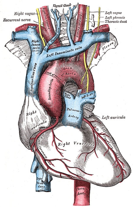 The arch of the aorta, and its branches.


External links
Template:Arteries of chest Template:Gray's
eu:Toraxeko aorta
hr:Prsna aorta
sr:Грудна аорта