Abdominal aorta
Template:WikiDoc Cardiology News Editor-In-Chief: C. Michael Gibson, M.S., M.D. [1]
The abdominal aorta is the largest artery in the abdominal cavity. As part of the aorta, it is a direct continuation of descending aorta (of the thorax).
Path
It begins at the level of the diaphragm, crossing it via the aortic hiatus at the vertebral level of T12. It travels down the posterior wall of the abdomen in front of the vertebral column. It thus follows the curvature of the lumbar vertebrae, that is, convex forward. The peak of this convexity is at the level of the third lumbar vertebra (L3).
It runs parallel to the inferior vena cava, which is located just to the right of the abdominal aorta, and becomes smaller in diameter as it gives off branches.
Branches
The abdominal aorta supplies blood to much of the abdominal cavity. It begins at T12, and usually has the following branches:
| Branch | Vertebra | Type | Paired? | A/P | Description |
| inferior phrenic | T12 | Parietal | yes | post. | originates just below the diaphragm, supplying it from below |
| celiac | T12 | Visceral | no | ant. | large anterior branch |
| superior mesenteric | L1 | Visceral | no | ant. | large anterior branch, arises just below celiac trunk |
| middle suprarenal | L1 | Visceral | yes | post. | to adrenal gland |
| renal | L2 | Visceral | yes | post. | large artery, each arising from the side of the aorta; supplies corresponding kidney |
| gonadal | L2 | Visceral | yes | post. | ovarian artery in females; testicular artery in males |
| lumbar | L1-L4 | Parietal | yes | post. | four on each side that supply the abdominal wall and spinal cord |
| inferior mesenteric | L3 | Visceral | no | ant. | large anterior branch |
| median sacral | L4 | Parietal | no | post. | artery arising from the middle of the aorta at its lowest part |
| common iliac | L4 | Terminal | yes | post. | branches (bifurcates) to supply blood to the lower limbs and the pelvis, ending the abdominal aorta |
Note that the bifurcation (union) of the inferior vena cava is at L5 and therefore below that of the bifurcation of the aorta.
Relations
The abdominal aorta lies slightly to the left of the midline of the body. It is covered, anteriorly, by the lesser omentum and stomach, behind which are the branches of the celiac artery and the celiac plexus; below these, by the lienal vein, the pancreas, the left renal vein, the inferior part of the duodenum, the mesentery, and aortic plexus.
Posteriorly, it is separated from the lumbar vertebræ and intervertebral fibrocartilages by the anterior longitudinal ligament and left lumbar veins.
On the right side it is in relation above with the azygos vein, cisterna chyli, thoracic duct, and the right crus of the diaphragm—the last separating it from the upper part of the inferior vena cava, and from the right celiac ganglion; the inferior vena cava is in contact with the aorta below.
On the left side are the left crus of the diaphragm, the left celiac ganglion, the ascending part of the duodenum, and some coils of the small intestine.
Relationship with inferior vena cava
The abominal aorta's venous counterpart, the inferior vena cava (IVC), travels parallel to it on its right side.
- Above the level of the umbilicus, the aorta is somewhat posterior to the IVC, sending the right renal artery travelling behind it. The IVC likewise sends its opposite side counterpart, the left renal vein, crossing in front of the aorta.
- Below the level of the umbilicus, the situation is generally reversed, with the aorta sending its right common iliac artery to cross its opposite side counterpart (the left common iliac vein) anteriorly.
Collateral circulation
The collateral circulation would be carried on by the anastomoses between the internal thoracic artery and the inferior epigastric artery; by the free communication between the superior and inferior mesenterics, if the ligature were placed between these vessels; or by the anastomosis between the inferior mesenteric artery and the internal pudendal artery, when (as is more common) the point of ligature is below the origin of the inferior mesenteric artery; and possibly by the anastomoses of the lumbar arteries with the branches of the internal iliac artery.
Additional images
-
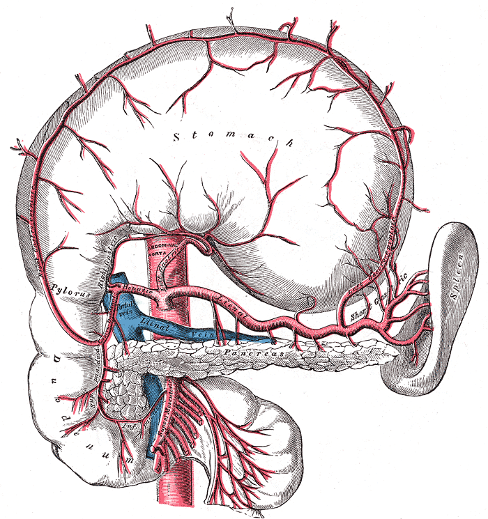 The celiac artery and its branches; the stomach has been raised and the peritoneum removed.
 -
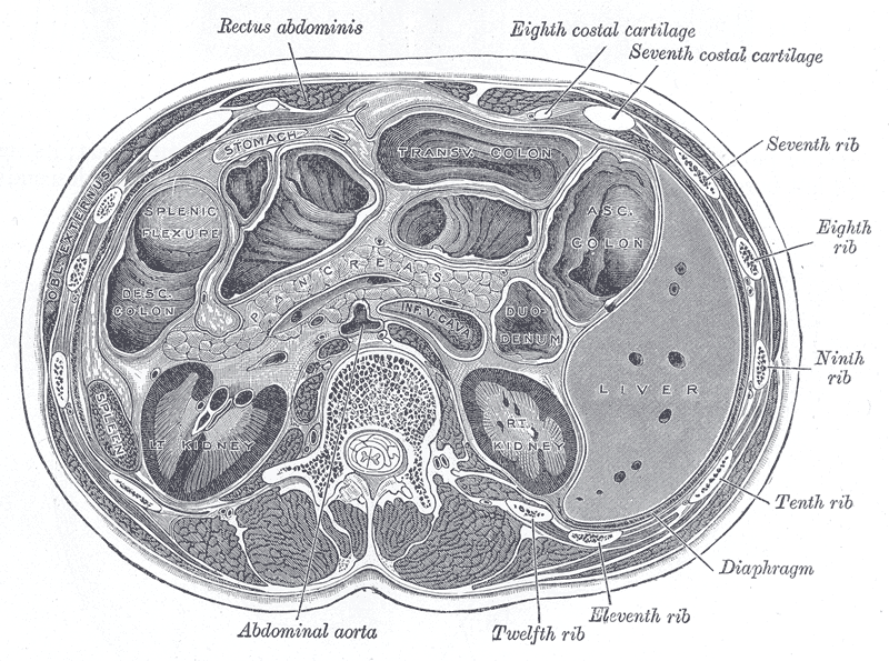 Transverse section through the middle of the first lumbar vertebra, showing the relations of the pancreas.


See also
External links
Template:Arteries of thorax and abdomen
ca:Aorta abdominal
eu:Abdomeneko aorta
it:Aorta addominale