Persistent left superior vena cava
For patient information click here
| Persistent left superior vena cava | |
 | |
|---|---|
| Persistent Left Superior Vena Cava: Gross natural color heart in situ dissected to show persistent left superior vena cava Image courtesy of Professor Peter Anderson DVM PhD and published with permission © PEIR, University of Alabama at Birmingham, Department of Pathology | |
| ICD-10 | Q26.1 |
| ICD-9 | 747.49 |
Editor-In-Chief: C. Michael Gibson, M.S., M.D. [1];Associate Editor: Cafer Zorkun, M.D., Ph.D. [2]
Overview
A persistent left superior vena cava (PLSVC) is the most common variation of the thoracic venous system,[1][2] is prevalent in 0.3% of the population,[3] and an embryologic reminant that results from a failure to involute.
In PLSVC, the left brachiocephalic vein does not develop fully and the left upper limb and head & neck drain into the right atrium via the coronary sinus. The variation, in insolation, is considered benign, but is very frequently associated with cardiac abnormalities (e.g. ventricular septal defect, atrioventricular septal defect) that have a significant mortality and morbidity.[4] It is more frequent in patients with congenital heart defects.[5]
Diagnosis
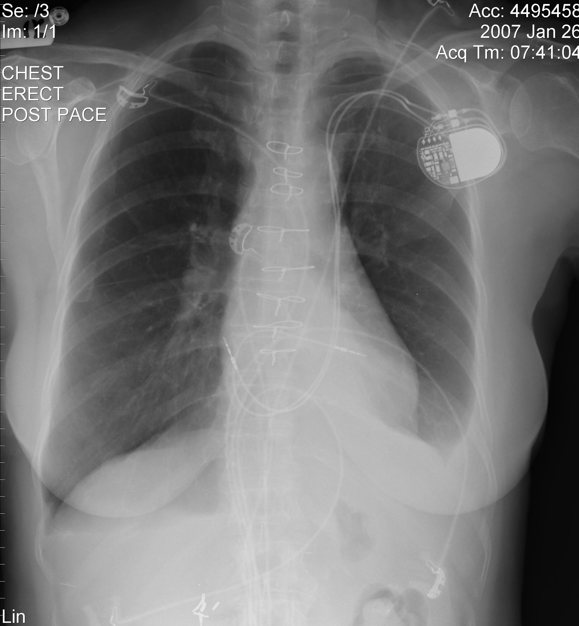
Chest x-ray
-
Chest x-ray of a patient with persistent SVC (Courtesy of RadsWiki and copylefted)
-
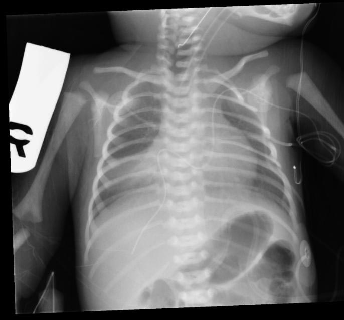
The atrial and ventricular pacemaker leads passing through a persistent left SVC.
-
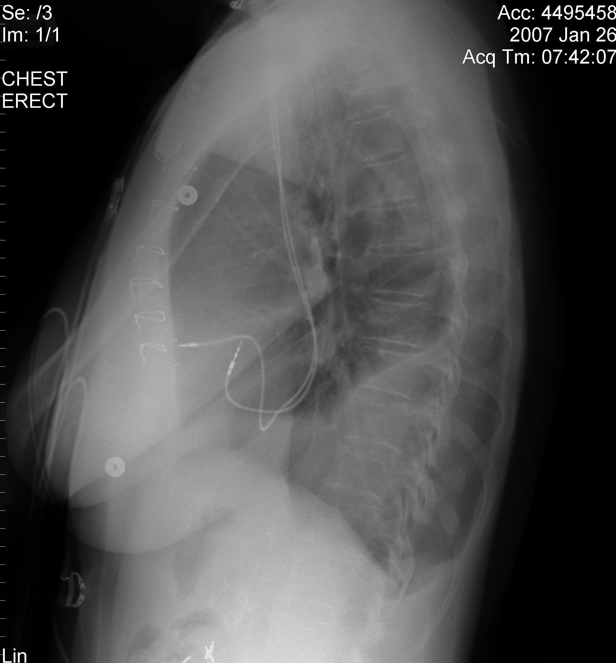
The atrial and ventricular pacemaker leads passing through a persistent left SVC.


CT
Images shown below are courtesy of RadsWiki and copylefted


-
![Image 1 - Duplication of inferior vena cava. This is a not uncommon variation. The left sided IVC (black arrow) most commonly ends in the normal left renal vein. Image 2 - Duplication of superior vena cava. This is a rare variation of the SVC. The left sided SVC (white arrow) is draining into the left atrium, thereby bypassing the pulmonary circulation. The right SVC is normal. [6] [7] (Image courtesy of Dr Abhijit Datir)](/images/d/d6/Vena_cava_anomalies.jpg)
Image 1 - Duplication of inferior vena cava. This is a not uncommon variation. The left sided IVC (black arrow) most commonly ends in the normal left renal vein. Image 2 - Duplication of superior vena cava. This is a rare variation of the SVC. The left sided SVC (white arrow) is draining into the left atrium, thereby bypassing the pulmonary circulation. The right SVC is normal. [6] [7] (Image courtesy of Dr Abhijit Datir)
![Image 1 - Duplication of inferior vena cava. This is a not uncommon variation. The left sided IVC (black arrow) most commonly ends in the normal left renal vein. Image 2 - Duplication of superior vena cava. This is a rare variation of the SVC. The left sided SVC (white arrow) is draining into the left atrium, thereby bypassing the pulmonary circulation. The right SVC is normal. [6] [7] (Image courtesy of Dr Abhijit Datir)](/index.php/File:Vena_cava_anomalies.jpg)
Cardiac Catheterization
Images shown below are courtesy of RadsWiki and copylefted


<googlevideo>-1621953796774001204&hl=en</googlevideo>
<googlevideo>-6286747543454457354&hl=en</googlevideo>
Pathologic Findings
Image shown below is courtesy of Professor Peter Anderson DVM PhD and Published with permission. © PEIR, University of Alabama at Birmingham, Department of Pathology
-
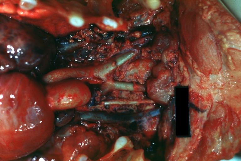
Persistent Left Superior Vena Cava: Gross natural color heart in situ dissected to show persistent left superior vena cava
References
- ↑ Pahwa R, Kumar A. Persistent left superior vena cava: an intensivist's experience and review of the literature. South Med J. 2003 May;96(5):528-9. PMID 12911199.
- ↑ Gonzalez-Juanatey C, Testa A, Vidan J, Izquierdo R, Garcia-Castelo A, Daniel C, Armesto V. Persistent left superior vena cava draining into the coronary sinus: report of 10 cases and literature review. Clin Cardiol. 2004 Sep;27(9):515-8. PMID 15471164.
- ↑ Freedom RM, Culham JAG, Moes CAF. Angiography of Congenital Heart Disease. Macmillan Publishing Co., New. York, 1984. URL: http://www.learningradiology.com/archives03/COW%20061-Persistent%20Left%20SVC/leftsvccorrect.htm. Accessed on March 24, 2006.
- ↑ Berg C, Knuppel M, Geipel A, Kohl T, Krapp M, Knopfle G, Germer U, Hansmann M, Gembruch U. Prenatal diagnosis of persistent left superior vena cava and its associated congenital anomalies. Ultrasound Obstet Gynecol. 2006 Mar;27(3):274-80. PMID 16456841.
- ↑ Bjerregaard P, Laursen HB. Persistent left superior vena cava. Incidence, associated congenital heart defects and frontal plane P-wave axis in a paediatric population with congenital heart disease. Acta Paediatr Scand. 1980 Jan;69(1):105-8. PMID 7368902.
- ↑ J. Edward Bass, Michael D. Redwine et al. Spectrum of Congenital Anomalies of the Inferior Vena Cava: Cross-sectional Imaging Findings. RadioGraphics 2000; 20: 639.
- ↑ GM Kellman, MB Alpern, MA Sandler, and BM Craig. Computed tomography of vena caval anomalies with embryologic correlation. RadioGraphics 1988; 8: 533.
External links
- Radiograph of a persistent left SVC - learningradiology.com.