Vertebral artery
Template:WikiDoc Cardiology News Editor-In-Chief: C. Michael Gibson, M.S., M.D. [1]
Overview
The vertebral arteries are branches of the subclavian arteries.
Cervical
They arise, one on each side of the body, then enter deep to the transverse process of the level of the 6th cervical vertebrae (C6).
It then proceeds superiorly, under the transverse process of each cervical vertebra until C1.
This path is largely parallel to, but distinct from, the route of the carotid artery ascending through the neck.
At the C1 level the vertebral arteries travel across the posterior arch of the atlas before entering the foramen magnum.
Cranial
Inside the skull, the two vertebral arteries join up to form the basilar artery at the base of the medulla oblongata.
The basilar artery is the main blood supply to the brainstem and connects to the Circle of Willis to potentially supply the rest of the brain if there is compromise to one of the carotids.
At each cervical level, the vertebral artery sends branches to the surrounding musculature via anterior spinal arteries.
Division into four parts
The vertebral artery may be divided into four parts:
First part
The first part runs upward and backward between the Longus colli and the Scalenus anterior.
In front of it are the internal jugular and vertebral veins, and it is crossed by the inferior thyroid artery; the left vertebral is crossed by the thoracic duct also.
Behind it are the transverse process of the seventh cervical vertebra, the sympathetic trunk and its inferior cervical ganglion.
Second part
The second part runs upward through the foramina in the transverse processes of the upper six cervical vertebræ, and is surrounded by branches from the inferior cervical sympathetic ganglion and by a plexus of veins which unite to form the vertebral vein at the lower part of the neck.
It is situated in front of the trunks of the cervical nerves, and pursues an almost vertical course as far as the transverse process of the atlas, above which it runs upward and lateralward to the foramen in the transverse process of the atlas.
Third part
The third part issues from the latter foramen on the medial side of the Rectus capitis lateralis, and curves backward behind the superior articular process of the atlas, the anterior ramus of the first cervical nerve being on its medial side; it then lies in the groove on the upper surface of the posterior arch of the atlas, and enters the vertebral canal by passing beneath the posterior atlantoöccipital membrane.
This part of the artery is covered by the Semispinalis capitis and is contained in the suboccipital triangle—a triangular space bounded by the Rectus capitis posterior major, the Obliquus superior, and the Obliquus inferior.
The first cervical or suboccipital nerve lies between the artery and the posterior arch of the atlas.
Fourth part
The fourth part pierces the dura mater and inclines medialward to the front of the medulla oblongata; it is placed between the hypoglossal nerve and the anterior root of the first cervical nerve and beneath the first digitation of the ligamentum denticulatum.
At the lower border of the pons it unites with the vessel of the opposite side to form the basilar artery.
Asymmetry
The left vertebral artery is usually larger and carries more blood.[1]
See also
References
- ↑ "Doppler sonography evaluation of flow velocity and volume of the extracranial internal carotid and vertebral arteries in healthy adults". J Clin Ultrasound. 35 (1): 27–33. 2007. PMID 17149761.
Additional images
-
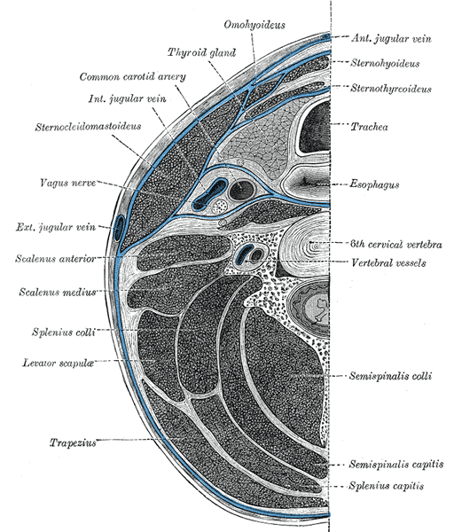 Section of the neck at about the level of the sixth cervical vertebra.
 -
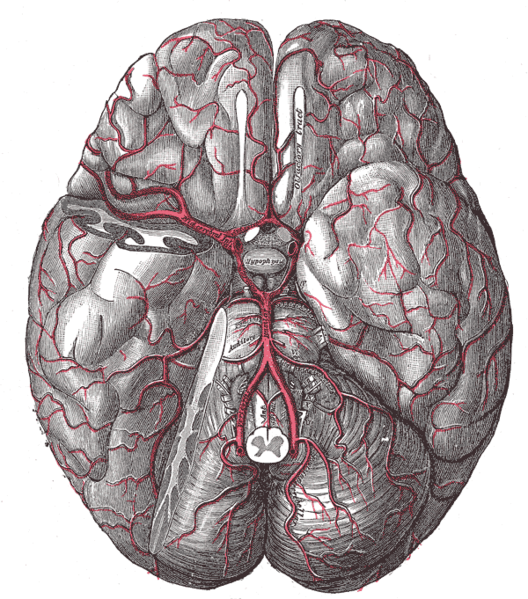 The arteries of the base of the brain.
 -
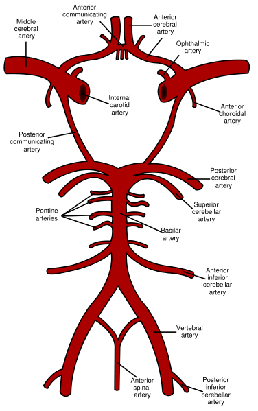 Diagram of the arterial circulation at the base of the brain.
 -
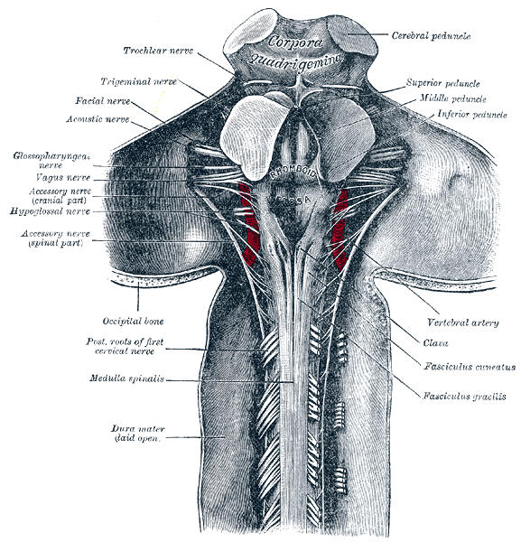 Upper part of medulla spinalis and hind- and mid-brains; posterior aspect, exposed in situ.
 -
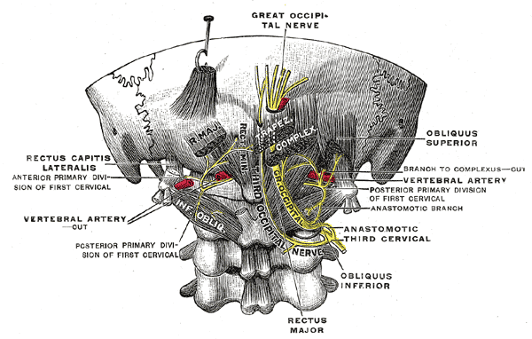 Posterior primary divisions of the upper three cervical nerves.


External links
- Template:SUNYAnatomyLabs
- Template:LoyolaMedEd
- Template:UMichAtlas
- Template:EMedicineDictionary
- Template:NormanAnatomy (Template:NormanAnatomyFig)
- Template:RocheLexicon
- Template:RocheLexicon
Template:Arteries of head and neck