Soleus muscle
Overview
In humans and some other mammals, the soleus is a powerful muscle in the back part of the lower leg (the calf). It runs from just below the knee to the heel, and is involved in standing and walking. It is closely connected to the gastrocnemius muscle and some anatomists consider them to be a single muscle, the triceps surae. Its name is derived from the solefish whose shape it resembles.
The soleus is located in the superficial posterior compartment of the leg. Not all mammals have a soleus muscle; one notable species that lacks the soleus is the dog.
Origin and insertion
It originates from the posterior (back) surfaces of the head of the fibula and its upper third, as well as the middle third of the internal border of the tibia.
Its other end forms a common tendon with the gastrocnemius muscle; this tendon is known as the calcaneal tendon or Achilles tendon and inserts onto the posterior surface of the calcaneus, or heel bone.
Relations
Superficial to the soleus (closer to the skin) is the gastrocnemius muscle.
The plantaris muscle and a portion of its tendon run between the two muscles. Deep to it (farther from the skin) is the transverse intermuscular septum, which separates the superficial posterior compartment of the leg from the deep posterior compartment.
On the other side of the fascia are the tibialis posterior muscle, the flexor digitorum longus muscle, and the flexor hallucis longus muscle, along with the posterior tibial artery and posterior tibial vein and the tibial nerve.
Since the anterior compartment of the leg is lateral to the tibia, the bulge of muscle medial to the tibia on the anterior side is actually the posterior compartment. The soleus is superficial midshaft of the tibia.
Function
The action of the calf muscles, including the soleus, is to plantar flex the foot (that is, they increase the angle between the foot and the leg).
They are powerful muscles and are vital in walking, running, and dancing.
The soleus specifically plays an important role in standing; if not for its constant pull, the body would fall forward.
Also, in upright posture, it is responsible for pumping venous blood back into the heart from the periphery, and is often called the peripheral heart or the sural (tricipital) pump [1].
Additional images
-
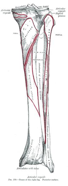 Bones of the right leg. Posterior surface.
 -
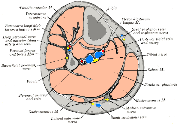 Cross-section through middle of leg.
 -
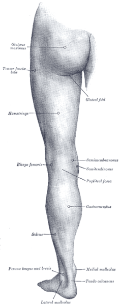 Back of left lower extremity.

References
- Gray, Henry. Pick, T. Pickering, & Howden, Robert (Eds.) (1995). Gray's Anatomy (15th ed.). New York: Barnes & Noble Books.
External links
Template:Muscles of lower limb
de:Musculus soleus he:שריר הסוליה nl:Musculus soleus sv:Soleus Template:WikiDoc Sources