Gastrocnemius muscle
Template:Infobox Muscle Editor-In-Chief: C. Michael Gibson, M.S., M.D. [1]
Overview
The gastrocnemius muscle (pronounced Template:IPA) is a powerful superficial muscle that is in the back part of the lower leg (the calf). It runs from its 2 heads just above the knee to the heel, and is involved in standing and walking. Along with the soleus muscle it forms the calf muscle.
The gastrocnemius is located with the soleus in the superficial posterior compartment of the leg. It originates from the posterior (back) surfaces of the distal head of the femur. Its other end forms a common tendon with the soleus muscle; this tendon is known as the calcaneal tendon or Achilles tendon and inserts onto the posterior surface of the calcaneus, or heel bone.
Deep to the gastrocnemius (farther from the skin) is the soleus muscle, some anatomists consider both to be a single muscle, the triceps surae. The plantaris muscle and a portion of its tendon run between the two muscles, which is involved in "unlocking" the knee from the standing position. On the other side of the fascia are the tibialis posterior muscle, the flexor digitorum longus muscle, and the flexor hallucis longus muscle, along with the posterior tibial artery and posterior tibial vein and the tibial nerve. Since the anterior compartment of the leg is lateral to the tibia, the bulge of muscle medial to the tibia on the anterior side is actually the posterior compartment. The soleus is superficial midshaft of the tibia.
Additional Information
The gastrocnemius muscle is very prone to spasms; the painful, involuntarily, contraction of the muscle for up to several minutes. It is commonly known as a "charley horse".
This muscle is prone to injury called torn calf muscle which is disabling. See Calf muscle The Gastrocnemius muscle may also become inflamed due to overuse. Anti-inflammatory drugs and physical therapy may be necessary.
Additional images
-
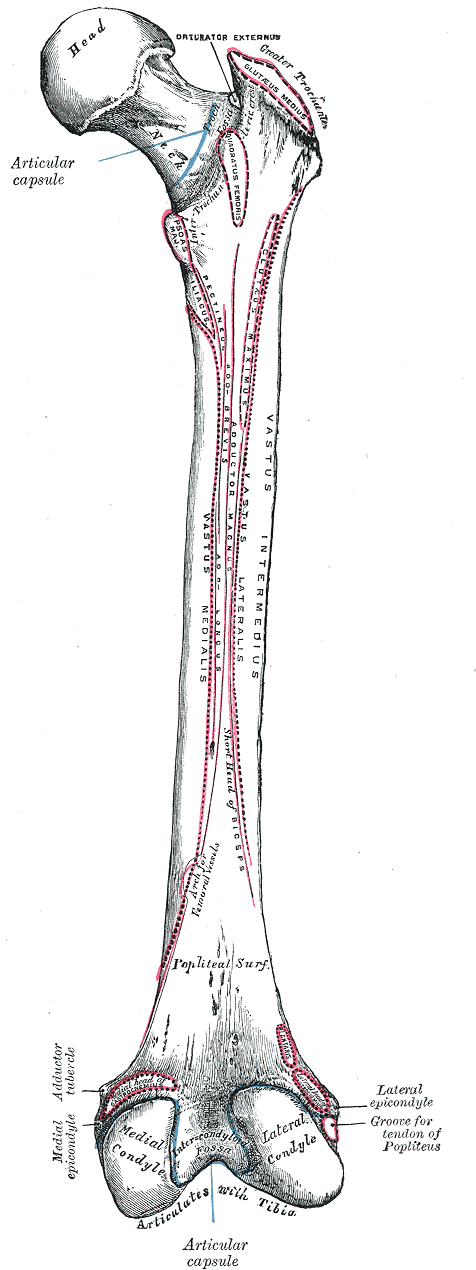 Right femur. Posterior surface.
 -
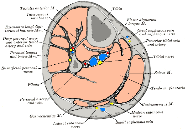 Cross-section through middle of leg.
 -
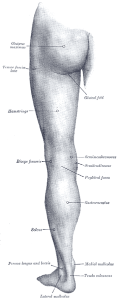 Back of left lower extremity.

External links
Template:Muscles of lower limb
de:Musculus gastrocnemius
eo:Suro
it:Gastrocnemio
he:שריר הסובך
nl:Musculus gastrocnemius
fi:Pohjelihas
sv:Gastrocnemius