Calcaneus
|
WikiDoc Resources for Calcaneus |
|
Articles |
|---|
|
Most recent articles on Calcaneus |
|
Media |
|
Evidence Based Medicine |
|
Clinical Trials |
|
Ongoing Trials on Calcaneus at Clinical Trials.gov Clinical Trials on Calcaneus at Google
|
|
Guidelines / Policies / Govt |
|
US National Guidelines Clearinghouse on Calcaneus
|
|
Books |
|
News |
|
Commentary |
|
Definitions |
|
Patient Resources / Community |
|
Patient resources on Calcaneus Discussion groups on Calcaneus Directions to Hospitals Treating Calcaneus Risk calculators and risk factors for Calcaneus
|
|
Healthcare Provider Resources |
|
Causes & Risk Factors for Calcaneus |
|
Continuing Medical Education (CME) |
|
International |
|
|
|
Business |
|
Experimental / Informatics |
Overview
The calcaneus is the largest bone of the human foot. The skeleton of the human foot is made up of three groups of bones: the tarsus, the metatarsus and the phalanges. The tarsal bones consist of the calcaneus, talus, cuboid, navicular, and the first, second, and third cuneiforms. The calcaneus forms part of the tarsi and constitutes the heel of the human foot or the point of an animal's hock . It is also known as the heel bone.
Human
It articulates with two other tarsal bones, the talus above and the cuboid toward the midfoot. In addition to receiving the weight of the body with each step, the calcaneus is the anchor for the plantar fascia, which supports the arch of the foot.
Calcaneal tuberosity
The posterior-most portion of the calcaneus is the calcaneal tuberosity, a large, non-articulating process that is the insertion point for the calcaneal tendon (or Achilles tendon). In addition, this process is the origin for some of the muscles and tendons of the foot.
Horse
The calcaneus has two articulations, being part of the Proximal intertarsal joint and the Talocalcaneal joint. As in humans it is the insertion of the gastrocnemius and superficial digital flexor tendons. The point of the calcaneus is covered by the calcanean bursa.
See also
- Bone terminology
- Calcaneal fracture, also known as Lover's fracture and Don Juan fracture
- Terms for anatomical location
Additional images
-
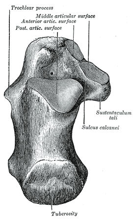
Left calcaneus, superior surface
-
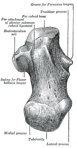
Left calcaneus, inferior surface
-
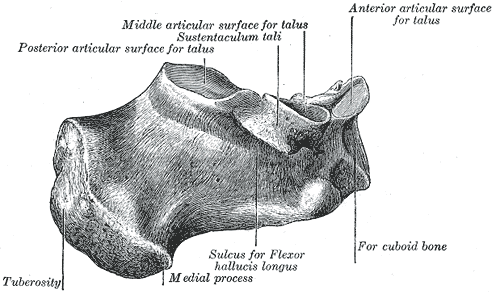
Left calcaneus, medial surface
-

Bones of the right foot. Dorsal surface.
-
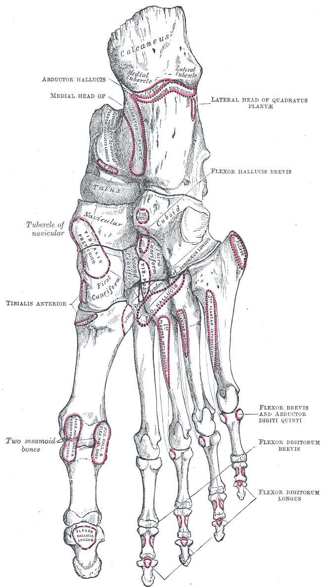
Bones of the right foot. Plantar surface.
-
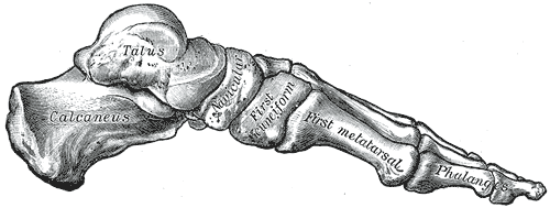
Skeleton of foot. Medial aspect.
-
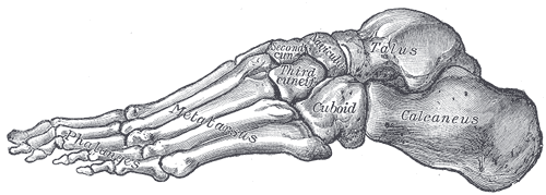
Skeleton of foot. Lateral aspect.
-
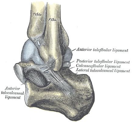
Capsule of left talocrura articulation (distended). Lateral aspect.
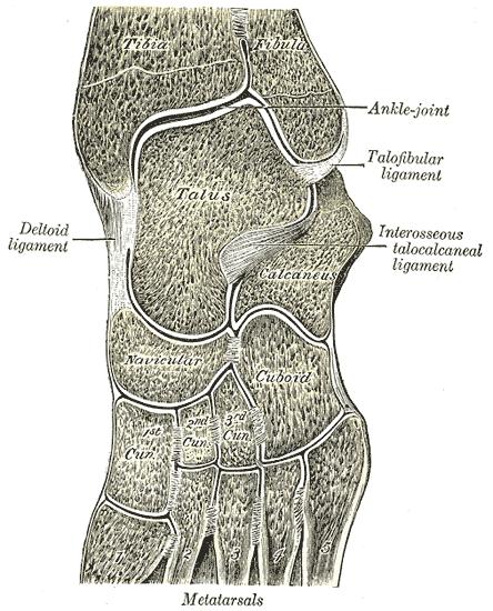
-
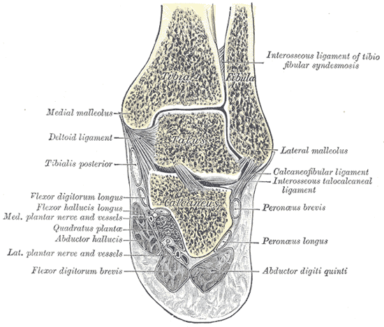
Coronal section through right talocrural and talocalcaneal joints.
-
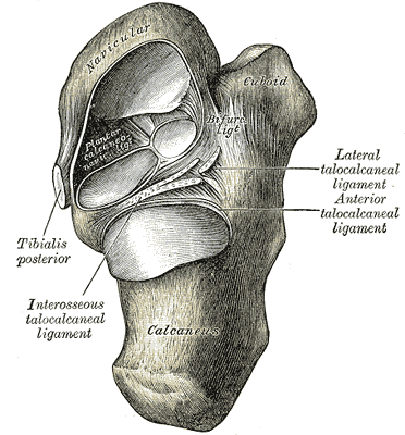
Talocalcaneal and talocalcaneonavicular articulations exposed from above by removing the talus.
-
Oblique section of left intertarsal and tarsometatarsal articulations, showing the synovial cavities.
-
Calcaneus fracture X-ray


External links
Template:Bones of lower extremity