Tibia
Template:Infobox Bone Editor-In-Chief: C. Michael Gibson, M.S., M.D. [1]
The tibia is the larger of the two bones in the leg below the knee in vertebrates.
In humans
The tibia or shin bone, in human anatomy, is found medial (towards the middle) and anterior (towards the front) to the other such bone, the fibula. It is the second-longest bone in the human body, the largest being the femur. The tibia articulates with the femur and patella superiorly, the fibula laterally and with the ankle inferiorly.
Gender differences
In the male, its direction is vertical, and parallel with the bone of the opposite side, but in the female it has a slightly oblique direction downward and lateralward, to compensate for the greater obliquity of the femur.
Structure
It is prismoid in form, expanded above, where it enters into the knee-joint, contracted in the lower third, and again enlarged but to a lesser extent below.
The tibia is connected to the fibula by an interosseous membrane, forming a type of joint called a syndesmoses.
Blood supply
The tibia derives its arterial blood supply from two sources:[1]
- the nutrient artery (main source)
- periosteal vessels derived from the anterior tibial artery
Additional images
-
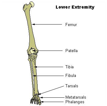
Lower extremity
-
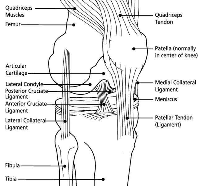
Knee diagram
-
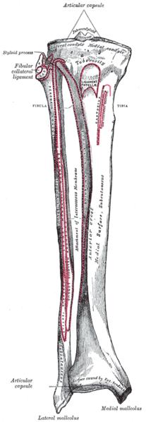
Bones of the right leg. Anterior surface.
-
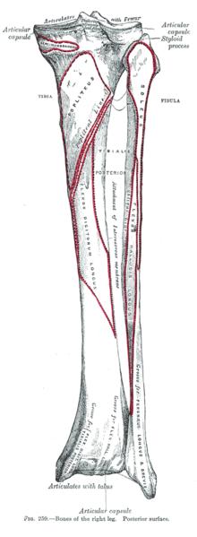
Bones of the right leg. Posterior surface.
-
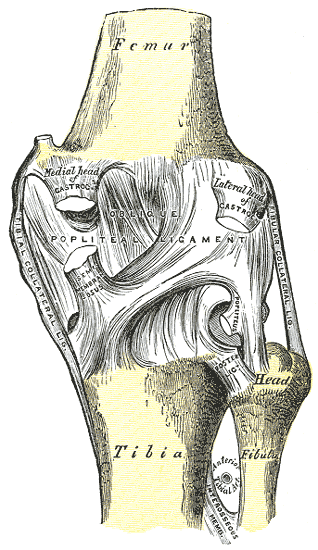
Right knee-joint. Posterior view.
-
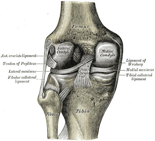
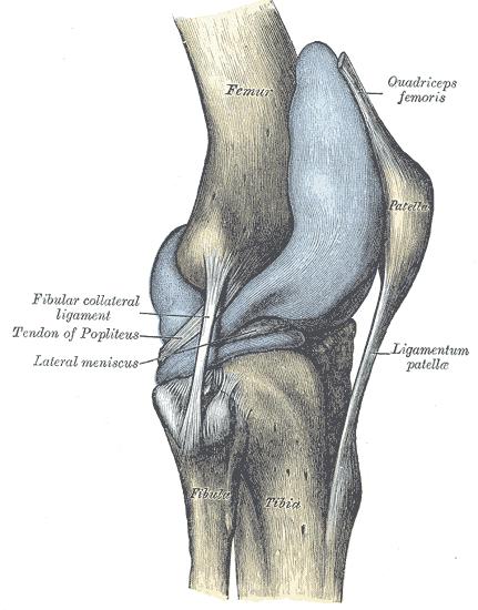
Right knee-joint, from the front, showing interior ligaments.
-
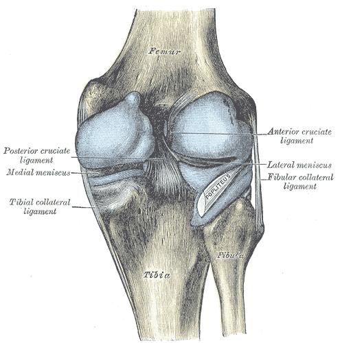
Left knee-joint from behind, showing interior ligaments.
-
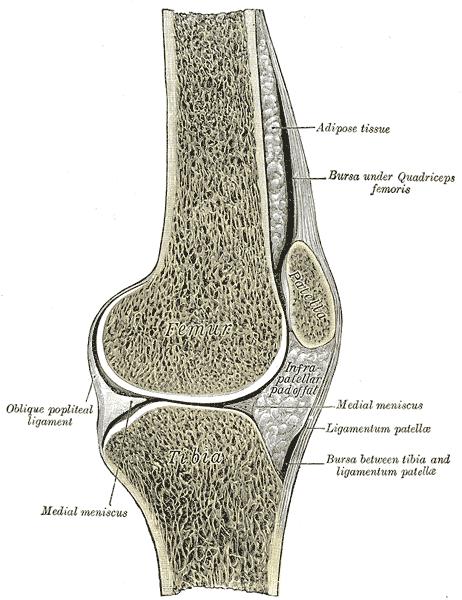
Sagittal section of right knee-joint.
-
Capsule of right knee-joint (distended). Lateral aspect.
-
Capsule of right knee-joint (distended). Posterior aspect.
-
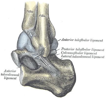
Capsule of left talocrura articulation (distended). Lateral aspect.
-
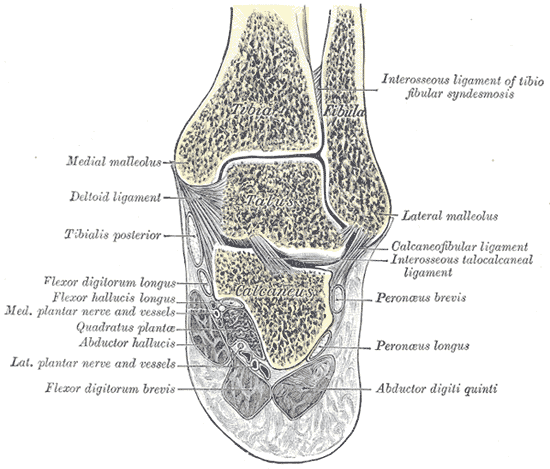
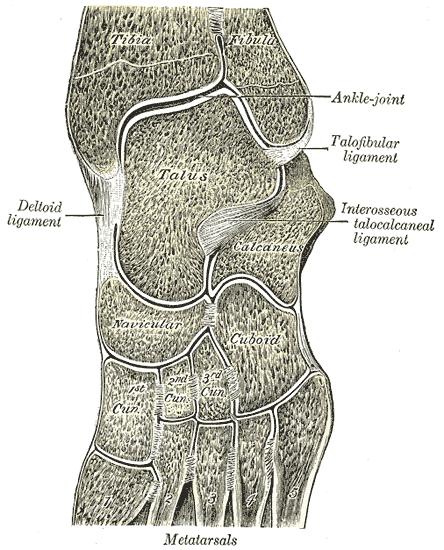
Coronal section through right talocrural and talocalcaneal joints.
-
Oblique section of left intertarsal and tarsometatarsal articulations, showing the synovial cavities.
-
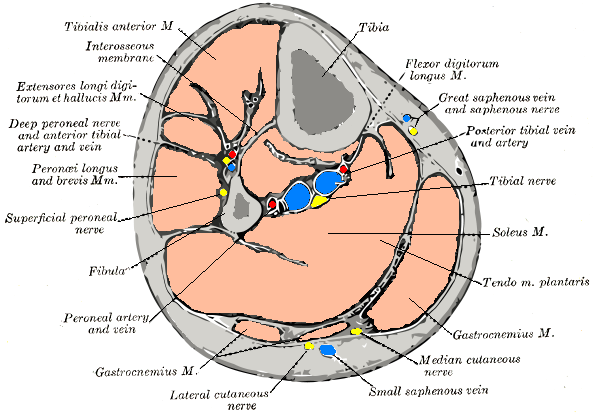
Cross-section through middle of leg.


See also
- Articulations between the tibia and fibula
- Bone terminology
- Terms for anatomical location
- Ossification of tibia
- Upper extremity of tibia
- Body of tibia
- Lower extremity of tibia
- Shin Splints
External links
References
Template:Bones of lower extremity
ca:Tíbia cs:Holenní kost de:Tibia (Wirbeltiere) eo:Tibio it:Tibia (osso) he:שוקה la:Tibia (os) lt:Blauzdikaulis nl:Scheenbeen no:Tibia nn:Tibia sl:Golenica fi:Sääriluu sv:Skenben tl:Lulod uk:Великогомілкова кістка Template:Jb1 Template:WH Template:WikiDoc Sources