Facial artery
Editor-In-Chief: C. Michael Gibson, M.S., M.D. [1]
The facial artery (external maxillary artery in older texts) is a branch of the external carotid artery that supplies structures of the face.
Course
The facial artery arises in the carotid triangle from the external carotid artery a little above the lingual artery and, sheltered by the ramus of the mandible, passes obliquely up beneath the digastric and stylohyoid muscles, over which it arches to enter a groove on the posterior surface of the submandibular gland.
It then curves upward over the body of the mandible at the antero-inferior angle of the masseter; passes forward and upward across the cheek to the angle of the mouth, then ascends along the side of the nose, and ends at the medial commissure of the eye, under the name of the angular artery.
This vessel, both in the neck and on the face, is remarkably tortuous: in the former situation, to accommodate itself to the movements of the pharynx in deglutition; and in the latter, to the movements of the mandible, lips, and cheeks.
Relations
In the neck, its origin is superficial, being covered by the integument, platysma, and fascia; it then passes beneath the digastric and stylohyoid muscles and part of the submandibular gland, but superficial to the hypoglossal nerve.
It lies upon the middle pharyngeal constrictor and the superior pharyngeal constrictor, the latter of which separates it, at the summit of its arch, from the lower and back part of the tonsil.
On the face, where it passes over the body of the mandible, it is comparatively superficial, lying immediately beneath the dilators of the mouth. In its course over the face, it is covered by the integument, the fat of the cheek, and, near the angle of the mouth, by the platysma, risorius, and zygomaticus major. It rests on the buccinator and levator anguli oris, and passes either over or under the infraorbital head of the levator labii superioris.
The anterior facial vein lies lateral/posterior to the artery, and takes a more direct course across the face, where it is separated from the artery by a considerable interval. In the neck it lies superficial to the artery.
The branches of the facial nerve cross the artery from behind forward.
Branches
The branches of the facial artery are:
- facial
- Inferior labial artery
- Superior labial artery
- Lateral nasal branch to nasalis muscle
- Angular artery - the terminal branch
Th lateral pterygoid muscle divides the facial artery into 3 parts.
Muscles
Muscles supplied by the facial artery include:
- buccinator
- levator anguli oris
- levator labii superioris
- levator labii superioris alaeque nasi
- levator veli palatini
- masseter
- mentalis
- mylohyoid
- nasalis
- palatoglossus
- palatopharyngeus
- platysma
- procerus
- risorius
- styloglossus
- transverse portion of the nasalis
Additional images
-
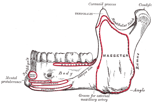 Mandible. Outer surface. Side view.
 -
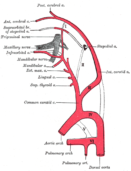 Diagram showing the origins of the main branches of the carotid arteries.
 -
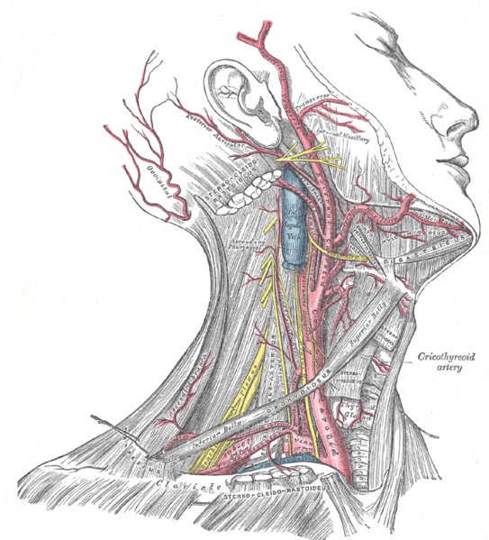 Superficial dissection of the right side of the neck, showing the carotid and subclavian arteries.
 -
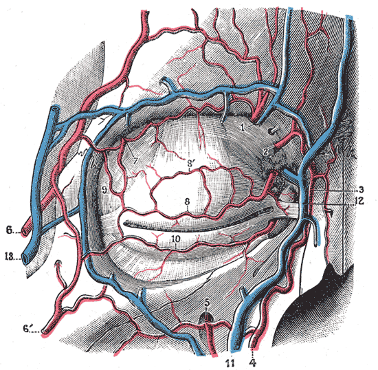 Bloodvessels of the eyelids, front view.
 -
 Side of neck, showing chief surface markings.


Template:WikiDoc Cardiology News
See also
External links
- Template:Medcyclopaedia
- Template:EMedicineDictionary
- Template:SUNYAnatomyLabs - "The Facial Artery and Vein"
- Template:SUNYAnatomyFigs - "Branches of the external carotid artery."
- Template:SUNYAnatomyLabs - "Common Carotid Artery and Branches of the External Carotid Artery"
- Template:MUNAnatomy