Ileum
Editor-In-Chief: C. Michael Gibson, M.S., M.D. [1]
Overview
|
WikiDoc Resources for Ileum |
|
Articles |
|---|
|
Media |
|
Evidence Based Medicine |
|
Clinical Trials |
|
Ongoing Trials on Ileum at Clinical Trials.gov Clinical Trials on Ileum at Google
|
|
Guidelines / Policies / Govt |
|
US National Guidelines Clearinghouse on Ileum
|
|
Books |
|
News |
|
Commentary |
|
Definitions |
|
Patient Resources / Community |
|
Directions to Hospitals Treating Ileum Risk calculators and risk factors for Ileum
|
|
Healthcare Provider Resources |
|
Continuing Medical Education (CME) |
|
International |
|
|
|
Business |
|
Experimental / Informatics |
In anatomy of the digestive system, the ileum is the final section of the small intestine. It is about 2-4 m long in humans, follows the duodenum and jejunum, and is separated from the cecum by the ileocecal valve (ICV). The pH in the ileum is usually between 7 and 8 (neutral or slightly alkaline).
Function
Its function is mainly to absorb vitamin B12 and bile salts and whatever products of digestion that were not absorbed by the jejunum. The wall itself is made up of folds, each of which has many tiny finger-like projections known as villi, on its surface. In turn, the epithelial cells which line these villi possess even larger numbers of microvilli. Therefore the ileum has an extremely large surface area both for the adsorption (attachment) of enzyme molecules and for the absorption of products of digestion. The DNES (diffuse neuroendocrine system)cells that line the ileum contain the protease and carbohydrase enzymes (gastrin, secretin, cholecystokinin) responsible for the final stages of protein and carbohydrate digestion. These enzymes are present in the cytoplasm of the epithelial cells. The villi contain large numbers of capillaries which take the amino acids and glucose produced by digestion to the hepatic portal vein and the liver.
Lacteals are small lymph vessels, and are present in villi. They absorb fatty acid and glycerol, the products of fat digestion. Layers of circular and longitudinal smooth muscle enable the digested food to be pushed along the ileum by waves of muscle contractions called peristalsis.
Differences between jejunum and ileum
There is no line of demarcation between the jejunum and the ileum. There are, however, subtle differences between the two.
- The ileum has more fat inside the mesentery than the jejunum.
- The ileum is a paler color, and tends to be of a smaller caliber as well.
- While the length of the intestinal tract contains lymphoid tissue, only the ileum has abundant Peyer's patches.
These unencapsulated lymphoid nodules contain large amounts of lymphocytes and other cells of the immune system.
Embryology
In the fetus the ileum is connected to the navel by the vitelline duct. In roughly 3% of humans, this duct fails to close during the first seven weeks after birth, causing a condition called Meckel's diverticulum.
Veterinary anatomy
In veterinary anatomy, the ileum is distinguished from the jejunum by being that portion of the jejunoileum that is connected to the caecum by the ileocaecal fold.
Additional images
-
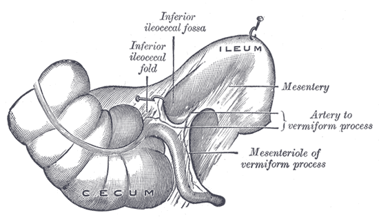 Inferior ileocecal fossa.
 -
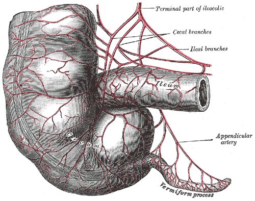 Arteries of cecum and vermiform process.
 -
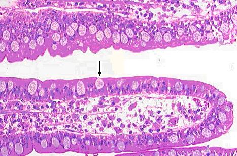 Goblet cell in ileum


References
External links
- Template:SUNYAnatomyLabs - "Abdominal Cavity: The Jejunum and the Ileum"
- Template:SUNYAnatomyImage
- Template:SUNYAnatomyImage
- Histology image: 12001oca – Histology Learning System at Boston University
- Ileal Villi at endoatlas.com
- Ileum Microscopic Cross Section at nhmccd.edu
- Ileum 20x at deltagen.com
- Peyer's Patches at thehealthnews.org
de:Ileum eo:Ileo fa:درازروده id:Usus penyerapan it:Ileo (intestino) lt:Klubinė žarna hu:Csípőbél nl:Kronkeldarm no:Ileum simple:Ileum sk:Bedrovník sl:Vito črevo sv:Krumtarm