Vitelline duct
|
WikiDoc Resources for Vitelline duct |
|
Articles |
|---|
|
Most recent articles on Vitelline duct Most cited articles on Vitelline duct |
|
Media |
|
Powerpoint slides on Vitelline duct |
|
Evidence Based Medicine |
|
Clinical Trials |
|
Ongoing Trials on Vitelline duct at Clinical Trials.gov Trial results on Vitelline duct Clinical Trials on Vitelline duct at Google
|
|
Guidelines / Policies / Govt |
|
US National Guidelines Clearinghouse on Vitelline duct NICE Guidance on Vitelline duct
|
|
Books |
|
News |
|
Commentary |
|
Definitions |
|
Patient Resources / Community |
|
Patient resources on Vitelline duct Discussion groups on Vitelline duct Patient Handouts on Vitelline duct Directions to Hospitals Treating Vitelline duct Risk calculators and risk factors for Vitelline duct
|
|
Healthcare Provider Resources |
|
Causes & Risk Factors for Vitelline duct |
|
Continuing Medical Education (CME) |
|
International |
|
|
|
Business |
|
Experimental / Informatics |
Overview
At the end of the fourth week the yolk-sac presents the appearance of a small pear-shaped vesicle (umbilical vesicle) opening into the digestive tube by a long narrow tube, the vitelline duct.
The vesicle can be seen in the after-birth as a small, somewhat oval-shaped body whose diameter varies from 1 mm. to 5 mm.; it is situated between the amnion and the chorion and may lie on or at a varying distance from the placenta.
As a rule the duct undergoes complete obliteration during the seventh week, but in about two per cent of cases its proximal part persists as a diverticulum from the small intestine, Meckel's diverticulum, which is situated about two feet above the ileocolic junction, and may be attached by a fibrous cord to the abdominal wall at the umbilicus.
Sometimes a narrowing of the lumen of the ileum is seen opposite the site of attachment of the duct.
Additional images
-
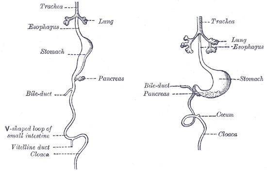 Front view of two successive stages in the development of the digestive tube.
