Femur
|
WikiDoc Resources for Femur |
|
Articles |
|---|
|
Media |
|
Evidence Based Medicine |
|
Clinical Trials |
|
Ongoing Trials on Femur at Clinical Trials.gov Clinical Trials on Femur at Google
|
|
Guidelines / Policies / Govt |
|
US National Guidelines Clearinghouse on Femur
|
|
Books |
|
News |
|
Commentary |
|
Definitions |
|
Patient Resources / Community |
|
Directions to Hospitals Treating Femur Risk calculators and risk factors for Femur
|
|
Healthcare Provider Resources |
|
Continuing Medical Education (CME) |
|
International |
|
|
|
Business |
|
Experimental / Informatics |
Editor-In-Chief: C. Michael Gibson, M.S., M.D. [1]
In humans, it is the longest, most voluminous, and strongest bone. The average human femur is 48 centimeters (19 in) in length and 2.34 cm (0.92 in) in diameter and can support up to 30 times the weight of an adult.[1] It forms part of the hip (at the acetabulum) and part of the knee.
The word femur is Latin for thigh. Theoretically in strict usage, femur bone is more proper than femur, as in classical Latin femur means "thigh", and os femoris means "the bone within it".
In medical Latin its genitive is always femoris, but in classical Latin its genitive is often feminis, and should not be confused with case forms of femina, which means "woman".
Fractures
Femur bone fractures, on occasion, are liable to cause permanent disability because the thigh muscles pull the fragments so they overlap, and the fragments re-unite incorrectly. To avoid this, femur fracture patients should be put into traction to keep the fragments pulled into proper alignment.
With modern medical procedures, such as the insertion of rods and screws by way of surgery (known as Antegrade [through the hip] or Retrograde [through the knee] femoral rodding), those suffering from femur fractures can now generally expect to make a full recovery, though one that generally takes 3 to 6 months due to the bone's size. Patients should not put weight on the leg without permission from an orthopedic surgeon since this can delay the healing process.
The thigh is generally not put in a cast since the surgical hardware does the job of straightening the bone and holding the fracture together while it heals. Permanent complications with this procedure include the risk of intra-articular sepsis, arthritis and knee stiffness. After the bone is healed, there is no further need for the hardware but, while it is left in some patients permanently, those who lead an active lifesytle may experience discomfort where the hardware projects into the leg muscle and, in such cases, the hardware can be removed, most commonly by means of out-patient surgery.
Hip fracture
If bone is weakened, the proximal end of the femur bone near the hip joint is prone to fragility fracture. Most at risk are European descent, post-menopausal women, and osteoporosis severely increases this risk. Out of all the bones in the skeleton, the femur takes the longest to heal. This bone is the longest and strongest bone in the human body. When the average human being jumps this bone withstands a force of half a ton just a testament to its strength.
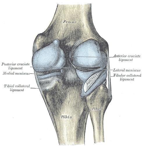
Intercondylar Fossa
The intercondylar fossa is present between the condyles at the distal end of the femur. In addition to the intercondylar eminence on the tibial plateau, there is both an anterior and posterior intercondylar fossa (area), the sites of anterior cruciate and posterior cruciate ligament attachment, respectively.
In other animals
Parallel structures by the same name exist in other complex animals, such as the bone inside a ham or a leg of lamb. The name femur is also given to the most proximal full-length jointed segment of an arthropod's leg.
References
- ↑ "The Longest Human Bone". thelongestlistofthelongeststuffatthelongestdomainnameatlonglast.com. Retrieved 2007-12-14.
External links
- Image with major components labeled at v
- Femoral fractures at aofoundation.org
- Template:ViennaCrossSection
Additional images
-
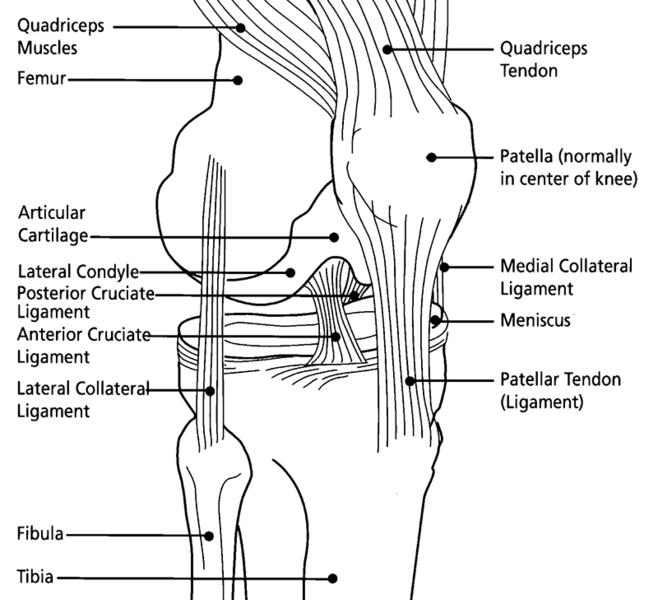
Knee diagram
-
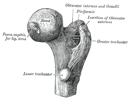
Upper extremity of right femur viewed from behind and above.
-
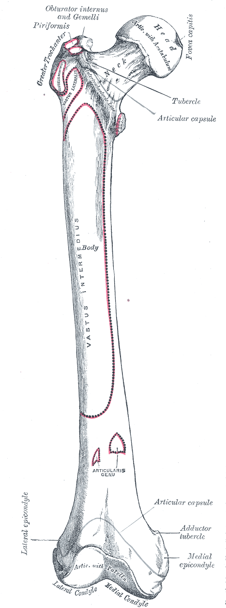
Right femur. Anterior surface.
-
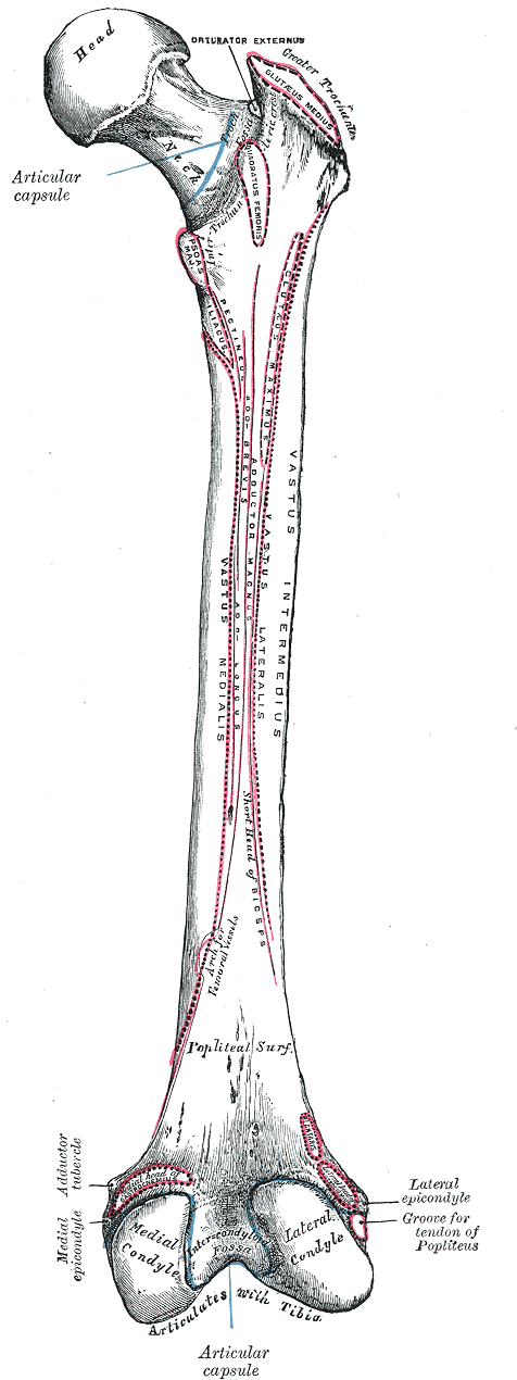
Right femur. Posterior surface.
-
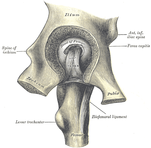
Left hip-joint, opened by removing the floor of the acetabulum from within the pelvis.
-
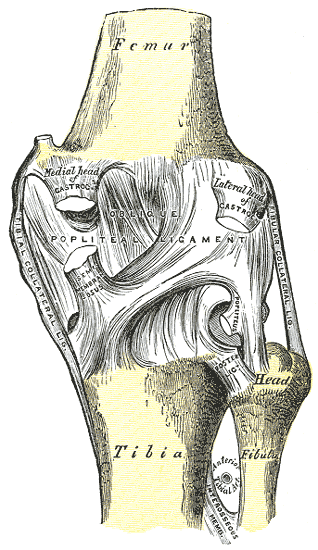
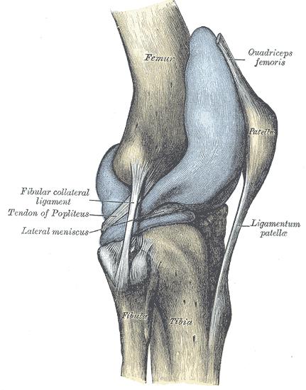
Right knee-joint. Posterior view.
-
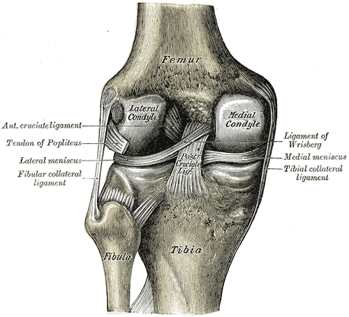
Left knee-joint from behind, showing interior ligaments.
-
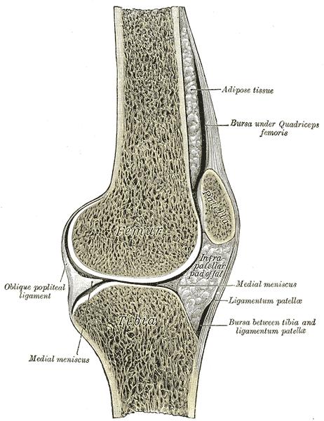
Sagittal section of right knee-joint.
-
Capsule of right knee-joint (distended). Lateral aspect.
-
Capsule of right knee-joint (distended). Posterior aspect.
-
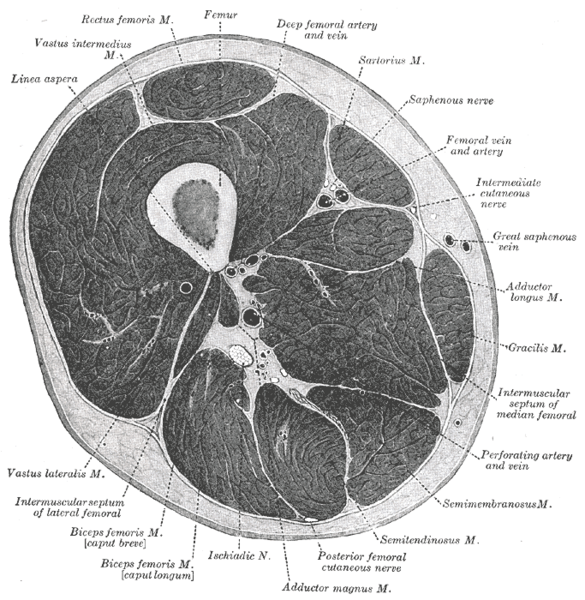
Cross-section through the middle of the thigh.
-
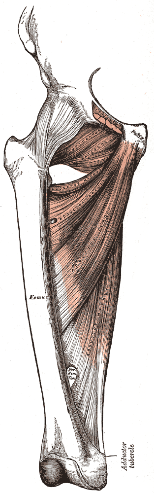
Deep muscles of the medial femoral region.


Template:Bones of lower extremity
cs:Stehenní kost da:Lårbensknogle de:Oberschenkelknochen eo:Femuralo id:Tulang paha it:Femore he:עצם הירך la:Femur lt:Šlaunikaulis nl:Dijbeen no:Femur fi:Reisiluu sv:Lårben uk:Стегнова кістка wa:Oxhea del coxhe