Small intestine cancer pathophysiology
|
Small intestine cancer Microchapters |
|
Diagnosis |
|---|
|
Treatment |
|
Case Studies |
|
Small intestine cancer pathophysiology On the Web |
|
American Roentgen Ray Society Images of Small intestine cancer pathophysiology |
|
Risk calculators and risk factors for Small intestine cancer pathophysiology |
Editor-In-Chief: C. Michael Gibson, M.S., M.D. [1]
Overview
Duodenal cancer


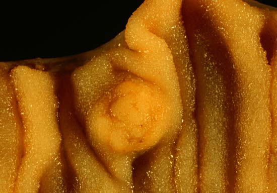
Duodenal cancer is a cancer in the beginning section of the small intestine. It is relatively rare compared to gastric cancer and colorectal cancer. Its histology is usually adenocarcinoma. Familial adenomatous polyposis (FAP) is a risk factor for developing this cancer.
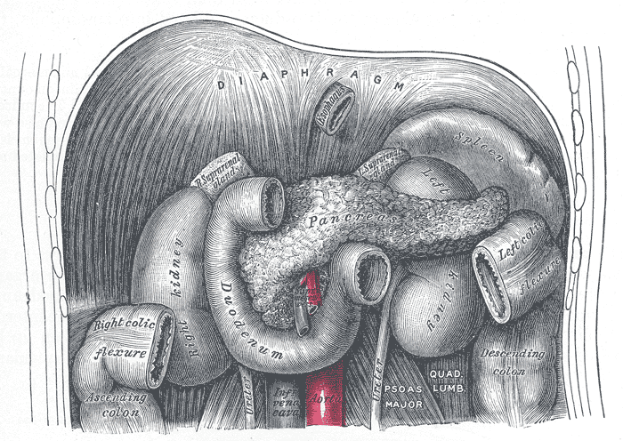
The duodenum is the first part of the small intestine. It is located between the stomach and the jejunum. After foods combine with stomach acid, they descend into the duodenum where they mix with bile from the gall bladder and digestive juices from the pancreas.
Duodenal cancer is difficult to remove surgically because of the area that it resides in, there are many blood vessels supplying the lower body. Chemotherapy is sometimes used to try and shrink the canceous mass. Other times intestinal bypass surgery is tried to reroute the stomache to intestine connection around the blockage. A 'Whipple' is a possible surgery that is tried sometimes with this cancer.
The cancerous mass tends to block food from getting to the small intestine. If food can not get to the intestines, it will cause pain, acid reflux, and weight loss because the food can not get to where it is supposed to be processed and absorbed by the body.
Some patients are fitted with tubes to either add nutrients (feeding tubes) or drainage tubes to remove excess processed food that can not pass the blockage.
Patients with duodenal cancer may experience abdominal pain, weight loss, nausea, vomiting, and chronic GI bleeding.