Meningioma pathophysiology: Difference between revisions
No edit summary |
|||
| Line 4: | Line 4: | ||
==Overview== | ==Overview== | ||
Meningioma arises from the [[arachnoid]] "cap" cells, which are normally involved in the protection of the [[central nervous system]] by forming a thick envelope of meninges around the [[brain]] and [[spinal cord]].<ref name="wiki">Meningioma. Wikipedia(2015) https://en.wikipedia.org/wiki/Meningioma#cite_note-pmid7731706-9 Accessed on September, 25th 2015</ref><ref name="C">Meningioma. Canadian Cancer Society http://www.cancer.ca/en/cancer-information/cancer-type/brain-spinal/brain-and-spinal-tumours/meningioma/?region=mb September, 25th 2015</ref> The majority of meningiomas are [[benign]].<ref name="wiki">Meningioma. Wikipedia(2015) https://en.wikipedia.org/wiki/Meningioma#cite_note-pmid7731706-9 Accessed on September, 25th 2015</ref><ref name="C">Meningioma. Canadian Cancer Society http://www.cancer.ca/en/cancer-information/cancer-type/brain-spinal/brain-and-spinal-tumours/meningioma/?region=mb September, 25th 2015</ref> Development of meningioma is the result of multiple genetic mutations.<ref name="wiki">Meningioma. Wikipedia(2015) https://en.wikipedia.org/wiki/Meningioma#cite_note-pmid7731706-9 Accessed on September, 25th 2015</ref> On gross pathology, a gray, well-circumscribed, dome-shaped mass is a characteristic finding of meningioma.<ref name="wiki">Meningioma. Wikipedia(2015) https://en.wikipedia.org/wiki/Meningioma#cite_note-pmid7731706-9 Accessed on September, 25th 2015</ref> On microscopic [[histopathological]] analysis, whorled appearance, [[calcification]], and [[psammoma]] bodies are characteristic findings of of meningioma.<ref name="liber">Meningioma. Liberpathology(2015) http://librepathology.org/wiki/index.php/Meningioma#Quick_overview accessed on September, 25th 2015</ref> | |||
==Pathogenesis== | ==Pathogenesis== | ||
Meningioma arises from [[arachnoid]] "cap" cells | Meningioma arises from the [[arachnoid]] "cap" cells, which are normally involved in the protection of the [[central nervous system]] by forming a thick envelope of meninges around the [[brain]] and [[spinal cord]]. The majority of meningiomas are [[benign]]; however, a minority of cases may develop a [[malignant]] nature. The exact pathophysiology of meningioma depends on the histological subtype of the tumor.<ref name="wiki">Meningioma. Wikipedia(2015) https://en.wikipedia.org/wiki/Meningioma#cite_note-pmid7731706-9 Accessed on September, 25th 2015</ref> Meningiomas are frequently found attached to the [[dura]] surrounding the brain tissue in several locations that include:<ref name="wiki">Meningioma. Wikipedia(2015) https://en.wikipedia.org/wiki/Meningioma#cite_note-pmid7731706-9 Accessed on September, 25th 2015</ref> | ||
* [[Frontal lobe]] | * [[Frontal lobe]] | ||
* [[Parietal lobe]] | * [[Parietal lobe]] | ||
| Line 21: | Line 21: | ||
==Genetics== | ==Genetics== | ||
*Development of meningioma is the result of multiple genetic mutations.<ref name="wiki">Meningioma. Wikipedia(2015) https://en.wikipedia.org/wiki/Meningioma#cite_note-pmid7731706-9 Accessed on September, 25th 2015</ref> | *Development of meningioma is the result of multiple genetic mutations.<ref name="wiki">Meningioma. Wikipedia(2015) https://en.wikipedia.org/wiki/Meningioma#cite_note-pmid7731706-9 Accessed on September, 25th 2015</ref> | ||
*The | *The majority of meningioma cases involve a mutation in the neurofibromatosis 2 gene located on chromosome 22.<ref name="wiki">Meningioma. Wikipedia(2015) https://en.wikipedia.org/wiki/Meningioma#cite_note-pmid7731706-9 Accessed on September, 25th 2015</ref> | ||
*Other genes involved in the pathogenesis of meningioma include:<ref name="wiki">Meningioma. Wikipedia(2015) https://en.wikipedia.org/wiki/Meningioma#cite_note-pmid7731706-9 Accessed on September, 25th 2015</ref> | *Other genes involved in the pathogenesis of meningioma include:<ref name="wiki">Meningioma. Wikipedia(2015) https://en.wikipedia.org/wiki/Meningioma#cite_note-pmid7731706-9 Accessed on September, 25th 2015</ref> | ||
:*Protein kinase [[AKT1 gene]] located on human [[chromosome 14]] | :*Protein kinase [[AKT1 gene]] located on human [[chromosome 14]] | ||
| Line 65: | Line 65: | ||
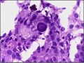
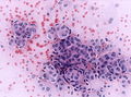
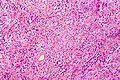
* | *Demonstrated below is a series of microscopic smears which feature specific findings observed in each subtype of meningioma:<ref name="liber">Meningioma. Liberpathology(2015) http://librepathology.org/wiki/index.php/Meningioma#Quick_overview accessed on September, 25th 2015</ref> | ||
<gallery> | <gallery> | ||
Image:Meningeotheliomatous meningeoma 1.jpg| | Image:Meningeotheliomatous meningeoma 1.jpg|Meningothelial meningioma with syncytial appearance and whorl formation | ||
Image:Meningothelial Meningioma showing 2.jpg|Meningothelial meningioma with [[psammoma]] body | |||
Image:Meningothelioal Meningioma 3.jpg| | Image:Meningothelioal Meningioma 3.jpg|Meningothelial meningioma with onion bulb formation | ||
Image:Meningothelial Meningioma 4.jpg| | Image:Meningothelial Meningioma 4.jpg|Meningothelial meningioma on [[HPS stain]] | ||
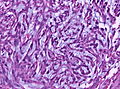
Image:Meningioma fibromatous variant.jpg| | Image:Meningioma fibromatous variant.jpg|[[Fibrous]] meningioma with [[spindle cell]]s in parallel bundles | ||
Image:Meningiom fibrous variant2.JPG| | Image:Meningiom fibrous variant2.JPG|Fibrous meingioma on EMA stain | ||
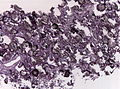
Image:Miningioma (2) transitional type.jpg| | Image:Miningioma (2) transitional type.jpg|Transitional meningioma with coexisting [[lobular]] and fasicular growth patterns | ||
Image:Psammomatous meningioma.jpg| | Image:Psammomatous meningioma.jpg|Psammomatous meningioma with numerous psammoma bodies | ||
Image:Angiomatous meningioma.jpg| | Image:Angiomatous meningioma.jpg|Angiomatous meningioma with hyalinized [[vessel]]s | ||
Image:Microcystic meningeoma.jpg| | Image:Microcystic meningeoma.jpg|Microcystic meningioma with [[cystic]] appearance and increased [[pleomorphism]] of the elongated cells | ||
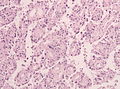
Image:Secretory meningioma.jpg| | Image:Secretory meningioma.jpg|Secretory meningioma with secretory [[granule]]s | ||
Image:Secretory meningioma PAS.jpg| | Image:Secretory meningioma PAS.jpg|Secretory meningioma with [[PAS stain]] positive secretory granules | ||
Image:Chordoid meningoma.jpg| | Image:Chordoid meningoma.jpg|Chordoid meningioma with myxoid appearance | ||
Image:Papillary meningioma.jpg| | Image:Papillary meningioma.jpg|[[Papillary]] meningioma with discohesive [[tumor]] cells located around a fibrovascular core | ||
Image:Rhabdoid meningioma.jpg| | Image:Rhabdoid meningioma.jpg| Rhabdoid meningioma with abundant [[cytoplasm]] and cross-striations | ||
</gallery> | </gallery> | ||
Revision as of 11:59, 25 September 2015
|
Meningioma Microchapters |
|
Diagnosis |
|---|
|
Treatment |
|
Case Studies |
|
Meningioma pathophysiology On the Web |
|
American Roentgen Ray Society Images of Meningioma pathophysiology |
|
Risk calculators and risk factors for Meningioma pathophysiology |
Editor-In-Chief: C. Michael Gibson, M.S., M.D. [1] Associate Editor(s)-in-Chief: Haytham Allaham, M.D. [2]
Overview
Meningioma arises from the arachnoid "cap" cells, which are normally involved in the protection of the central nervous system by forming a thick envelope of meninges around the brain and spinal cord.[1][2] The majority of meningiomas are benign.[1][2] Development of meningioma is the result of multiple genetic mutations.[1] On gross pathology, a gray, well-circumscribed, dome-shaped mass is a characteristic finding of meningioma.[1] On microscopic histopathological analysis, whorled appearance, calcification, and psammoma bodies are characteristic findings of of meningioma.[3]
Pathogenesis
Meningioma arises from the arachnoid "cap" cells, which are normally involved in the protection of the central nervous system by forming a thick envelope of meninges around the brain and spinal cord. The majority of meningiomas are benign; however, a minority of cases may develop a malignant nature. The exact pathophysiology of meningioma depends on the histological subtype of the tumor.[1] Meningiomas are frequently found attached to the dura surrounding the brain tissue in several locations that include:[1]
- Frontal lobe
- Parietal lobe
- Brain convexity
- Sphenoid ridge
- Sella turcica
- Posterior fossa
- Olfactory groove
- Meckel's cave
- Tentorium cerebelli
- Confluence of sinuses
Genetics
- Development of meningioma is the result of multiple genetic mutations.[1]
- The majority of meningioma cases involve a mutation in the neurofibromatosis 2 gene located on chromosome 22.[1]
- Other genes involved in the pathogenesis of meningioma include:[1]
- Protein kinase AKT1 gene located on human chromosome 14
- Meningioma 1 MN1 gene located on human chromosome 22
- Phosphatase and tensin homolog PTEN gene located on both human chromosomes 10 and 19
- SMO gene located on human chromosome 6 and 7
Gross Pathology
- On gross pathology, a gray, well-circumscribed, dome-shaped mass is a characteristic finding of meningioma.[1]
Microscopic Pathology
- On microscopic histopathological analysis, whorled appearance, calcification, and psammoma bodies are characteristic findings of of meningioma.[3]
- The table below differentiates between the three main groups of meningioma according to WHO histological classification:[3][2][4]
| Grade | Histologic features | Image |
|---|---|---|
|
Benign (Grade I) meningioma |
 | |
|
Atypical (Grade II) meningioma |
Brain invasion, 4 or more mitosis/10 HPF, necrosis, increased cell count, high nucleus:cytoplasm ratio, increased nucleoli size, presence of sheeting |
 |
|
Anaplastic (Grade III) meningioma |
20 or more mitoses/10 HPF, histology similar to carcinoma or sarcoma |
 |
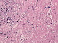
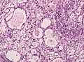
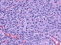
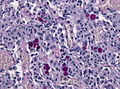
- Demonstrated below is a series of microscopic smears which feature specific findings observed in each subtype of meningioma:[3]
-
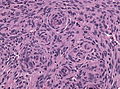
Meningothelial meningioma with syncytial appearance and whorl formation
-
Meningothelial meningioma with psammoma body
-
Meningothelial meningioma with onion bulb formation
-
Meningothelial meningioma on HPS stain
-
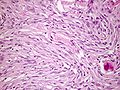
Fibrous meningioma with spindle cells in parallel bundles
-
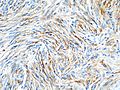
Fibrous meingioma on EMA stain
-
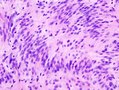
Transitional meningioma with coexisting lobular and fasicular growth patterns
-
Psammomatous meningioma with numerous psammoma bodies
-
Angiomatous meningioma with hyalinized vessels
-
Microcystic meningioma with cystic appearance and increased pleomorphism of the elongated cells
-
Secretory meningioma with secretory granules
-
Secretory meningioma with PAS stain positive secretory granules
-
Chordoid meningioma with myxoid appearance
-
-
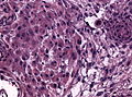
Rhabdoid meningioma with abundant cytoplasm and cross-striations


_transitional_type.jpg)


References
- ↑ 1.0 1.1 1.2 1.3 1.4 1.5 1.6 1.7 1.8 1.9 Meningioma. Wikipedia(2015) https://en.wikipedia.org/wiki/Meningioma#cite_note-pmid7731706-9 Accessed on September, 25th 2015
- ↑ 2.0 2.1 2.2 Meningioma. Canadian Cancer Society http://www.cancer.ca/en/cancer-information/cancer-type/brain-spinal/brain-and-spinal-tumours/meningioma/?region=mb September, 25th 2015
- ↑ 3.0 3.1 3.2 3.3 Meningioma. Liberpathology(2015) http://librepathology.org/wiki/index.php/Meningioma#Quick_overview accessed on September, 25th 2015
- ↑ Meningeoma. Radiopaedia(2015)http://radiopaedia.org/articles/meningioma Accessed on September, 25th 2015