Hip fracture

Template:DiseaseDisorder infobox
|
WikiDoc Resources for Hip fracture |
|
Articles |
|---|
|
Most recent articles on Hip fracture Most cited articles on Hip fracture |
|
Media |
|
Powerpoint slides on Hip fracture |
|
Evidence Based Medicine |
|
Clinical Trials |
|
Ongoing Trials on Hip fracture at Clinical Trials.gov Clinical Trials on Hip fracture at Google
|
|
Guidelines / Policies / Govt |
|
US National Guidelines Clearinghouse on Hip fracture
|
|
Books |
|
News |
|
Commentary |
|
Definitions |
|
Patient Resources / Community |
|
Patient resources on Hip fracture Discussion groups on Hip fracture Patient Handouts on Hip fracture Directions to Hospitals Treating Hip fracture Risk calculators and risk factors for Hip fracture
|
|
Healthcare Provider Resources |
|
Causes & Risk Factors for Hip fracture |
|
Continuing Medical Education (CME) |
|
International |
|
|
|
Business |
|
Experimental / Informatics |
Editor-In-Chief: C. Michael Gibson, M.S., M.D. [1]; Associate Editor(s)-in-Chief: Mohammadmain Rezazadehsaatlou[2].
Overview
The "hip" joint is known as a ball-and-socket joint. It allows the femur bone to bend and rotate at the pelvis. Comparing to the injuries to the knee, ankle, and shoulder which are well documented, injuries to the hip, pelvis, and thigh get little attentions due to their lower prevalence. A hip fracture is a known as a fracture of the upper quarter of the femur bone while any other types of injuries to the socket, or acetabulum, itself is not considered a "hip fracture." Management of fractures to the socket is a completely different consideration. The hip fracture count as a serious problems associated with serious and life-threatening complications. Hip fractures most commonly occur due to the:
- Fall to the side of the hip
- A direct blow to the side of the hip
- Other medical conditions such as osteoporosis, cancer, or stress injuries affecting the strength.
During fracture the most common site of fracture are:
- The head of the femur
- The neck of the femur
- Between or below the greater trochanter and the lesser trochanters
Historical Perspective
There are no reliable information regarding the historical perspective of the hip bone fracture.
Causes
The main etiology of the hip fracture is thought to be a loading may be placed on a leg during falling or from a direct blow to the side of the hip. The main cause of hip fracture is trauma. Such as the most fractures the hip fracture is caused by a falling or automobile accident. Meanwhile, the normal healthy bones are extremely tough and resilient and can withstand most powerful impacts. As a person age, two factors cause higher risk of fractures:
- Weaker bones
- Greater risk of falling
Stress fractures as a common causes of fractures can be found due to the repeated stresses and strains. Importantly children having more physically active lifestyles than adults, are also prone to fractures. People with any underlying diseases such as osteoporosis, infection, or a tumor affecting their bones having a higher risk of fractures. As mentioned in previous chapters, this type of fracture is known as a pathological fracture. Stress fractures, which result from repeated stresses and strains, commonly found among professional sports people, are also common causes of fractures.
Life-threatening Causes
- There are no life-threatening causes of hip fracture , however complications resulting from hip fracture is common.
Common Causes
Common causes of hip fracture may include:
- Trauma (Fall on an outstretched hand)
Less Common Causes
Less common causes of hip fracture include conditions that predisposes to fracture:
Causes by Organ System
| Cardiovascular | No underlying causes |
| Chemical/Poisoning | No underlying causes |
| Dental | No underlying causes |
| Dermatologic | No underlying causes |
| Drug Side Effect | No underlying causes |
| Ear Nose Throat | No underlying causes |
| Endocrine | No underlying causes |
| Environmental | No underlying causes |
| Gastroenterologic | No underlying causes |
| Genetic | No underlying causes |
| Hematologic | No underlying causes |
| Iatrogenic | No underlying causes |
| Infectious Disease | No underlying causes |
| Musculoskeletal/Orthopedic | Osteoporosis and osteopenia. |
| Neurologic | No underlying causes |
| Nutritional/Metabolic | Osteoporosis and osteopenia. |
| Obstetric/Gynecologic | No underlying causes |
| Oncologic | No underlying causes |
| Ophthalmologic | No underlying causes |
| Overdose/Toxicity | No underlying causes |
| Psychiatric | No underlying causes |
| Pulmonary | No underlying causes |
| Renal/Electrolyte | No underlying causes |
| Rheumatology/Immunology/Allergy | No underlying causes |
| Sexual | No underlying causes |
| Trauma | Fall on an outstretched hand. |
| Urologic | No underlying causes |
| Miscellaneous | No underlying causes |
Causes in Alphabetical Order
List the causes of the disease in alphabetical order:
Pathophysiology
Mechanism
The hip fracture is caused by a fall or from a direct blow to the side of the hip. The form and severity of this fracture depends on the position of the hip joint at the moment of hitting the ground. The width of this mentioned angle affects the localization of the fracture. Pronation, supination and abduction positions leads the direction of the force and the compression of carpus and different appearances of injury. Its known that the hip fracture in normal healthy adults can be caused due to the high-energy trauma (e.g., motor vehicle accidents), sport related injuries, falling from height. But it should be noted that the most important Risk factors for insufficiency fractures is chronic metabolic disease such as steoporosis, osteopenia, eating-disordered behavior, higher age, prolonged corticosteroid usage, female gender, lower BMI, history of a recent falling, and prior fracture. Its been said that If the elbow is flexed during the falling, the chance of a type II or III lesion is high.
Pathophysiology
The pattern of bone fracture and severity of injury depends on variety of factors such as:
- Patients age
- Patients Weight
- Patients past medical history specifically any bone diseases affecting the quality of bone (such as osteoporosis, malignancies)
- Energy of trauma
- Bone quality
- Position of the specific organ during the trauma
- The below-mentioned processes cause decreased bone mass density:
- Autophagy is the mechanism through which osteocytes evade oxidative stress.
- The capability of autophagy in cells decreases as they age, a major factor of aging.
- As osteocytes grow, viability of cells decrease thereby decreasing the bone mass density
Differentiating Hip fracture from other Diseases
In the orthopedic medicine its important to know that the Hip fracture should be evaluated using radiography for both confirming diagnosis and also for evaluating the surrounding tissues. Other injuries such as possible femoral fracture-dislocation or collateral ligament injury, might be seen in Hip fracture. If the mechanism of injury suggests particularly low energy then the Osteoporosis should be considered. The pathological Fractures occurring in a bone with a tumor or Paget's disease) are rare but possible[3]. Also it should be noted that the both bone fractures can be complicated by acute compartment syndrome. Signs suggesting compartment syndrome are pain on extension of digits, and marked edema[3]. As another important fact in orthopedic fracture is if both-bone fractures were found in pediatric which is common after accidental trauma, but it may also be the due to the of child abuse; and in these cases a careful attention and evaluation should be considered if a child abuse is suspected.
- Hip Pointer
- Hip Tendonitis and Bursitis
- Acetabular fracture
- Snapping Hip Syndrome
- Slipped Capital Femoral Epiphysis
- Femoral Neck Stress Fracture
- Femoral head fracture
- Femoral Head Avascular Necrosis
- Femoral Neck Fracture
- Femoral Injuries and Fractures
- Femoral Neck Fracture
- Femoral shaft Fracture
- Iliopsoas Tendinitis
- Slipped Capital Femoral Epiphysis
- Pubic rami fracture
- Osteitis Pubis
- Septic hip
Epidemiology and Demographics
In the United States the hip fractures occurred in 280,000 Americans per year at a rate of over 5000 per week and it was estimated that its incidence will rise to over 500,000 annually over the next 40 years. And, this type of fracture cost the US approximately $7.2 billion annually and it is estimated that this cost will rise up to 16 billion USD annually by the year 2041. It should be noted that in the United States, the hip fracture and related mortality among persons older than 65 years of age is declining, while related comorbidities have increased.
Risk Factors
There are different risk factors that presidpose patient for the Hip fracture that include::
- High-risk contact sports
- Higher age (elderly adults are higher prone to such fractures)
- Osteoporosis
- Direct trauma
- Road / traffic accidents
- Falling
- Gunshot wounds
- Domestic violence
- Falling
- Glucocorticoid use
- Anabolic steroid use
- Paget disease of the bone
- low BMI
- Female sex
- Alcohol and tobacco
- Medical problems
- Physical inactivity
Classification
The most common types of the hip fractures :
- Femoral neck fracture: common among older adults and related to osteoporosis. Complication: cuts off the blood supply to the head of the femur which forms the hip joint.
- Intertrochanteric hip fracture: An intertrochanteric hip fracture occurs three to four inches from the hip joint. Complication: Normally does not interrupt the blood supply to the hip joint.
Hip fractures classified into:
Intracapsular fractures (known as femoral neck fractures):
45% of all acute hip fractures in the elderly
Usually due to a low-impact fall from a standing position or from twisting on a planted foot
Femoral neck fractures are susceptible to malunion and avascular necrosis of the femoral head
further Classification based on radiographic findings
- Type 1: undisplaced and incomplete fracture
- Type 2: undisplaced complete fracture
- Type 3: complete fracture but incompletely displaced
- Type 4: complete fracture and completely displaced
Extracapsular fractures
A: Intertrochanteric fracture:
- Between the greater and the lesser trochanter
- Common in the osteoporotic elderly population
- Due to a fall from a standing height with direct contact of the lateral thigh or torsion of the lower extremity.
- avascular necrosis or nonunion is rare.
B: Subtronchanteric fracture:
- Below the lesser trochanter, approximately 2.5 inches below
- 5% due to tumor, bone cysts, or Paget’s disease.
- 50% due to osteoporosis
- almost all of cases have osteopenia
| Area of invovlement | Classification | ||
|---|---|---|---|
| femoral head | Pipkin classification |
| |
| femoral neck | Subcapital | Garden classification |
|
| Transcervical | Pauwel's classification |
| |
| Basicervical | |||
| Trochanteric | Intertrochanteric (between the greater and lesser trochanter) | Ramadier's classification
Ramadier's classification (1956) Boyd and Griffin's classification (1949) Decoulx & Lavarde's classification (1969) Ender's classification (1970) Tronzo's classification (1973) Evans-Jensen classification (1975) Deburge's classification (1976) Briot's classification (1980) |
Evans-Jensen classification (1975)
B: 2-part displaced
|
| Pertrochanteric (through the trochanters) | |||
| Subtrochanteric | Seinsheimer classification |
C: 2-part spiral fracture with lesser trochanter attached to the distal fragment
| |
| American Academy of Orthopedic Surgeons (AAOS) classification |
|
Fractures are classified accordingly:
|
|
| Cooke and Newman (modified Bethea) classification |
|
||
| Johansson classification |
|
||
| Vancouver classification |
|
Screening
Osteoporosis is an important risk factor for human affecting human bone especially in men with the age of older than 50 years old and postmenopausal and women.
Based on the US Preventive Services Task Force (USPSTF) there are three groups of patients need to be screened for the osteoporosis:
- · Men with no history of osteoporosis
- · Women with the age of 65≤ year old, with no previous history of pathological fracture due to the osteoporosis
- · Women with the age of <65 years, with 10-year fracture risk of not less than a 65-year-old white woman (who has not any other risk factor)
Accordingly women older than age of 50 are the main target for the osteoporosis screening. There is no specific recommendation to screen men for the osteoporosis.
The USPSTF recommendations from 2002 included:
Meanwhile, there are two major modalities for the osteoporosis screening:
- · Dual energy x-ray absorptiometry (DXA) of the hip and lumbar spine bones
- · Quantitative ultrasonography of the calcaneus
*It should be noted of the two above mentioned modalities for screening the ultrasonograhy is preferred to the DXA due to its lower cost, lower ionizing radiation, more availability.
After the primary evaluation of the osteoporosis, the further evaluation are required in some cases such as:
· Women with normal bone density or mild osteopenia: T-score of greater than −1.50 – should have screening for 15 years.
· Women with moderate osteopenia: T-score of −1.50 to −1.99 – should have screening for 5 years.
· Women with advanced osteopenia: T-score of −2.00 to −2.49 - should have screening for 1 year.
Natural History, Complications and Prognosis
Natural History
- In cases with untreated hip fractures the malunion and deformity of HIP joint can be occurred.
Complication
The type and frequency of complications of the hip fractures varies. The main complication of the hip fractures is the high risk of re-fracture due to their instability. In general the following are the most common complications of the hip fractures :
- Infection of the bone
- Infection of soft tissue
- Compartment syndrome
- Non-union of the fracture
- Mal-union
- Arm shortening
- Nerve Damage
- Vascular injury
- Vascular bleeding
- Re-fracture
- Decreased Motion
- Painful movement
Prognosis
Generally, the hip fractures carry an approximately 30% risk of mortality at 1 year also around 25% to 75% of adults may not reach their pre-fracture functional level. Most of the hip fractures heal well with no functional or gross problems in the appearance of the injured bone. But, proper immobilized and proper orthopedic follow-up are required due to the higher risk of re-fracture, complete fracture and displacement of the fracture. The followoing mentioned factores affect the patients mortality rate: comorbid disease, low pre-injury cognitive function, abnormal preoperative ECG, age >85 years, and decreased pre-fracture mobility.
History and Symptoms
The diagnosis of a hip fracture is based on: patient history, physical examination, and radiography.
The related signs and symptoms include:
- Skin lacerations
- Weak pulse
- Open fractures
- Bruising
- Swelling
- Stiffness
- Inability to move
- Pain in touch
- Loss of function of the leg
- Difficulties in detection of pulses
- An antalgic gait pattern
In the physical exam the orthopedic surgeon should check the vascular status and amount of swelling in the leg. In MULTI-trauma patients or a tense compartment with neurological signs or stretch pain should be considered as the compartment syndrome, and the compartment pressures should be measured and monitored.
Physical Examination
The related signs and symptoms include:
- Inability to move
- Pain in hip or groin
- Inability to put weight on the involved leg
- Stiffness in and around injured hip area
- bruising in and around injured hip area
- swelling in and around injured hip area
- Shorter leg on the side of injured hip
- Edema around injured hip area
- Most of the time the edema will be a non-pitting edema
- Depends on the edema extent, it may even lead to compartment syndrome in the anterior and internal compartment of leg
- Bruising
- As a manifestation of internal injury to the local vessels by trauma or fractures bone
- Decrease in range of motion
- Movement of the fractures limb will be painful if possible at all
- Tenderness
- Deformity
- Fractured bone deformity may be touchable in the internal side of the leg if the fracture is displaced
Physical examination of patients with hip fractures is usually remarkable for swelling, tenderness, bruises, ecchymosis, deformity and restricted range of motion of the wrist.
Appearance of the Patient
- Patients with hip fractures usually appears normal unless the patients had a high energy trauma causing the open wound fracture.
Vital Signs
- Weak pulse may be seen when associated with polytrauma.
- Low blood pressure with normal pulse pressure may be present due to compound fracture with blood loss.
Skin
- Skin examination of patients with hip fractures includes:
HEENT
- HEENT examination of patients withhip fractures is usually normal.
Neck
- Neck examination of patients with hip fractures is normal.
Lungs
- Pulmonary examination of patients with hip fractures is usually normal.
Heart
- Cardiovascular examination of patients with hip fractures is usually normal.
Abdomen
- Abdominal examination of patients with hip fractures is usually normal.
Back
- Back examination of patients with hip fractures is usually normal.
Genitourinary
- Genitourinary examination of patients with hip fractures is usually normal.
Neuromuscular
- Neuromuscular examination of patients with hip fractures is usually normal.
- However, some patients may develop neuropraxia of the branch of the Ulnar nerve resulting in decreased sensation of fingers.
Laboratory Findings
There is a limited laboratory tests useful in the diagnosis of bone fractures such as hip fractures Meanwhile, aged men and women may have some abnormalities in their laboratory findings suggestive of osteoporosis.
- Laboratory tests for the diagnosis of osteoporosis are:
- Complete blood count (CBC)
- Serum total calcium level
- Serum Ionized calcium level
- Serum phosphate level
- Serum alkaline phosphatase level
- Serum 25-(OH)-vitamin D level
X Ray
The orthopedic surgeon should consider to have at least two radiographic projections (ie, anteroposterior [AP] and lateral) of the hip. These show the fracture, the extent of displacement, and the extent of comminution. The orthopedic surgeon should pay serious attention toward finding any foreign bodies in open fractures and gunshot injuries. Also imperative is to include the elbow and wrist joint in the radiographs of hip fractures to ensure that the distal radioulnar joint injuries are not missed
A tuberosity view is helpfull ascertain the rotational displacement of the fracture. Also, it would be helpful for the orthopedic surgeon in planning how much supination or pronation is needed to achieve accurate anatomic reduction. The ulna is laid flat on the cassette with its subcutaneous border in contact with the cassette; the x-ray tube is tilted toward the olecranon by 20°. This radiograph is then compared with a standard set of diagrams that show the prominence of the radial tuberosity in various degrees of pronation and supination in order to determine the scope of the rotational deformity
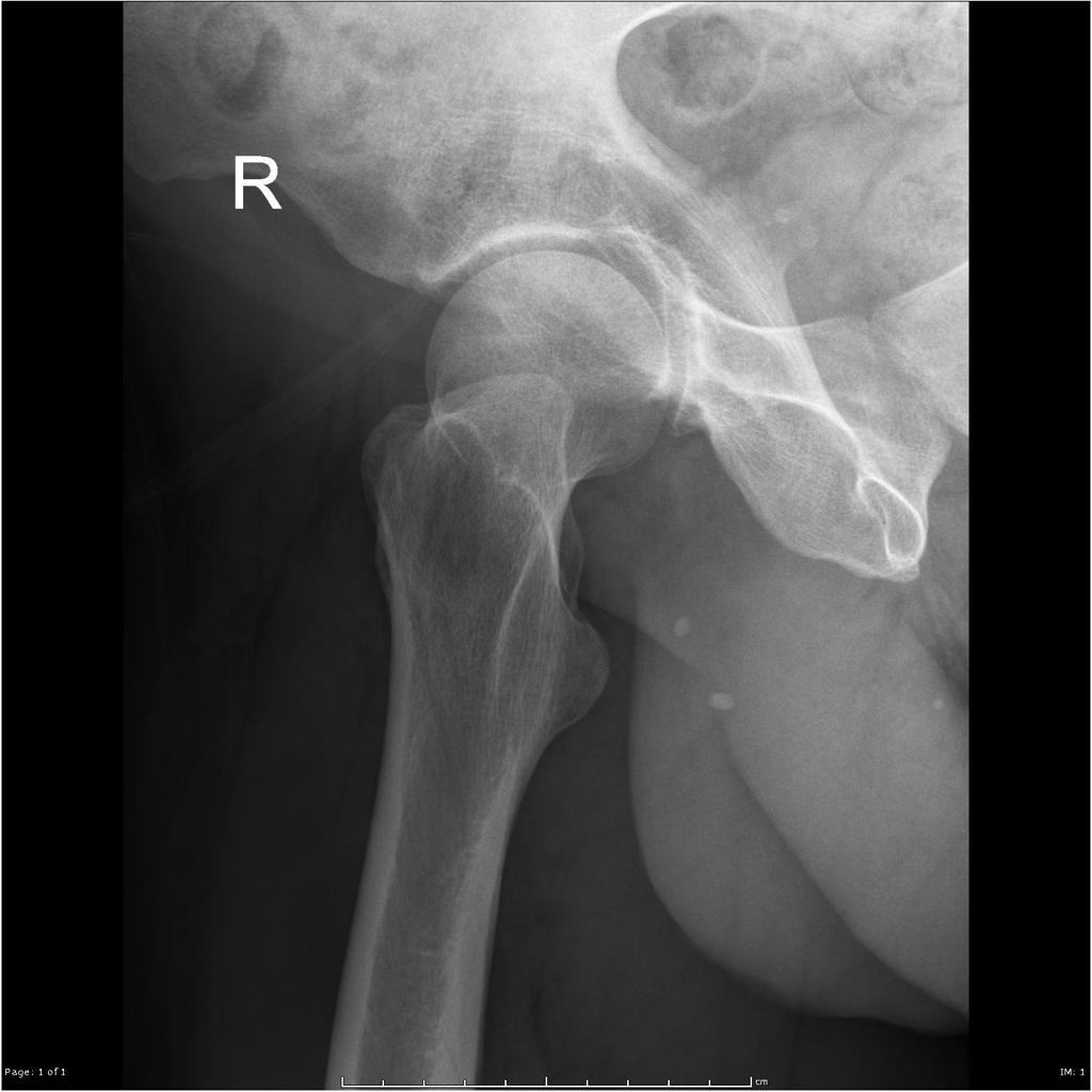
-
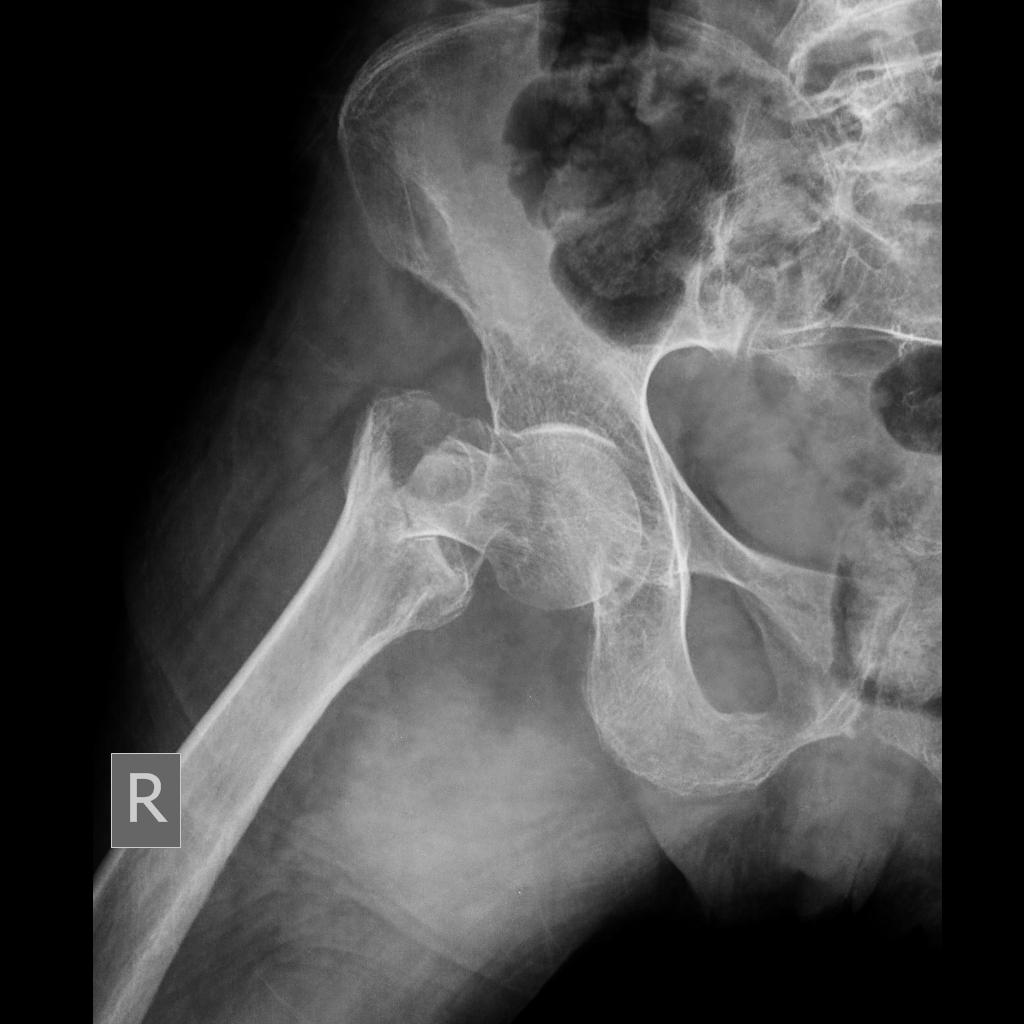
Fracture at the base of the right femoral neck.
-
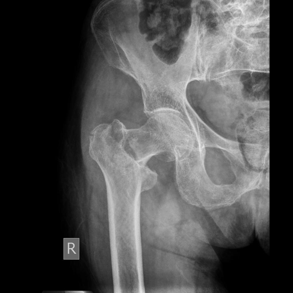
Fracture at the base of the right femoral neck.
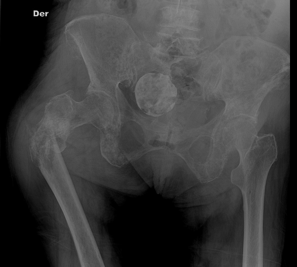
-
Fracture-dislocation of hip.
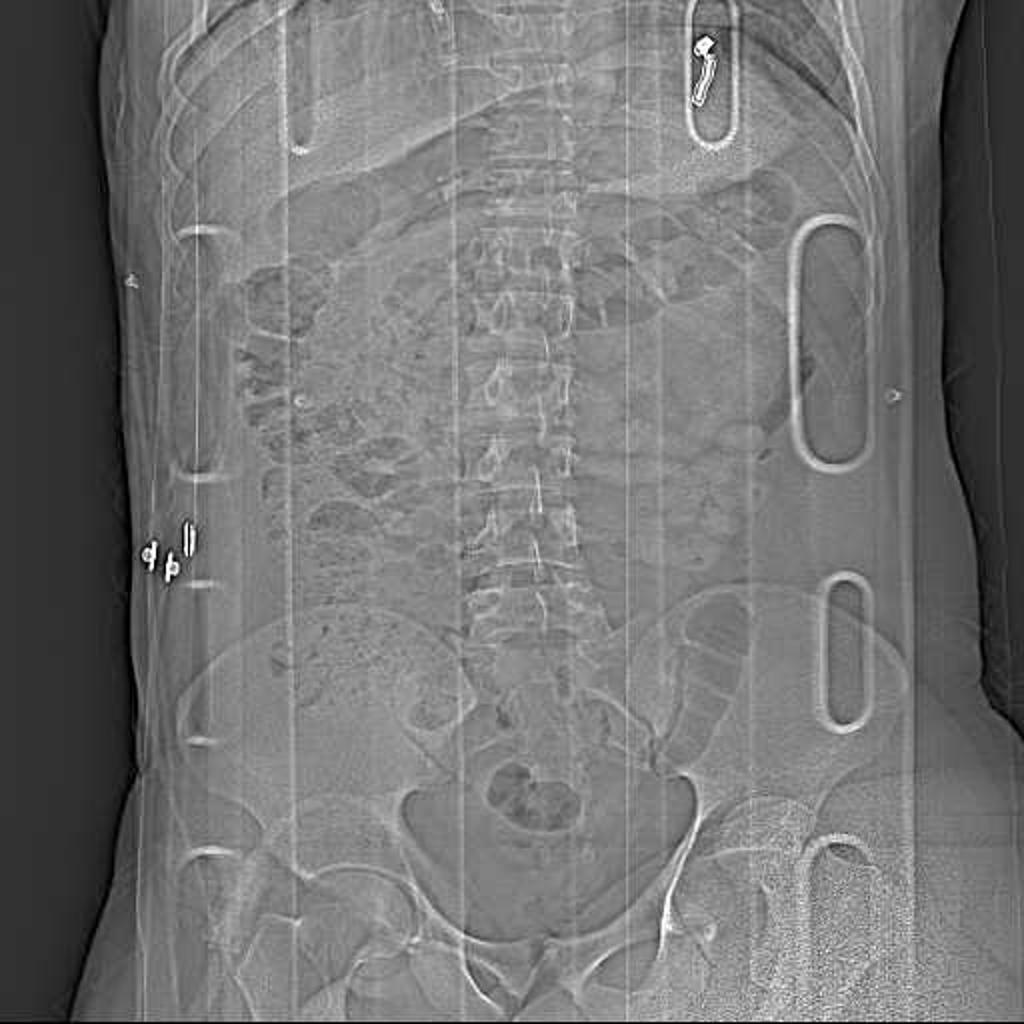
-
Pathologic fracture of the left proximal femur. Note the extensive bone metastases. A rounded calcified mass in the right side of the pelvis is consistent with an old calcified uterine fibroid.
-
No abnormality detected! Right hip pain.
-
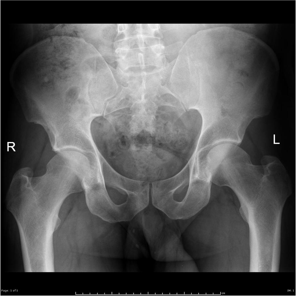
No abnormality detected! Right hip pain.

.JPEG)


.jpg)

CT
- CT-scan in the case of Hip fracture is the best modality if you can not have an exclusive diagnosis by X-ray itself can not be made.
- Its been reported that the articular fractures of the distal radius were statistically more likely to occur at the intervals between the ligament attachments than at the ligament attachments.
- The most common fracture sites were the center of the sigmoid notch, between the short and long radiolunate ligaments, and the central and ulnar aspects of the scaphoid fossa dorsally. These results suggest that CT may be used to identify the subsequent propagation of the fracture and the likely site of the impaction of the carpus on the distal radius articular surface.
-
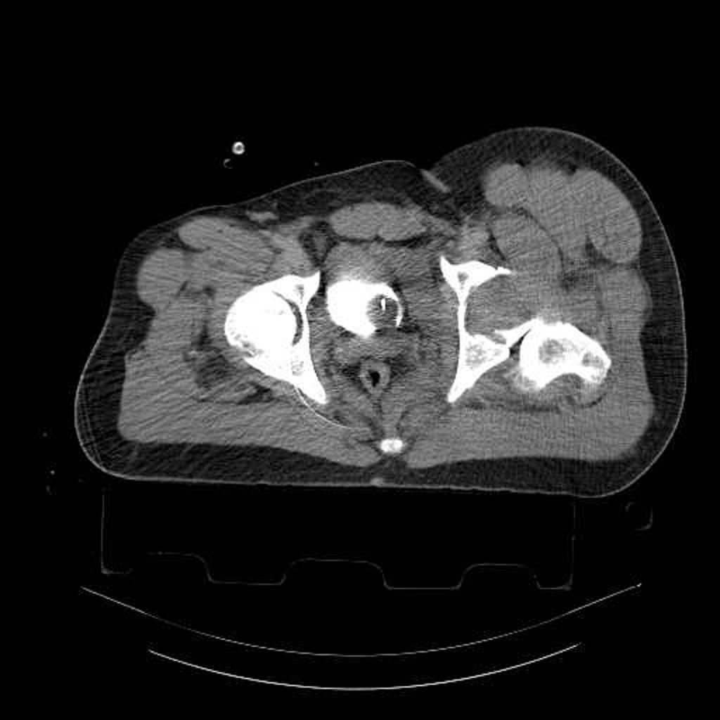
Fracture-dislocation of hip. CT scan shows posterior dislocation of femoral head associated with fracture of both anterior and posterior columns of acetabulum.
.jpg)
MRI
Magnetic resonance imaging (MRI) is an expensive technique that should not be used routinely. MRI is a powerful diagnostic tool to assess the abnormalities of the bone, ligaments and soft tissues associated with the Hip fractures, but it is known as a limited utility in radioulnar injuries and is not indicated in uncomplicated hip fractures. Meanwhile, the MRI can be useful in in following mentioned evaluations: Evaluation of occult fractures Evaluation of the post-traumatic or avascular necrosis of bones Evaluation of tendons Evaluation of nerve Evaluation of carpal tunnel syndrome
-
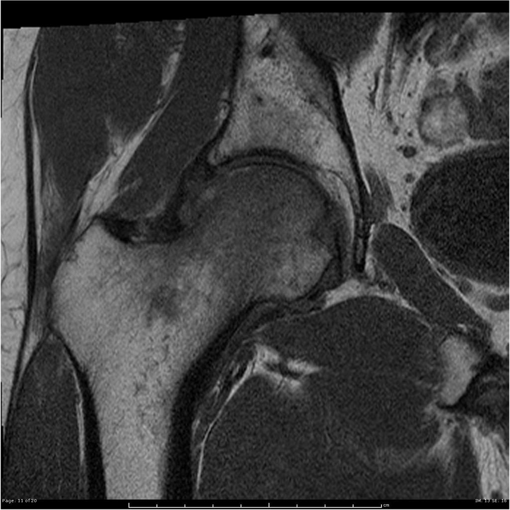
Coronal T1 Extensive marrow edema within the femoral head and neck. There is a subchondral fracture within the femoral head.
-
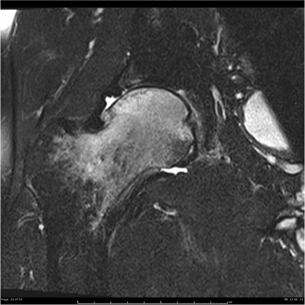
Coronal T2 fat sat Extensive marrow edema within the femoral head and neck. There is a subchondral fracture within the femoral head.
-
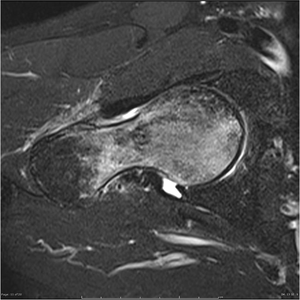
Coronal Axial T2 fat sat Extensive marrow edema within the femoral head and neck. There is a subchondral fracture within the femoral head.
-
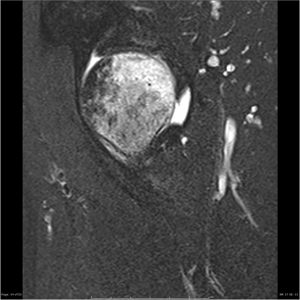
Coronal Sagittal T2 fat sat Extensive marrow edema within the femoral head and neck. There is a subchondral fracture within the femoral head.
.jpg)
.jpg)
.jpg)
.jpg)
Other Imaging Findings
There are no other imaging findings associated with Hip fracture.
Other Diagnostic Studies
There are no other imaging findings associated with Hip fracture.
Treatment
The first step in managing a patient with a fracture is to stabilize the patient if he/she is unstable due to blood loss, etc by giving them intravenous fluids and giving them some painkillers if the pain is severe. If only one bone is broken, using cast or brace might be a possible treatment option.
Non-surgical therapy
- The first step in managing a patient with a fracture is to stabilize the patient if he/she is unstable due to blood loss, etc by giving them intravenous fluids and giving them some painkillers if the pain is severe.
- In children, the usual plan is to attempt closed reduction followed by cast immobilization. In adults, treatment with immobilization in a molded long arm cast can be used in those rare occasions of a non-displaced fracture of the leg bone. If the fracture shifts in position, it may require surgery to put the bones back together.
- Rigid immobilization is suggested in preference to removable splints in nonoperative treatment for the management of Hip bone fractures.
- For all patients with Hip fractures, a post-reduction true lateral radiograph is suggested .
- Patients probably do not need to begin early leg motion routinely after stable fracture fixation.
- Adjuvant treatment of Hip fractures with vitamin C is suggested for the prevention of disproportionate pain.
- If operative treatment is refused or the risks of surgery are considered to be too high the main emphasis of treatment is on pain relief. Skeletal traction may be considered for long term treatment.
Complications of Non-surgical therapy
Failure of non-surgical therapy is common:
- Re-displacement to its original position
- Stiffness
- Post traumatic osteoarthritis leading to wrist pain and loss of function
- Other risks specific to cast treatment include:
- Compression of the swollen leg causing compartment syndrome
- Reflex sympathetic dystrophy is a serious complication
- Stiffness is universal following a prolonged period of immobilization and swelling
- DVT/pulmonary embolism
- Pressure sores and
Surgery
- There are a variety of methods and implants useful to stabilize the Monteggia fracture, ranging from closed reduction and percutaneous pin fixation to the use of intra-medullary devices.
- However, the most common fixation methods to treat complex Greenstick fracture include external fixation, and open reduction and internal fixation.
External Fixation With or Without Percutaneous Pin Fixation
- Wrist spanning external fixation employs ligamentotaxis to restore and maintain length, alignment, and rotation of ulnar bone.
- Reduction is typically obtained through closed or minimally open methods and preserves the fracture biology.
- The addition of percutaneous pins enhances the ability to reduce and stabilize fracture fragments.
Complications of External Fixation
- Pin tract infection
- Injury to the superficial branch of the nerve
- Complex regional pain syndrome
Open reduction and internal fixation with plates and screws
- This is the most common type of surgical repair for Monteggia fracture
- During this type of procedure, the bone fragments are first repositioned (reduced) into their normal alignment.
- The bones held together with special screws and metal plates attached to the outer surface of the bone.
Complications of open reduction and internal fixation with plates and screws
- Infection
- Damage to nerves and blood vessels
- Synostosis
- Nonunion
Pain Management
Pain after an injury or surgery is a natural part of the healing process.
Medications are often prescribed for short-term pain relief after surgery or an injurysuch as:
- opioids
- non-steroidal anti-inflammatory drugs (NSAIDs)
- local anesthetics
Be aware that although opioids help relieve pain after surgery or an injury, they are a narcotic and can be addictive. It is important to use opioids only as directed by doctor.
Interventions
The following options can be helpful for patients to rehabilitate after their fracture :
- Joints mobilization
- compression bandage
- Soft tissue massage
- Exercises and Activity modification
- Forearm taping
- Forearm bracing
Postoperative Rehabilitation
- Complex Monteggia fracture warrant individualized immobilization and rehabilitation strategies.
- Similarly, the addition of a thumb spica cast or orthosis with positioning of the wrist in slight ulnar deviation for management of a comminuted radial column fracture may prevent loss of reduction.
- Because some Monteggia fractures are the result of high-energy injuries, a prolonged period of wrist immobilization and soft-tissue rest may be beneficial and has not been shown to affect clinical outcomes.
- The wrist is typically immobilized for 2 weeks post-operatively in a sugar tong splint with neutral forearm rotation.
- At 6 weeks post-operatively, the wrist is placed into a removable orthosis, and active and passive range of motion (ROM) is initiated.
- Full weight bearing commences at approximately 3 months post-operatively after consolidation of the fracture is noted on radiographs.
- The presence of varying degrees of hand, wrist, and elbow stiffness is inevitable and may result from poor pain control, lack of effort in controlled mobilization, edema, concomitant ipsilateral upper extremity fractures, or peripheral nerve injuries.
- Early stretching and mobilization of the intrinsic and extrinsic tendons of the hand is important to prevent finger stiffness.
- Edema control can be initiated with compression gloves, digital massage, and active and passive ROM of the hand.
- A home exercise program or outpatient occupational therapy is started immediately post-operatively to maintain full range of motion of the hand and limit the development of intrinsic muscle tightness
Operation
The most common surgical techniques include:
Open reduction internal fixation (ORIF)
External fixation
Percutaneous pinning
Combination of the above-mentioned techniques
The proper operative choice is often depends on the type of fracture and it can be categorized into three groups:
partial articular fractures
displaced articular fractures
metaphyseal unstable extra- or metaphyseal minimal articular involvement.
When both bones of the forearm (radius and ulna) are fractured, they are both exposed and provisionally reduced before fixation of either bone is completed. The fracture with the least comminution (usually the ulna) is fixed first. After reduction and provisional fixation of both bones, pronation and supination are examined; if normal, definitive fixation is performed. The general rule is that bone grafting is recommended when more than one third of the circumference of the bone is comminuted.
The common therapeutic options in orthopedic medicine for ulnar fractures are:
- Open reduction and internal fixation with plates and screws
Known as the most common type of surgical repair for forearm fractures.
- Open reduction and internal fixation with rods
- External fixation
The indications for intramedullary nailing are :
- Segmental fractures
- Poor skin condition
- Selected nonunions or failed compression platings
- Multiple injuries
- Diaphyseal fractures in osteopenic patients
Postoperative Rehabilitation
- Complex Galeazzi fracture-dislocation warrant individualized immobilization and rehabilitation strategies.
- Similarly, the addition of a thumb spica cast or orthosis with positioning of the wrist in slight ulnar deviation for management of a comminuted radial column fracture may prevent loss of reduction. *Because most multifragmentary Galeazzi fracture-dislocation are the result of high-energy injuries, a prolonged period of wrist immobilization and soft-tissue rest may be beneficial and has not been shown to affect clinical outcomes.
- The wrist is typically immobilized for 2 weeks post-operatively in a sugar tong splint with neutral forearm rotation.
- At 6 weeks post-operatively, the wrist is placed into a removable orthosis, and active and passive range of motion (ROM) is initiated.
- Full weight bearing commences at approximately 3 months post-operatively after consolidation of the fracture is noted on radiographs.
- The presence of varying degrees of hand, wrist, and elbow stiffness is inevitable and may result from poor pain control, lack of effort in controlled mobilization, edema, concomitant ipsilateral upper extremity fractures, or peripheral nerve injuries.
- Early stretching and mobilization of the intrinsic and extrinsic tendons of the hand is important to prevent finger stiffness.
- Edema control can be initiated with compression gloves, digital massage, and active and passive ROM of the hand.
- A home exercise program or outpatient occupational therapy is started immediately post-operatively to maintain full range of motion of the hand and limit the development of intrinsic muscle tightness
Primary Prevention
There are various preventive options to reduce the incidence of the Galeazzi fracture-dislocation
- Using forearm and wrist guards during practicing sports (skating, biking)
- Using forearm and wrist guards during driving motorbikes
- Avoid falls in elderly individuals
- Prevention and/or treatment of osteoporosis
- Healthy diet
Secondary Prevention
It should be noted that the Post-menopausal women specially older than the age of 65 are at the higher risk of osteoporosis consequently these type of patients at greater risk for the pathological fractures .
So the Calcium and vitamin D supplementation play important role in increasing the bone mineral density (BMD) consequently decrease the risk of fracture in these type of patients. Also, avoiding excessive alcohol and quitting smoking play important role in this regard.
Detecting osteoporosis
- DEXA(dual-energy x-ray absorptiometry) scan
- Serum calcium and vitamin D levels
- Ultrasonography of the calcaneus
Pharmacological therapy
- The primary goal for the treatment of osteoporosis is to reduce longtime fracture risk in patients. Increasing bone mineral density (BMD) in response to the treatment is far less important than improvement of clinical aspects of osteoporosis, i.e., osteoporoticfracture. Therefore, most of the drugs efficacy is measured by the extent they improve the fracture risk instead of increasing BMD.
- During the treatment, if a single fracture happens, it does not necessarily indicate treatment failure or the need to be started on an alternative treatment or patient referral to a specialist.
- Calcium and vitamin D supplementation have been found to be effective in reducing the long term fracture risk, significantly. In order to suggest the people to use vitamin D and calcium supplements, the physician needs to make sure that patient is not able to obtain the nutrients through the daily intake. The available supplemental ions of calcium include calcium carbonate, calcium citrate, and vitamin D3 in various dosage forms.
Life style modifications [1]
- Exercise: Exercise promotes the mineralization of bone and bone accumulation particularly during growth. High impact exercise, in particular, has been shown to prevent the development of osteoporosis. However, it can have a negative effect on bone mineralization in cases of poor nutrition, such as anorexia nervosa and celiac disease.
- Nutrition: A diet high in calcium and vitamin D prevents bone loss. Patients at risk for osteoporosis, such as persons with chronic steroid use are generally treated with vitamin D and calcium supplementation. In renal disease, more active forms of vitamin D, such as 1,25-dihydroxycholecalciferol or calcitriol are used; as the kidney cannot adequately generate calcitriol from calcidiol (25-hydroxycholecalciferol), which is the storage form of vitamin D.
- By quitting smoking, osteoporosis as well as other diseases can be prevented.
- Avoiding excessive alcohol intake or drinking only in moderation (1–2 alcoholic beverages/day).
- Taking least possible dosages of certain medications that are associated with osteoporosis (anticonvulsants or corticosteroids).
External links
- [http://www.wheelessonline.com/ortho/monteggias_fracture
- [https://orthoinfo.aaos.org/en/diseases--conditions/forearm-fractures-in-children/
- [https://orthoinfo.aaos.org/en/diseases--conditions/elbow-fractures-in-children/
Related Chapters
References
Overview
A hip fracture is a fracture in the proximal end of the femur (the long bone running through the thigh), near the hip joint.
The term "hip fracture" is commonly used to refer to four different fracture patterns and is often due to osteoporosis; in the vast majority of cases, a hip fracture is a fragility fracture due to a fall or minor trauma in someone with weakened osteoporotic bone. Most hip fractures in people with normal bone are the result of high-energy trauma such as car accidents.
The mortality following a hip fracture is between 20% and 35% within one year in patients aged 82 ± 7 years old, of which 80% were women.
Types
Many subtypes of fractures about the hip joint are colloquially known as 'hip fractures'. Although a true hip fracture involves the joint, the following four proximal femur fractures are commonly referred to as 'hip fractures'. The differences between them are important because each is treated differently.
- Femoral head fracture denotes a fracture involving the femoral head. This is usually the result of high energy trauma and a dislocation of the hip joint often accompanies this fracture.
- Femoral neck fracture (sometimes Neck of Femur (NOF), subcapital, or intracapsular fracture) denotes a fracture adjacent to the femoral head in the neck between the head and the greater trochanter. These fractures have a propensity to damage the blood supply to the femoral head, potentially causing avascular necrosis.
- Intertrochanteric fracture denotes a break in which the fracture line is between the greater and lesser trochanter on the intertrochanteric line. It is the most common type of 'hip fracture' and prognosis for bony healing is generally good if the patient is otherwise healthy.
- Subtrochanteric fracture actually involves the shaft of the femur immediately below the lesser trochanter and may extend down the shaft of the femur.
Incidence
Approximately 320,000 hospitalizations occur each year due to hip fractures in the US.
Pathogenesis/risk factors
Most hip fractures occur as a result of low-energy falls in elderly patients. Falls are uncommon in young adults due to better balance and strength and when they do occur, they usually do not cause the "hip fracture" pattern of injury that is commonly seen in the elderly. It was formerly thought, but Harvard medical scientists disproved, that benzodiazepine use increased the risk. A person with normal hips will not fracture following a fall from standing. Hip fracture following a fall is likely to be a pathological fracture. The most common causes of weakness in bone are:
- Osteoporosis. Hip fractures are one of the most serious complications of osteoporosis; in fact a measure of success or failure of treatment of osteoporosis is the proportion of patients who sustain a hip fracture. There was a 43% reduction in hip fractures by vitamin D and calcium supplementation, and vitamin D defiency being a common problem [[2]].
- Homocysteine, a toxic 'natural' amino acid linked to the cause of heart disease, stroke and bone fractures, reduced by B-vitamins in this study [3], it reduced the amount of hip fractures by 80% after 2 years. This was despite no differences in bone density and in the number of falls between the vitamin and the placebo groups.
- Other metabolic bone diseases such as Paget's disease, osteomalacia, osteopetrosis and osteogenesis imperfecta. Stress fractures may occur in the hip region with metabolic bone disease.
- Benign or malignant primary bone tumours are rare causes of hip fractures.
- Metastatic cancer deposits in the proximal femur may weaken the bone and cause a pathological hip fracture.
- Infection in the bone is a rare cause of hip fracture.
Another element in the risk of sustaining a hip fracture is the risk of falling. Fall prevention is an area if interest with concerns in the area of providing a safe environment for people at risk, custodial care, walking aids, medication issues etc. Hip protectors are padded plastic shields that can be placed over the trochanters of people at risk of falling or of sustaining a fragility fracture. However, they are not effective in reducing the likelihood of a hip fracture and compliance is poor.
Natural history
Hip fractures are very dangerous episodes especially for elderly and frail patients. The risk of dying from the stress of the surgery and the injury in the first few days is about 10%. If the condition is untreated the pain and immobility imposed on the patient increase that risk. Problems such as pressure sores and chest infections are all increased by immobility. The prognosis of untreated hip fractures is very poor.
Clinical features
The classic clinical presentation of a hip fracture is an elderly patient who sustained a low-energy fall and now has pain and is unable to bear weight. On examination, the affected extremity is often shortened and externally rotated.
Investigations
X-rays of the affected hip usually make the diagnosis obvious; AP and lateral views should be obtained.
In situations where a hip fracture is suspected but is not obvious on x-ray, a CT scan with 3D reconstruction may be helpful. MRI has gained importance in the diagnosis of occult fractures of the femoral neck. Within 24 hours changes can be seen on MRI. Bone scan is less useful because it may take up to 1 week to demonstrate changes especially in the elderly.
As the patients most often require an operation, full pre-operative general investigation is required. This would normally include blood tests, ECG and chest x-ray.
Femoral neck fracture
Femoral neck fractures involve the narrow neck between the round head of the femur and the shaft. This fracture often disrupts the blood supply to the head of the femur.
Garden classified this fracture into four types:
- Type 1 is a stable fracture with impaction in valgus.
- Type 2 is complete but non-displaced.
- Type 3 is displaced (often rotated and angulated) with varus displacement but still has some contact between the two fragments.
- Type 4 is completely displaced and there is no contact between the fracture fragments.
The blood supply of the femoral head is much more likely to be disrupted in Garden types 3 or 4 fractures.
Surgeons may treat these types of fracture by replacing the fractured bone with a prosthesis arthroplasty. Alternatively the treatment is to reduce the fracture (manipulate the fragments back into a good position) and fix them in place with three metal screws.
A serious but common complication of a fractured femoral neck is avascular necrosis. The vasculature to the femoral head is easily disturbed during fractures or from swelling inside the joint capsule. This can lead to strangulation of the blood supply to the femoral head and death of the bone and cartilage.
Intertrochanteric fracture
Intertrochanteric fractures occur between the greater and lesser trochanters. They are usually fixed with a sliding hip screw and plate. Healing is usually good when the patient is healthy.
See also
References
External links
- Orthopedics.com article on hip fractures
- American Academy of Orthopedic Surgeons article on hip fractures
- Fractures of the Femoral Neck Wheeless Textbook of Orthopaedics
- Intertrochanteric Fractures Wheeless' Textbook of Orthopaedics
- Proximal femoral fracture Musculoskeletal Radiology of Fractures
- Hip fractures