Endocarditis natural history, complications and prognosis: Difference between revisions
No edit summary |
|||
| Line 30: | Line 30: | ||
The [[electrocardiogram]] shows sinus rhythm and a [[QRS]] with a left axis deviation, a [[QRS]] duration of 118 milliseconds and a tall are wave in the first precordial lead V1 with an [[R wave]] height of approximately 21 mm. The prolonged [[QRS duration]] and the [[S waves]] that are seen as lead one and lead the six suggest a right on the Branch block and the a left axis deviation suggests a left anterior hemi-block . Finally the tall R wave in V1 lead suggests right to ventricular hypertrophic | The [[electrocardiogram]] shows sinus rhythm and a [[QRS]] with a left axis deviation, a [[QRS]] duration of 118 milliseconds and a tall are wave in the first precordial lead V1 with an [[R wave]] height of approximately 21 mm. The prolonged [[QRS duration]] and the [[S waves]] that are seen as lead one and lead the six suggest a right on the Branch block and the a left axis deviation suggests a left anterior hemi-block . Finally the tall R wave in V1 lead suggests right to ventricular hypertrophic | ||
[[Image:Endocarditis complications.jpg|center|800px]] | [[Image:Endocarditis complications.jpg|center|800px]] | ||
==Cutaneous== | ==Cutaneous== | ||
# [[Petechiae]] of the [[conjunctiva]], [[oropharynx]], [[skin]], and legs | # [[Petechiae]] of the [[conjunctiva]], [[oropharynx]], [[skin]], and legs | ||
Revision as of 14:01, 15 October 2012
|
Endocarditis Microchapters |
|
Diagnosis |
|---|
|
Treatment |
|
2014 AHA/ACC Guideline for the Management of Patients With Valvular Heart Disease |
|
Case Studies |
|
Endocarditis natural history, complications and prognosis On the Web |
|
FDA on Endocarditis natural history, complications and prognosis |
|
CDC onEndocarditis natural history, complications and prognosis |
|
Endocarditis natural history, complications and prognosis in the news |
|
Blogs on Endocarditis natural history, complications and prognosis |
|
to Hospitals Treating Endocarditis natural history, complications and prognosis |
|
Risk calculators and risk factors for Endocarditis natural history, complications and prognosis |
Editor-In-Chief: C. Michael Gibson, M.S., M.D. [1]
Overview
Complications of endocarditis can occur as a result of the locally destructive effects of the infection. These complications include perforation of valve leaflets causing congestive heart failure, abscesses, disruption of the heart's conduction system, and embolization to the brain (causing a stroke), to the coronary artery (causing a heart attack), to the lung (causing pulmonary embolism), to the spleen (causing a splenic infarct) and to the kidney (causing a renal infarct).
Monitoring for Complications of Infectious Endocarditis
Among those patients at high risk, careful monitoring should be undertaken to detect the early development of complications such as:
- Valvular dysfunction, usually insufficiency of the mitral or aortic valves;
- Myocardial or septal abscesses
- Congestive heart failure
- Metastatic infection
- Embolic phenomenon
A complete list of complications of infective endocarditis include the following:
Cardiac
- Murmur
- A new aortic diastolic murmur suggests dilatation of the aortic annulus or eversion, rupture, or fenestration of an aortic leaflet
- The sudden onset of a loud mitral pansystolic murmur suggests rupture of chorda tendineae or fenestration of a mitral valve leaflet
- Congestive heart failure
- Cardiac rhythm disturbances including AV block
- Occasionally, pericarditis
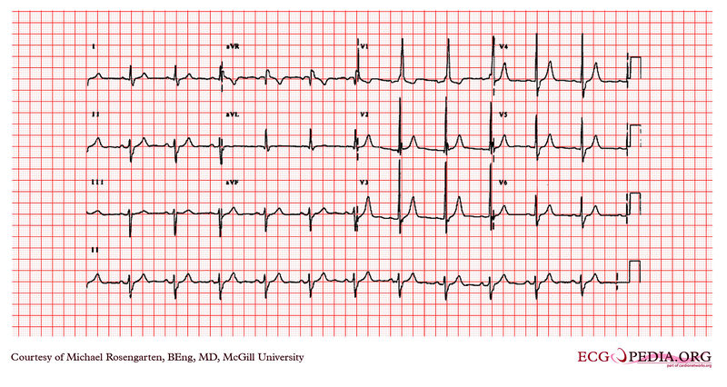
This is an electrocardiogram from a man in his 80's. The patient has severe lung disease, has mitral regurgitation secondary to bacterial endocarditis , and is taking digoxin, Lasix and potassium.
The electrocardiogram shows sinus rhythm and a QRS with a left axis deviation, a QRS duration of 118 milliseconds and a tall are wave in the first precordial lead V1 with an R wave height of approximately 21 mm. The prolonged QRS duration and the S waves that are seen as lead one and lead the six suggest a right on the Branch block and the a left axis deviation suggests a left anterior hemi-block . Finally the tall R wave in V1 lead suggests right to ventricular hypertrophic

Cutaneous
- Petechiae of the conjunctiva, oropharynx, skin, and legs
- Linear subungual splinter haemorrhages of the lower or middle nail bed
- Oslers nodes
- Janeway lesions
Musculoskeletal
- Myalgias
- Arthralgias
- Arthritis
- Low back pain
- Rheumatoid factor is elevated in up to 50% of patients with endocarditis for > 6 wk
- Clubbing of fingers is present in < 15% of patients
Ocular
- Petechial hemorrhages
- Flame-shaped hemorrhages
- Roth's spots,
- Cotton-wool exudates in the retina
Embolic
- Significant arterial emboli occur in 30%–50% of patients, causing the following:
- Stroke
- Monocular blindness
- Acute abdominal pain, ileus, and melena
- Pain and gangrene in the extremities
- CNS emboli are common
- Coronary emboli, often asymptomatic, can cause myocardial infarction
- Pulmonary emboli common in right-sided endocarditis, causing pulmonary infarcts or focal pneumonitis
Splenic
- Splenomegaly is observed in 15%–30% of patients
- Splenic infarcts occur in up to 40% of patients
- Splenic abscesses occur in ~ 5% of patients
Renal
- Microscopic hematuria occurs in ~ 50% of patients
- Embolic renal infarction
- Diffuse membranoproliferative glomerulonephritis
Mycotic aneurysms
Occur in any artery in 2%–8% of patients, causing the following:
- Pain or headache
- Pulsatile mass
- Fever
- Sudden expanding hematoma
- Signs of major blood loss
Neurologic
- Neurologic complications occur in 25%–40% of cases
- Strokes caused by cerebral embolisms in ~ 15% of cases, causing the following:
- Altered level of consciousness
- Seizures
- Fluctuating focal neurologic signs
- Cerebral aneurysms occur in 1%–5% of cases, causing the following:
- Headache
- Focal signs
- Acute intracerebral or subarachnoid hemorrhage caused by rupture
- Mild meningeal irritation resulting from slow leakage
- Brain abscesses may occur in acute endocarditis caused by Staphylococcus aureus
- Seizures